www.jped.com.br
ORIGINAL ARTICLE
Molecular characterization of rotavirus genotypes in
immunosuppressed and non-immunosuppressed
pediatric patients
夽
Luciane A. Pereira
a, Carla E.O. Ferreira
a, Giovana D. Turchetto
b,
Meri B. Nogueira
b, Luine R. Vidal
b, Cristina R. Cruz
c,
Maria C. Debur
d, Sergio M. de Almeida
e, Sonia M. Raboni
f,∗aBiologist, Laboratório de Virologia, Universidade Federal do Paraná (UFPR), Curitiba, PR, Brazil bBiochemist, Laboratório de Virologia, UFPR, Curitiba, PR, Brazil
cAssociate Professor, Departamento de Pediatria, UFPR, Curitiba, PR, Brazil
dBiochemist, Laboratório de Saúde Pública do Estado, Secretaria Estadual de Saúde do Paraná, Curitiba, PR, Brazil eMD, Laboratório de Virologia, UFPR, Curitiba, PR, Brazil
fAssociate Professor, Laboratório de Virologia e Departamento de Doenc¸as Infecciosas, UFPR, Curitiba, PR, Brazil
Received 24 July 2012; accepted 19 November 2012 Available online 26 April 2013
KEYWORDS
Gastroenteritis; Genotypes;
Pediatric infections; Rotavirus
Abstract
Objective: To describe the genotypic variability of group A rotavirus (RVA) found in immuno-suppressed and non-immunoimmuno-suppressed pediatric patients treated at the Hospital de Clínicas da Universidade Federal do Paraná (HC-UFPR), Curitiba, Paraná.
Methods: A cross-sectional study was conducted with 1,140 stool samples collected from April, 2001 to December, 2008 in outpatients and hospitalized patients with acute gastroenteri-tis referred to the hospital. RVA diagnosis was performed through the latex agglutination method and enzyme immunoassay. Reverse transcription followed by multiplex hemi-nested polymerase chain reaction (PCR) and nucleotide sequencing were used for genotype charac-terization. Genotype combinations, clinical, epidemiological, laboratory data, and presence of hospital-acquired infections were reported.
Results: A total of 80 rotavirus-positive stool samples were analyzed. The most frequent associations between genotypes G and P were: G4 P[8] (38.9%), G1 P[8] (30.5%), G9 P[8] (13.9%), G2 P[4] (6.9%), and G3 P[8] (1.4%). G2 P[4] was the most prevalent genotype after
夽 Please cite this article as: Pereira LA, Ferreira CE, Turchetto GD, Nogueira MB, Vidal LR, Cruz CR, et al. Molecular characterization of rotavirus genotypes in immunosuppressed and non-immunosuppressed pediatric patients. J Pediatr (Rio J). 2013;89:278---85.
∗Corresponding author.
E-mail:sraboni@ufpr.br (S.M. Raboni).
the vaccine implementation in the years 2006 and 2008. A total of 62,5% of infected children were aged less than 12 months. Of these, 55.6% had severe dehydration and 26.7% needed intensive care. A frequency of 12.5% of nosocomial infections was found. No correlation was observed between genotype and severity of infection in the study patients.
Conclusion: RVA infections can be associated with severe clinical manifestations, and the surveillance of genotypic variability of this virus is crucial to monitor the emergence of new strains and the impact of the immunization in these patients.
© 2013 Sociedade Brasileira de Pediatria. Published by Elsevier Editora Ltda. All rights reserved.
PALAVRAS-CHAVE
Gastroenterite; Genótipos;
Infecc¸ões pediátricas; Rotavírus
Caracterizac¸ão molecular dos genótipos do rotavírus em pacientes pediátricos imunossuprimidos e não imunossuprimidos
Resumo
Objetivo: Descrever a variabilidade genotípica do rotavírus grupo A (RVA) encontrado em pacientes pediátricos imunocompetentes e imunocomprometidos tratados no Hospital de Clíni-cas/Universidade Federal do Paraná (HC/UFPR), Curitiba, Paraná.
Métodos: Foi realizado um estudo transversal com 1.140 amostras de fezes coletadas, de abril de 2001 a dezembro de 2008, em pacientes ambulatoriais e pacientes hospitalizados com gastroenterite aguda encaminhados ao hospital. As técnicas usadas foram o método da aglutinac¸ão do látex e imunoensaio enzimático para diagnóstico de RVA. Foi realizada transcric¸ão reversa, seguida por PCR multiplexsemi-nestede sequência de nucleotídeos para caracterizac¸ão do genótipo. Foram relatadas as combinac¸ões genotípicas, dados clínicos, epi-demiológicos,laboratoriais e a presenc¸a de infecc¸ões hospitalares.
Resultados: Foi analisado um total de 80 amostras de fezes positivas para rotavírus. As associac¸ões mais frequentes entre os genótipos G e P foram: G4 P[8] (38,9%), G1 P[8] (30,5%), G9 P[8] (13,9%), G2 P[4] (6.9%) e G3 P[8] (1.4%). O genótipo prevalente foi G2 P[4] depois da implementac¸ão da vacina nos anos de 2006 e 2008. Verificou-se que um total de 62,5% das crianc¸as infectadas tinham idade abaixo de 12 meses. Destas, 55,6% tinham desidratac¸ão grave, e 26,7% precisaram de cuidados intensivos. Encontrou-se uma frequência de 12,5% de infecc¸ões hospitalares. Não se observou correlac¸ão entre o genótipo e a gravidade da infecc¸ão nos pacientes estudados.
Conclusão: As infecc¸ões por RVA podem associar-se a manifestac¸ões clínicas graves e é crucial a vigilância da variabilidade genotípica desse vírus para monitorizar a emergência de novas cepas e o impacto da imunizac¸ão nesses pacientes.
© 2013 Sociedade Brasileira de Pediatria. Publicado por Elsevier Editora Ltda. Todos os direitos reservados.
Introduction
Group A rotaviruses (RVA) are the major etiologic agents of acute watery diarrhea in children aged less than 5 years worldwide. On a global scale, they are responsible for approximately 611,000 deaths per year, mostly in low-income countries.1
RVA infections remain an important cause of pediatric hospitalization, particularly in developing countries, where demographic and socio-economic factors are associated with increased mortality rates. Vaccination has a significant impact on the frequency of disease; nevertheless, severe infections persist, and the possible emergence of new geno-types must be considered. The diversity of rotavirus strains underscores the need for intensive strain surveillance; thus, the implementation of laboratory surveillance is critical to prevent outbreaks.2
Rotaviruses are classified into seven major groups (A through G), but most of infections are associated to rotavirus A, although groups B and C have been found in human ill-ness. Among RVA, distinct genotypes (G and P outer capsid antigen) have been described, with G1P[8], G2P[4], G3P[8],
G4P[8], and G9P[8] the most commonly identified worldwide and in Brazil.3
Several reports have demonstrated the importance of this pathogen as responsible for hospitalization of children with acute gastroenteritis (AGE). In Brazil, epidemiological find-ings suggest detection rates ranging from 12% to 42%.4The Hospital de Clínicas da Universidade Federal do Paraná (HC-UFPR) is a tertiary center that receives patients referred from Curitiba and the metropolitan region. Analyzes of the cause of all cases of gastroenteritis admitted to the HC-UFPR have shown that RVA is the most frequently found pathogen (20%) in the studied population.5
In 2006, two rotavirus vaccines became available, a monovalent rotavirus vaccine (Rotarix®, GlaxoSmithKline Biologicals Inc) and a pentavalent rotavirus vaccine (RV5; RotaTeq®, Merck & Co., Inc.). Both vaccines are recom-mended by the World Health Organization (WHO) and have been used in several countries, and they have demonstrated a significant reduction of hospitalization and mortality due to rotavirus gastroenteritis.6,7
free through the public health system since March of 2006. The vaccine coverage in all the country in 2006 and 2007 was 60% and 75%, respectively.8However, the South and South-east Regions had the highest vaccine coverage, and it the largest reduction in the rate of hospitalization of children due to AGE was observed there.9
Previous analysis conducted in 2009 at this hospital to assess the impact of vaccination against RVA showed a reduc-tion of 54.2% and 39.4% in medical consultareduc-tions for children less than 12 months old and between 12 and 60 months, respectively. Furthermore, there was a reduction of 43.9% and 33.3% in the number of hospitalizations for gastroen-teritis in children under 12 months and aged 12---60 months, respectively, considering the coverage of around 80% in the abovementioned period (unpublished data). Epidemiologi-cal surveillance for RVA diarrheal illness was established in the country to monitor the genotypic diversity of circulating RVA, as well as the rise of emerging and re-emerging RVA strains .10
Several studies involving the genetic variability of RVA have been published in Brazil; nonetheless, the major-ity of these were conducted in the Central and Southeast Regions, and the information about other regions is scarce. This study aimed to describe RVA genotypic variability over an eight-year period, and to assess clinical and epi-demiological features of infected patients, as well as the impact of the immunization program on viral genetic diversification.
Methods
Samples and study design
This was a cross-sectional study that involved the examina-tion of 1,140 stool samples of outpatients and inpatients with acute gastroenteritis referred to HC---UFPR. The patients were admitted to pediatric wards or to the hematopoietic stem cells transplantation (HSCT) unit. The stool samples were collected from April of 2001 to Decem-ber of 2008, and were sent to the virology laboratory for RVA detection and posterior genotyping studies. Medical records of infected patients were reviewed, and the clinical data were collected using specific forms.
This study was approved by the Ethics of Research on Human Beings Committee of the HC-UFPR, under registra-tion No. 4441.023/2002-04.
Criteria for the classification of severity of dehydration
Dehydration was classified as mild, moderate, or severe, and evaluated on a clinical dehydration scale for children as previously reported.11
Detection of viral antigen
Fecal samples were initially tested for group A rotavirus antigen by screening tests - LA, (Virotect Rota kit---Omega Diagnostics or Rotascreen kit---Microgen Bioproducts) and EIA (EIARA kit---Biomanguinhos or Rotascreen II kit---Microgen
Bioproducts), according to the manufacturer’s instructions. The performance of these methods was analyzed and their results were compared to those previously reported.12 Pos-itive samples were sequentially analyzed by molecular methods.
Viral genomic RNA extraction and multiplex hemi-nested RT- PCR
Genomic RNA was extracted using aliquots of 200L of fecal
suspension (10% wt/vol) and silica filter, in accordance with the process previously described.13 The RNA obtained was analyzed by a multiplex hemi-nested real time polymerase chain reaction (RT-PCR) to define the viral genotype, using previously described methods.13Briefly, the RNA was reverse transcribed and amplified by using specific primers corre-sponding to a conserved nucleotide sequence of the VP4 and VP7 genes, fragments of 876 bp and 904 bp, respectively.13 The amplified fragment was used as a template to a second PCR, using a combined typing scheme of the pool of primers to identify VP7: pool A (G1, G2, G3, G4, and G5), pool B (G8, G9, and G10), and pool C (G6 and G11); and to identify VP4: pool A [P4], [P6], [P8], [P9], and pNCDV [P1] genotypes. The results obtained were confirmed with individual primers for the identified genotype.
Sequencing, assembling and nucleotide sequence comparison
Samples that were positive in the first-step PCR, and which could not be genotyped by multiplex nested PCR were ana-lyzed by nucleotide sequencing. The PCR products were purified using Invisorb® Spin PCRapid kit (Invitek Inc---USA), after both DNA strands were directly sequenced as described in the Thermo Sequenase kit (USB Inc --- Ohio, USA) man-ual. BigDye® Terminator method was used on an ABI 3100 (Applied Biosystems Inc --- USA). Specific primers from the first and second PCR were used to detect the RVA.
The BioEdit Sequence Alignment Editor was used to assemble the fragments into the most likely sequence.14 A set of VP4 and VP7 segment sequence was retrieved from Genbank,15 comprising representative RVA geno-types. Nucleotide differences were quantified using the MegaAlignTMsoftware (DNASTAR®, Inc - USA).
All molecular analysis and sequencing reactions were per-formed at the virology laboratory of HC/UFPR.
Statistical analyses
Statistical analysis was performed using the chi-squared test or Fisher’s exact test, as appropriate. The tests were performed using GraphPad Prism version 5.0 for Windows (GraphPad Software --- San Diego, California, USA). Only two-tailed tests were used. A p-value of < 0.05 was considered statistically significant.
Results
60
50
40
30
20
10
0
30
25
20
15
10
5
0
Apr/01 Jan/02 Jan/03 Jan/04
Jul Oct Apr Jul Oct Apr Jul Oct Apr Jul Oct
Jan/05
Oct
Jul
Apr
Jan/06
Oct
Jul
Apr
Jan/07
Oct
Jul
Apr
Jan/08
Oct
Jul
Apr
Months/Years
T
e
mperature/rainfall
Collected samples
Collected samples Positive RVA Temperature Rainfall
Figure 1 Seasonality of rotavirus-positive specimens according to temperature (◦C) and rainfall (mm) in the period from April,
2001 to December, 2008, Curitiba, Brazil.
enough samples for further analysis, and were selected for the performance of multiplex hemi-nested RT-PCR for genotypes determination and nucleotide sequence, when necessary. 72 samples (72/80 - 90%) were RT-PCR posi-tive for RVA; of these, 78% (56/72) were from hospitalized patients.
Fig. 1 shows the distribution of RVA during the eight-year study and its relation to monthly average temperature (◦C)
and rainfall (mm).
G and P combinations
Sixty-six (66/72 --- 91.6%) samples were genotyped. The genotypes found were G4 P [8] (28/72 --- 38.9%), G1 P [8] (22/72 --- 30.5%), G9 P [8] (10/72 --- 13.9%), G2 P [4] (5/72 --- 6.9%), and G3 P[8] (1/72 --- 1.4%). Six samples could not be sequenced, probably because the primers used did not correspond to genotype investigated; also, these samples presented a weak band in the agarose gel, undermining the quality of sequencing reactions performed. Differences in G and P genotype distribution were detected in distinct years, reflecting the yearly change in epidemiology of human rotavirus, with an alternation between genotypes every one or two years (fig. 2). No mixed RVA infections were detected.
G1 P[8]
G4 P[8]
G9 P[8]
G8 P[8]
G2 P[8]
GNT P[8]
GNT P[4] 2008
2006
2005
2004
2003
2002
2001
0 5 10 15 20 25 30
Figure 2 P/G genotype combinations among rotavirus identi-fied from 2001 to 2008, Brazil.
After the implementation of the vaccination program, only G2 P [4] and GNT P [8] genotypes were found.
Demographic and clinical data
A total of 69 (69/80 - 86.2%) medical records were reviewed. 65% of the patients were male. The median age of patients was nine months (IQR, 6 - 16.5 months), and most cases occurred in patients aged < 12 months. Despite the broad frequency of patients with underlying diseases, a total of 51% (37/72) of the patients were admitted primarily due to severity of diarrhea. Regarding the vaccination status of patients admitted after 2006, only two patients reported previous RVA immunization: one received the complete scheme and the other only the first dose; both were non-immunosuppressed patients.
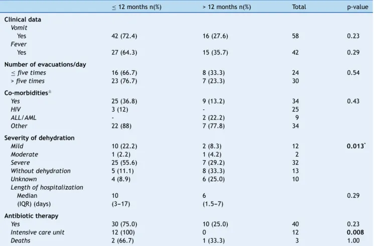
For clinical and laboratory data analysis, the patients were divided into two groups according to age: children≤12 months (65%, 45/69) and children > 12 months (35%, 24/69). Comparison of the clinical ward showed a greater frequency of patients < 12 months in the intensive care unit (ICU) (p = 0.008). A total of 64% of children presented dehydration, of which 78.3% were≤12 months (p = 0.01) (Table 1). Three (3.7%) children evolved to death, one related to gastroen-teritis. No statistically significant differences were observed in the comparison between genotype and presence of dehy-dration (p = 0.86).
RVA and hospital-acquired infections
Table 1 Clinical and laboratory data of patients (n = 69 patients) with acute gastroenteritis due to group A rotavirus, Hospital de Clínicas/Universidade Federal do Paraná, 2001 to 2008.
≤12 months n(%) > 12 months n(%) Total p-value
Clinical data Vomit
Yes 42 (72.4) 16 (27.6) 58 0.23
Fever
Yes 27 (64.3) 15 (35.7) 42 0.29
Number of evacuations/day
≤five times 16 (66.7) 8 (33.3) 24 0.54
>five times 23 (76.7) 7 (23.3) 30
Co-morbidities±
Yes 25 (36.8) 9 (13.2) 34 0.43
HIV 3 (12) - 25
ALL/AML - 2 (22.2) 9
Other 22 (88) 7 (77.8) 34
Severity of dehydration
Mild 10 (22.2) 2 (8.3) 12 0.013*
Moderate 1 (2.2) 1 (4.2) 2
Severe 25 (55.6) 7 (29.2) 32
Without dehydration 5 (11.1) 8 (33.3) 13
Unknown 4 (8.9) 6 (25.0) 10
Length of hospitalization
Median 10 6 0.29
(IQR) (days) (3---17) (1.5---7)
Antibiotic therapy
Yes 30 (75.0) 10 (25.0) 40 0.23
Intensive care unit 12 (100) 0 12 0.008
Deaths 2 (66.7) 1 (33.3) 3 1.00
Data are shown as number (percentage), unless otherwise indicated.
ALL, acute lymphocytic leukemia; AML, acute myeloid leukemia; HIV, human immunodeficiency virus; IQR, interquartile range; n, number of samples.
* p-value calculated comparing presence or absence of dehydration.
Discussion
The introduction of the RVA vaccine in the national immu-nization schedule contributed to a significant reduction in the frequency of this infection in the pediatric population. However, this pathogen can be associated with severe dis-ease, and the surveillance of its genotypic variability is crucial to monitor the emergence of new strains circulating in humans.
The variation of G and P genotypes observed in dif-ferent years highlights the mechanisms used by the RVA to escape immune selective pressure and thus maintain the pathogen in nature. Analysis of combinations of geno-types G and P type have demonstrated that genotype G1 P[8] was predominant in 2001 and 2003, similar to pre-vious findings.13 The genotype G3 P[8] was identified in only one sample in 2003. Regarding genotype G4 P[8], its occurrence was detected in 2001, as the second most fre-quent genotype, and in 2002 it prevailed; similar results were reported in Paraguay.16 The genotype G9 P[8] was identified in this study in the years 2002, 2004, and 2005.
According to some reports,17 this genotype is now circulat-ing more widely. Genotype G2 P[4] has re-emerged since 2006, and it has become predominant in the samples ana-lyzed in this study, as well as in other studies in Brazil and in other countries.18 In year 2007, no RVA was detected in the studied samples; however, some reports in Brazil showed the continuity of the detection of G2 P[4] genotype until 2008.19
T able 2 Group A rotavirus and hospital-acquired infections, 2001---2008. Year Age (months) Genotype Onset of diarrhea (day) Medical diagnosis Prevalent genotype * 2001 31 G1P[8] D7 BACT M G1P[8] 2 G1P[8] D4 BCL 2002 10 G4P[8] D6 BCN G4P[8] and G9P[8] 5 G9P[8] D3 SCD 17 G4P[8] D12 AML 2003 5 G1P[8] D13 TCB+Down S G1P[8] 2004 7 G4P[8] D7 HIV+CMV+ARI G4P[8] and G9P[8] 17 G4P[8] D4 MEN 2008 11 G2P[4] D63 TCB+BCN G2P[4] AML, acute myeloid leukemia; ARI, acute respiratory infection; BACT M, bacterial meningitis; BCL, bronchiolitis; BCN, bronchopneumonia; CMV , cyt omegalovirus; D, day of hospitalization; Down S, Down syndrome; MEN, meningoccemia; SCD, sickle cell disease; TCB, tracheobronchitis. *Most prevalent genotype in that year .
without RVA immunization,18,22and thus its circulation was probably associated to the natural reemergence of this genotype.3,23
Similar to previous findings, in this study a significant decrease in the frequency of RVA infections in the pedi-atric population was observed since the implementation of immunization, and the positive cases observed concern patients with incomplete immunization status or immuno-suppressed patients, who may not develop a complete immune response. After 2006, no RVA-positive samples were detected in outpatients.
RVA infections are most common in the wintertime in temperate regions, and year-round in tropical areas.24In the present study, an increase in positive cases was observed in certain years, particularly during the colder months, in agreement with other findings.13However, it has been found that the frequency of the disease varied throughout the year, suggesting that factors other than weather can influence the seasonality of this pathogen.25 Furthermore, in 2008, it was observed that RVA activity was spread throughout the entire year, peaking in the spring, which represented a delay of almost five months when compared to pre-immunization period. This was probably a result of a less susceptible pop-ulation and, consequently, the virus required more time to spread.23
RVA infections were predominant in children aged 0---12 months according previous reports,26 and the clinical man-ifestations varied in intensity according to age and host immunity. The classical clinical picture of RVA infections is reported as the abrupt onset of vomiting, fever, followed by diarrhea, and leading to dehydration.25,27It is worth not-ing that seven of the 12 patients affected by RVA infection who required hospitalization were admitted to the ICU with severe dehydration, did not have underlying diseases, and were younger than six months old.
A total of 49.2% of the hospitalized children was found to have moderate or severe dehydration, which corroborates the severity of this infection. However, no asso-ciation between disease severity and genotype was found, demonstrating that other factors (mainly previous clinical conditions) may be associated with the severity and intensity of infections caused by RVA.28
It is worth mentioning the importance of RVA associ-ated to hospital-acquired infections among children. Several factors, such as age, immune status, underlying disease, diagnostic and therapeutic interventions, season of the year, and duration of hospitalization may influence the acquisition of these infections. In addition to morbidity, these infections cause a major economic impact on developed and devel-oping countries.29 The incidence of nosocomial infections in this study was 12.5%; other reports found rates rang-ing from 8% to 33%.30 All patients had serious underlying diseases and this infection may have contributed to the increase in severity. The genotypes found in these patients reflected the same genotype circulating in the community, highlighting the importance of measures for hospital infec-tion control to prevent the spread of the pathogen in this environment.31
patients is important to prevent nosocomial transmission, to understand the clinical impact of these infections, to guide therapeutic measures to prevent the inappropriate use of antibiotics, and to indicate the need for specific immuniza-tion.
In conclusion, this study has demonstrated the genetic variability of RVA during an extensive period of monitoring and the severity of these infections in pediatric patients. It has also emphasized the importance of ongoing laboratory surveillance to detect the emergence of new genotypes and to determine whether this is a consequence of the global program of immunization, and to assess its impact on pedi-atric health.
Funding
Fundac¸ão Araucária/State of Paraná, Brazil.
Conflicts of interest
The authors declare no conflicts of interest.
References
1. Parashar UD, Gibson CJ, Bresee JS, Glass RI. Rotavirus and severe childhood diarrhea. Emerg Infect Dis. 2006;12: 304---6.
2. Ramani S, Iturriza-Gomara M, Jana AK, Kuruvilla KA, Gray JJ, Brown DW, et al. Whole genome characterization of reassor-tant G10P[11] strain (N155) from a neonate with symptomatic rotavirus infection: identification of genes of human and animal rotavirus origin. J Clin Virol. 2009;45:237---44.
3. Carvalho-Costa FA, Volotão E, de M, de Assis RM, Fialho AM, de Andrade J, da S, Rocha LN, et al. Laboratory-based rotavirus surveillance during the introduction of a vaccina-tion program, Brazil, 2005-2009. Pediatr Infect Dis J. 2011;30: S35---41.
4. Linhares AC. Rotavirus infection in Brazil: epidemiology and challenges for its control. Cad Saude Publica. 2000;16: 629---46.
5. Raboni SM, Nogueira MB, Hakim VM, Torrecilha VT, Lerner H, Tsuchiya LR. Comparison of latex agglutination with enzyme immunoassay for detection of rotavirus in fecal specimens. Am J Clin Pathol. 2002;117:392---4.
6. Lopman BA, Payne DC, Tate JE, Patel MM, Cortese MM, Parashar UD. Post-licensure experience with rotavirus vaccination in high and middle income countries; 2006 to 2011. Curr Opin Virol. 2012;2:434---42.
7. Soares-Weiser K, Maclehose H, Bergman H, Ben-Aharon I, Nagpal S, Goldberg E, et al. Vaccines for preventing rotavirus diarrhoea: vaccines in use. Cochrane Database Syst Rev. 2012;2:CD008521. 8. Brasil. Ministério da Saúde. Saúde Brasil 2008: 20 anos do Sis-tema Único de Saúde (SUS) no Brasil. Brasília: Ministério da Saúde; 2009.
9. Lanzieri TM, Costa I, Shafi FA, Cunha MH, Ortega-Barria E, Linhares AC, et al. Trends in hospitalizations from all-cause gas-troenteritis in children younger than 5 years of age in Brazil before and after human rotavirus vaccine introduction, 1998-2007. Pediatr Infect Dis J. 2010;29:673---5.
10. Santos N, Hoshino Y. Global distribution of rotavirus serotypes/genotypes and its implication for the develop-ment and impledevelop-mentation of an effective rotavirus vaccine. Rev Med Virol. 2005;15:29---56.
11. Gorelick MH, Shaw KN, Murphy KO. Validity and reliability of clinical signs in the diagnosis of dehydration in children. Pedi-atrics. 1997;99:E6.
12. Pereira LA, Raboni SM, Nogueira MB, Vidal LR, Almeida SM, Debur MC, et al. Rotavirus infection in a tertiary hospital: laboratory diagnosis and impact of immunization on pediatric hospitalization. Braz J Infect Dis. 2011;15: 215---9.
13. Santos JS, Alfieri AF, Leite JP, Skraba I, Alfieri AA. Molec-ular epidemiology of the human group A rotavirus in the Paraná State, Brazil. Braz Arch Biol Technol. 2008;51: 287---94.
14. Hall TA. BioEdit: a user-friendly biological sequence alignment editor and analysis program for Windows 95/98/NT. Nucl Acids Symp Ser. 1999;41:95---8.
15. National Center for Biotechnology Information. Bethesda (MD): U.S. National Library of Medicine. GenBank. [accessed 2011 Nov 27]. Available from: http://www.ncbi.nlm.nih.gov/
16. Coluchi N, Munford V, Manzur J, Vazquez C, Escobar M, Weber E, et al. Detection, subgroup specificity, and genotype diversity of rotavirus strains in children with acute diarrhea in Paraguay. J Clin Microbiol. 2002;40:1709---14.
17. Carmona RC, Timenetsky M, do C, Morillo SG, Richtzenhain LJ. Human rotavirus serotype G9, São Paulo, Brazil, 1996-2003. Emerg Infect Dis. 2006;12:963---8.
18. Ferrera A, Quan D, Espinoza F. Increased prevalence of genotype G2P (4) among children with rotavirus-associated gastroen-teritis in Honduras. In: 17th European Congress of Clinical
Microbiology and Infectious Diseases ICC; Mar 31---Apr 04, 2007. Munich. Hoboken (NJ): Wiley-Blackwell; 2007.
19. Cilli A, Luchs A, Morillo SG, Costa FF, Carmona R, de C, Timenetsky M, do C. Characterization of rotavirus and norovirus strains: a 6-year study (2004-2009). J Pediatr (Rio J). 2011;87: 445---9.
20. Carvalho-Costa FA, Assis RM, Fialho AM, Bóia MN, Alves DP, Mar-tins CM, et al. Detection and molecular characterization of group A rotavirus from hospitalized children in Rio de Janeiro, Brazil, 2004. Mem Inst Oswaldo Cruz. 2006;101:291---4. 21. Kirkwood CD, Boniface K, Barnes GL, Bishop RF. Distribution
of rotavirus genotypes after introduction of rotavirus vaccines, Rotarix®and RotaTeq®, into the National Immunization Program
of Australia. Pediatr Infect Dis J. 2011;30:S48---53.
22. Patel MM, de Oliveira LH, Bispo AM, Gentsch J, Parashar UD. Rotavirus P[4]G2 in a vaccinated population,Brazil. Emerg Infect Dis. 2008;14:863---5.
23. Sáfadi MA, Berezin EN, Munford V, Almeida FJ, de Moraes JC, Pinheiro CF, et al. Hospital-based surveillance to evaluate the impact of rotavirus vaccination in São Paulo. Brazil Pediatr Infect Dis J. 2010;29:1019---22.
24. Kane EM, Turcios RM, Arvay ML, Garcia S, Bresee JS, Glass RI. The epidemiology of rotavirus diarrhea in Latin Amer-ica. Anticipating rotavirus vaccines. Rev Panam Salud PublAmer-ica. 2004;16:371---7.
25. Nunes AA, de Mello LM, Parrode RN, Bittar JP, Domingues AL. Prevalence of rotavirus in acute diarrhea and its association with clinical signs and symptoms. J Trop Pediatr. 2010;56: 212---3.
26. Andreasi MS, Batista SM, Tozetti IA, Ozaki CO, Nogueira MM, Fiaccadori FS, et al. Rotavirus A among hospitalized infants, up to three years of age, with acute gastroenteritis in Campo Grande, State of Mato Grosso do Sul. Rev Soc Bras Med Trop. 2007;40:411---4.
27. Silva ML, Souza JR, Melo MM. Rotavirus prevalence in infants and children in the public healthcare system of the state of Pernambuco. Rev Soc Bras Med Trop. 2010;43: 548---51.
infected by G1 or G9 rotaviruses. J Clin Virol. 2009;46: 282---5.
29. Harrington M, Butler K, Cafferkey M. Rotavirus infection in hospitalised children: incidence and impact on healthcare resources. Ir J Med Sci. 2003;172:33---6.
30. Gianino P, Mastretta E, Longo P, Laccisaglia A, Sartore M, Russo R, et al. Incidence of nosocomial rotavirus infections,
symptomatic and asymptomatic, in breast-fed and non-breast-fed infants. J Hosp Infect. 2002;50:13---7.