Extranasopharyngeal Angio
fi
broma Originating
in the Inferior Turbinate: A Distinct Clinical Entity
at an Unusual Site
Marco Antonio Ferraz de Barros Baptista
1,2Fábio de Rezende Pinna
2Richard Louis Voegels
21Department of Otolaryngology, Hospital das Clínicas da Faculdade de
Medicina da Universidade de São Paulo, HCFMUSP, São Paulo, SP, Brazil
2Department of Otolaryngology, Faculdade de Medicina da
Universidade de São Paulo, São Paulo, Brazil
Int Arch Otorhinolaryngol 2014;18:403–405.
Address for correspondence Marco Antonio Ferraz de Barros Baptista, Departamento de Otorrinolaringologia, Hospital das Clínicas, Universidade de São Paulo, Av. Dr. Enéas de Carvalho Aguiar, 255 - 6 andar / sala 6167, São Paulo, SP 05403-000, Brazil
(e-mail: [email protected]).
Introduction
The extranasopharyngeal angiofibroma (ENPA) is histologi-cally similar to juvenile nasopharyngeal angiofibroma (JNA), differing from the latter in clinical and epidemiologic char-acteristics.1–3Prevalence, gender, age, affected site,
patho-genesis, clinical and computed tomography characteristics, and recurrence are completely different,1–3which leads some
authors to classify the ENPA as a disease different from the JNA.1
There are fewer than a hundred cases of ENPA described in the literature, and the maxillary sinus is the most frequently affected site, followed by the ethmoid; the entity is rare in the nasal septum and inferior turbinates.1–3The objective of this
study is to report a case of ENPA with a rare presentation in the inferior turbinate.
Review of Literature with Differential
Diagnosis
Angiofibromas that originate in or are localized in an area other than the nasopharynx are calledextranasopharyngeal angiofibromasoratypical angiofibromas.2,3
The JNA is the most common benign neoplasm of the nasopharynx, despite representing less than 0.05% of tumors of the head and neck.1,2,4,5It affects almost exclusively males between 12 and 14 years of age.1,2,4,5But the ENPA is even more unusual; it is more common in females between 17 and 22 years, Keywords
►
angio
fi
broma
►
differential diagnosis
►
inferior turbinate
Abstract
Introduction
The extranasopharyngeal angio
fi
broma is histologically similar to
juve-nile nasopharyngeal angio
fi
broma, differing from the latter in clinical and epidemiologic
characteristics.
Objectives
We present a case of extranasopharyngeal angio
fi
broma originating in the
inferior turbinate.
Resumed Report
The patient was a girl, 8 years and 6 months of age, who had
constant bilateral nasal obstruction and recurrent epistaxis for 6 months, worse on the
right side, with hyposmia and snoring. Nasal endoscopy showed a reddish lesion,
smooth, friable, and nonulcerated. Computed tomography showed a lesion with soft
tissue density in the right nasal cavity. We used an endoscopic approach and found the
lesion inserted in the right inferior turbinate. We did a subperiosteal dissection and
excision with a partial turbinectomy with a resection margin of 0.5 cm. Histopathology
reported it to be an extranasopharyngeal angio
fi
broma.
Conclusion
Although rare, extranasopharyngeal angio
fi
broma should be considered
in the diagnosis of vascular tumors of the head and neck.
received March 25, 2014 accepted after revision July 6, 2014
published online August 25, 2014
DOI http://dx.doi.org/ 10.1055/s-0034-1387811. ISSN 1809-9777.
Copyright © 2014 by Thieme Publicações Ltda, Rio de Janeiro, Brazil
THIEME
and its most common site is the maxillary sinus, followed by the ethmoid. It is very rare in the nasal septum and inferior turbinates.1–3Angiofibromas that originate in the inferior
turbi-nate are very rare; to the best of our knowledge, only nine cases of ENPA in nasal cavity have been previously reported in the English language literature. In seven cases, the tumor arose from the nasal septum, and only two from inferior turbinate.6–10
The origin of the JNA is at the top of the sphenopalatine foramen,1,2,5and its etiology is controversial.4ENPA’s etiolo-gy is associated with a migration error of the fascia basalis,1 justifying its presence in varied locations.2
The initial growth of the JNA follows a well-defined pattern in the nasal cavity, nasopharynx, and pterygopalatine fossa,4 leading to the triad of nasal obstruction, recurrent epistaxis, and nasopharyngeal tumor.1,2,5 The JNA has characteristic radiologic signs: Holman-Miller (anteriorization of the pos-terior wall of the maxillary sinus) and enlargement of the sphenopalatine foramen and pterygopalatine fossa.1,2,4,5
Histologically, the ENPA is similar to the JNA, with connec-tive tissue stroma and a matrix of dilated vessels without a muscular layer.2,3,5As for differential diagnosis, we have the hemangioma and the hemangiopericitoma.3 Although the JNA can be suspected based on known clinical and computed tomography (CT) chracteristics,2,4,5histopathologic exami-nation is essential to confirm the ENPA diagnosis.1
Treatment is surgical in both diseases.2Although the ENPA is nurtured by the maxillary artery4(just like the JNA), it may not cause excessive intraoperative bleeding due to the pre-dominance of fibrous stroma, unlike the JNA.1,2 Although benign, the JNA is locally aggressive, with recurrence rates of 6 to 27.5%2due to incomplete tumor removal.5
Case Report
The patient was a girl, 8 years and 6 months of age, who had constant bilateral nasal obstruction and recurrent epistaxis for 6 months, worse on the right side, with hyposmia and snoring. Nasal endoscopy showed a reddish lesion, smooth,
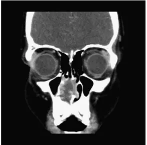
friable, and nonulcerated. The nasal mass obstructed the right nasal vestibule. The left nasal cavity and nasopharynx were normal, and no cervical lymphadenopathy was present. Middle meatuses and sphenoethmoidal recesses were free in the left side. CT showed a lesion with soft tissue density in the right nasal cavity (►Figs. 1,2, and 3). We suspected a vascular tumor and decided to remove the mass.
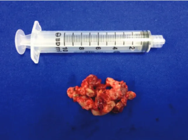
We used an endoscopic approach, identifying the lesion inserted in the right inferior turbinate, doing a subperiosteal dissection and excision with a partial turbinectomy with a resection margin of 0.5 cm. We did not perform preoperative embolization because the tumor was at an accessible location, and we expected that the bleeding could be easily controlled. However, the patient experienced excessive intraoperative bleeding (800 mL in 5 minutes). On gross inspection, the tumor was a 64-cm rubbery mass with smooth margins (►Fig. 4). On histopathology, it was composed offibrous stroma and numerous thin-walled blood vessels (►Fig. 5). Based on the
Fig. 1 Preoperative axial computed tomography scan showing the
tumor in the right nasal cavity with contrast enhancement.
Fig. 2 Preoperative coronal computed tomography scan showing the
tumor in the right nasal cavity with contrast enhancement.
Fig. 3 Preoperative sagittal computed tomography scan showing the
tumor in the right nasal cavity with contrast enhancement.
International Archives of Otorhinolaryngology Vol. 18 No. 4/2014
histopathologic findings, the tumor was diagnosed as an angiofibroma. The histopathologicfindings were revised and the ENPA diagnosis was confirmed by immunohistochemical
findings. The patient’s postoperative course was uneventful, and she showed no further symptoms. Over postoperative follow-up of 6 months, no recurrence was noted.
Discussion
ENPAs are usually found in the maxillary sinus.6Angiofi bro-mas that originate in the inferior turbinate are very rare; to the best of our knowledge, only nine cases of ENPA in the nasal cavity have been previously reported in the English language literature. In seven cases, the tumor arose from the nasal septum and only two arose from the inferior turbinate.6–10
The ENPA can evolve with a variety of symptoms and radiologic signs, depending on its site.1,2Our patient reported nasal obstruction due to a rare location in the right inferior turbinate.
The ENPA usually does not recur because its extraphar-yngeal location facilitates total ressection.1–3Our patient did
not complain of postoperative epistaxis, but had excessive intraoperative bleeding. She is now 9 years old; there has been no recurrence in 6 months.
Our patient had age and location different from most ENPAs, confirming the rarity of this case.
Therefore, although histologically similar, the ENPA and the JNA may be considered different diseases, due to totally different pathogenesis, epidemiology, and clinical and tomo-graphic presentations.1
This case challenges all the pathogenetic and evolutionary characteristics of JNA and ENPA. Cavernous hemangioma, hemangiopericytoma, or pyogenic granuloma is more plau-sible and this is more likely with the radiologic findings. Therefore, the histopathology was revised and the ENPA diagnosis was confirmed.
Final Comments
Although rare, ENPA should be considered in the diagnosis of vascular tumors of the head and neck. ENPA’s clinical and epidemiologic characteristics are different from those of JNA. In conclusion, ENPA differs significantly from JNA regard-ing clinical and radiologic presentations. ENPAs lack typical clinical and radiologic features as they develop in all age groups and in females. They arise from various sites, may be less vascularized, and produce a variety of symptoms depend-ing on the point of origin.
References
1 Windfuhr JP, Remmert S. Extranasopharyngeal angiofibroma: etiology, incidence and management. Acta Otolaryngol 2004; 124(8):880–889
2 Szymańska A, Szymański M, Morshed K, Czekajska-Chehab E, Szczerbo-Trojanowska M. Extranasopharyngeal angiofibroma: clinical and radiological presentation. Eur Arch Otorhinolaryngol 2013;270(2):655–660
3 Garcia-Rodriguez L, Rudman K, Cogbill CH, Loehrl T, Poetker DM. Nasal septal angiofibroma, a subclass of extranasopharyngeal angiofibroma. Am J Otolaryngol 2012;33(4):473–476
4 Lund VJ, Stammberger H, Nicolai P, et al; European Rhinologic Society Advisory Board on Endoscopic Techniques in the Manage-ment of Nose, Paranasal Sinus and Skull Base Tumours. European position paper on endoscopic management of tumours of the nose, paranasal sinuses and skull base. Rhinol Suppl 2010;(22):1–143 5 Ricardo LAC, Tiago RSL, Fava AS. Nasopharyngeal angiofibroma:
literature review. Rev Bras Otorrinolaringol 2003;69(3):394–403 6 Nomura K, Shimomura A, Awataguchi T, Murakami K, Kobayashi T. A case of angiofibroma originating from the inferior nasal turbi-nate. Auris Nasus Larynx 2006;33(2):191–193
7 Celik B, Erisen L, Saraydaroglu O, Coskun H. Atypical angiofi bro-mas: a report of four cases. Int J Pediatr Otorhinolaryngol 2005; 69(3):415–421
8 Alvi A, Myssiorek D, Fuchs A. Extranasopharyngeal angiofibroma. J Otolaryngol 1996;25(5):346–348
9 Gaffney R, Hui Y, Vojvodich S, Forte V. Extranasopharyngeal angiofibroma of the inferior turbinate. Int J Pediatr Otorhinolar-yngol 1997;40(2–3):177–180
10 Taggarshe D, Quraishi MS, Dugar JM. Inferior turbinate angiofi -broma: an atypical presentation [correction of preservation]. Rhinology 2004;42(1):45–47
Fig. 4 Extranasopharyngeal angiofibroma after endoscopic resection.
Fig. 5 Histologic examination: abundantfibrous component with
thin-walled vascular structures (hematoxylin-eosin staining, original magnification400).
International Archives of Otorhinolaryngology Vol. 18 No. 4/2014