TSC1
and
TSC2
gene mutations and their implications for treatment in
Tuberous Sclerosis Complex: a review
Clévia Rosset
1,2, Cristina Brinckmann Oliveira Netto
3and Patricia Ashton-Prolla
1,2,3,4 1Laboratório de Medicina Genômica, Centro de Pesquisa Experimental. Hospital de Clínicas de Porto
Alegre (HCPA), Porto Alegre, RS, Brazil.
2
Programa de Pós-Graduação em Genética e Biologia Molecular, Universidade Federal do Rio Grande do
Sul (UFRGS), Porto Alegre, RS, Brazil.
3Serviço de Genética Médica, Hospital de Clínicas de Porto Alegre (HCPA), Porto Alegre, RS, Brazil.
4Departamento de Genética, Universidade Federal do Rio Grande do Sul (UFRGS), Porto Alegre, RS,
Brazil.
Abstract
Tuberous sclerosis complex is an autosomal dominant disorder characterized by skin manifestations and formation of multiple tumors in different organs, mainly in the central nervous system. Tuberous sclerosis is caused by the
mu-tation of one of two tumor suppressor genes,TSC1 or TSC2. Currently, the development of novel techniques and
great advances in high-throughput genetic analysis made mutation screening of theTSC1 and TSC2 genes more
widely available. Extensive studies of theTSC1 and TSC2 genes in patients with TSC worldwide have revealed a
wide spectrum of mutations. Consequently, the discovery of the underlying genetic defects inTSC has furthered our understanding of this complex genetic disorder, and genotype-phenotype correlations are becoming possible, al-though there are still only a few clearly established correlations. This review focuses on the main symptoms and ge-netic alterations described in TSC patients from 13 countries in three continents, as well as on genotype-phenotype correlations established to date. The determination of genotype-phenotype correlations may contribute to the estab-lishment of successful personalized treatment for TSC.
Keywords: Tuberous sclerosis complex,TSCmutations, genotype-phenotype correlations,TSC1,TSC2.
Received: December 15, 2015; Accepted: March 1, 2016.
Tuberous Sclerosis Complex
Tuberous sclerosis, also known as Tuberous sclerosis complex (TSC) is an autosomal dominant neurocutaneous and progressive disorder, frequently characterized by the occurrence of multiple tumors in different organs. Pene-trance reaches 95% and is variable; expressivity also varies greatly even within a given family (Northrupet al., 1993). The incidence of TSC is 1/10,000 births, and its prevalence in the general population of Europe has been estimated to be 8.8/100,000 (Orphanet: Tuberous Sclerosis), affecting multiple ethnic groups (Joinsonet al., 2003).
Diagnosis and symptomatology
Tuberous sclerosis has been initially described by von Recklinghausen in 1862. In 1908, Heinrich Vogt estab-lished the diagnostic criteria for TSC as the so-called triad:
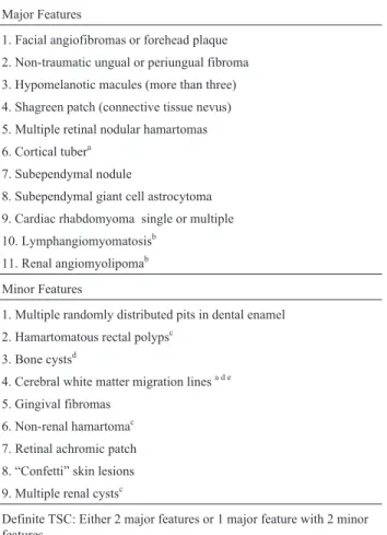
epilepsy, mental retardation and adenoma sebaceum. As none of these clinical signs were pathognomonic for TSC, clinical diagnostic criteria were revised by a consortium in 1998 (Roachet al., 1998), which proposed three diagnostic categories (definite, probable or possible TSC) based on the presence of major and/or minor features of the disease. Ta-ble 1 shows the revised and updated diagnostic criteria for TSC, established by the same consortium in 2012 (Northrup et al., 2013). A definite clinical diagnosis is made when two major features, or one major feature plus two minor features are present. Importantly, most major features are localized to the skin and central nervous sys-tem. Also, one must consider that the clinical manifesta-tions of TSC appear at distinct developmental points, and a person with suspected TSC may need multiple sequential evaluations before a definite clinical diagnosis can be made.
After skin and CNS findings, renal manifestations are the most common abnormalities associated with TSC. These include renal cell carcinoma, oncocytomas, angio-myolipomas (in 80% of patients) and renal cystic disease
Send correspondence to Patricia Ashton Prolla. Serviço de Gené-tica Médica, Hospital de Clínicas de Porto Alegre, Rua Ramiro Barcelos 2350. 90035-903 Porto Alegre, RS, Brazil. E-mail: [email protected].
(in 50% of the patients) (Dixonet al., 2011). Typically, re-nal manifestations in children with TSC are first seen in in-fancy and increase with age. Angiomyolipomas, one of the leading causes of death in TSC patients, are multiple and often bilateral. The associated mortality is due to complica-tions when these lesions become very large. Another conse-quence of angiomyolipomas is destruction of the normal renal parenchyma, resulting in renal failure and end-stage renal disease (Shepherdet al., 1991). Patients with clini-cally detectable renal cystic disease usually have a severe very early-onset polycystic phenotype (about 2% of TSC patients) (Sampsonet al., 1997).
Pulmonary involvement, specifically lymphangio-leiomyomatosis (LAM), is the third most common cause of TSC-associated morbidity, occurring in approximately 35% of female TSC patients. LAM is caused by prolifera-tion of atypical smooth muscle cells in the peribronchial, perivascular, and perilymphatic tissues of the lung (Kumasakaet al., 2004). LAM occurs almost exclusively in young women, typically presenting between 30 to 35 years of age. Symptoms have been reported to begin or worsen during pregnancy, suggesting that LAM may be hormonally influenced (Castroet al., 1995).
Skin lesions are detected in 70% of patients with TSC and include hypomelanotic macules, shagreen patches, confetti-like lesions, forehead fibrous plaque, facial angio-fibromas, and periungual and ungual fibromas (Schwartzet al., 2007). Depending on studied population, even as many as 100% of TSC patients younger than five years may pres-ent with hypopigmpres-ented macules. An aggregation of red-dish papules, appearing on the nose and cheeks in a charac-teristic butterfly distribution, belongs to the Vogt triad of signs. Although usually symmetrical, occasionally they may be found unilaterally (Jozwiak et al., 1998). Facial angiofibromas (adenoma sebaceum) are formed by hamar-tomatous growth of dermal connective tissue with rich vasculature and can result in decreased quality of life since they affect appearance, may cause disfigurement, and are prone to bleeding, which increases the possibility of infec-tion (Yates, 2006). Shagreen Patches are areas of thick, ir-regularly shaped, and elevated skin, usually found on the lower back. Mean age of appearance is about 8.1 years (Sun
et al., 2005). Ungual and subungual fibromas are small tu-mors that grow around and under toenails or fingernails. Their mean age of appearance is 14.9 years (Sun et al., 2005) and their prevalence in older patients (above 30 years) is close to 90%. Forehead plaques appear under the age of 14 years (Jozwiaket al., 1998), with mean age of ap-pearance being 2.6 years (Sunet al., 2005).
TSC is also associated with both retinal and non-retinal ocular findings (Rowleyet al., 2001). Hamartomas are the most common retinal manifestation of TSC and are identified in approximately 40 to 50% of individuals. For-tunately, they rarely compromise vision, although severe decreases in visual acuity and blindness has been reported in some cases due to hamartoma enlargement, macular in-volvement, retinal detachment, and vitreous hemorrhage (Robertson, 1999).
Multiple cardiac rhabdomyomas are cardiac tumors most frequently encountered during infancy and childhood and they occur in approximately 30% of TSC patients. On the other side, nearly 100% of fetuses with multiple rhabdo-myomas have TSC. Cardiac rhabdorhabdo-myomas usually do not cause symptoms or hemodynamic compromise, and the natural history for these lesions is spontaneous regression in the vast majority of cases. However, a minority of the cases may become symptomatic shortly after birth or in the
70 Rossetet al.
Table 1- Revised Diagnostic Criteria for Tuberous Sclerosis Complex *.
Major Features
1. Facial angiofibromas or forehead plaque 2. Non-traumatic ungual or periungual fibroma 3. Hypomelanotic macules (more than three) 4. Shagreen patch (connective tissue nevus) 5. Multiple retinal nodular hamartomas 6. Cortical tubera
7. Subependymal nodule
8. Subependymal giant cell astrocytoma 9. Cardiac rhabdomyoma single or multiple 10. Lymphangiomyomatosisb
11. Renal angiomyolipomab
Minor Features
1. Multiple randomly distributed pits in dental enamel 2. Hamartomatous rectal polypsc
3. Bone cystsd
4. Cerebral white matter migration linesa d e
5. Gingival fibromas 6. Non-renal hamartomac
7. Retinal achromic patch 8. “Confetti” skin lesions 9. Multiple renal cystsc
Definite TSC: Either 2 major features or 1 major feature with 2 minor features
Probable TSC: One major feature and one minor feature Possible TSC: Either 1 major feature or 2 or more minor features
*Revised Diagnostic Criteria for Tuberous Sclerosis Complex established
by a consortium in 2012 (Northrupet al., 2012).
a
When cerebral cortical dysplasia and cerebral white matter migration tracts occur together, they should be counted as one rather than two fea-tures of TSC.
bWhen both lymphangiomyomatosis and renal angiomyolipomas are
present, other features of TSC should be present before a definitive diag-nosis is assigned.
cHistologic confirmation is suggested. d
Radiographic confirmation is sufficient.
eOne panel member recommended three or more radial migration lines
first year of life. Finally, hamartomas may also occur in or-gans of the endocrine system and rare case reports exist of angiomyolipomas or fibroadenomas in the pituitary gland, pancreas, or gonads (O’Callaghan and Osborne, 2010).
Neurological involvement
Neurologic complications are the most common and often the most impairing aspect of TSC. Structural neuro-logical abnormalities include cortical tubers, subepen-dymal nodules (SENs) and subepensubepen-dymal giant cell tumors (SGCTs). Brain tumors in TSC are rare (2 to 10% of pa-tients with TSC and 1.1-1.4% of all pediatric brain tumors) (Frèrebeauet al., 1985). Cortical tubers are developmental abnormalities present in more than 88% of children with TSC (Cucciaet al., 2003), and the average number of tubers per patient ranges from 5 to 50 in different studies. Tubers lead to loss of the classical six-layered cyto-architecture of the cerebral cortex and are thought to be responsible for more than 75% of the epileptic disorders in patients with TSC (Orphanet: Tuberous Sclerosis). The second more fre-quent structural neurological lesions in children with TSC are SENs, which are small hamartomas that occur in the walls of the lateral ventricles. Only SENs located in the re-gion of the Monro foramina may have the potentiality to grow and to transform into SGCTs (5%-20% of patients). The last but not least important type of encephalic lesion is SGCT, affecting an average of 10% of children with TSC. SGCTs are benign, slow-growing tumors of mixed glio-neuronal cells including giant cells. They are typically lo-cated near the foramen of Monro, hence they can cause increased intracranial pressure, obstructive hydrocephalus, focal neurologic deficits and death (Orphanet: Tuberous Sclerosis, 2015). Approximately 90% of TSC patients ex-perience seizures and about 50% have documented cogni-tive impairment, autism, or other behavioral disorders.
Epilepsy is likely the most prevalent and challenging clinical manifestation of TSC, and virtually all subtypes of seizure have been reported. At least one third of patients de-velop refractory epilepsy; attention deficit-hyperactivity disorder and psychiatric comorbidities, such as mood disor-ders, anxiety, obsessive compulsive behavior and alcohol-ism are also frequently present. Among the different sites of tumor development, the brain remains undoubtedly the most problematic in terms of therapeutic management and screening. Brain tumors are the cause of more than 50% of deaths among children with TSC (Webbet al., 1996). Intel-lectual disability has a prevalence of 40%-50% in TSC; 30% are severely affected with IQs in the very low range, and 70% have IQs in the normal, yet slightly left-shifted range (Joinsonet al., 2003).
Molecular genetics of TSC: the
TSC1 and
TSC2 genes
Tuberous sclerosis is caused by mutations in one of two tumor suppressor genes: TSC1 (9q34) and TSC2
(16p13.3). TheTSC1 gene spans about 53kb of genomic DNA with 23 exons coding for hamartin, a hydrophilic pro-tein with 1164 amino acids and 130 kDa. Hamartin is ex-pressed in several adult tissues and plays a key role in the regulation of cell adhesion. This protein shows no homo-logy with any other vertebrate protein. The TSC2 gene comprises approximately 43kb of genomic DNA with 41 exons encoding a 5.5 kb transcript and a 1807 amino acid protein, tuberin, with 198 kDa. This protein contains a hy-drophilic N-terminal domain and a conserved 163 amino acid region encoded by exons 34-38, near the C-terminal portion, which has homology with the Ras superfamily GTPases proteins rap1GAP and mSpa1 (Maheshwaret al., 1997). Therefore, tuberin is a GTPase activating protein that regulates the GTP binding and hydrolysing activity of the Ras superfamily of proteins and helps to regulate cell growth, proliferation and differentiation. The other do-mains of tuberin are less conserved, and additional homo-logies between tuberin and other proteins have not been identified. Serfontein et al. (2011) used bioinformatics tools to examine the presence of conserved elements of
TSC1andTSC2across different organisms. The analyzed organisms showed a wide range in the degree to which resi-dues implicated in signalling are conserved (or present at all) in comparison to the humanTSC1andTSC2sequences. Not surprisingly, the mammalian proteins of Rattus norvegicusandMus musculusshared the largest number of residues with the human proteins.
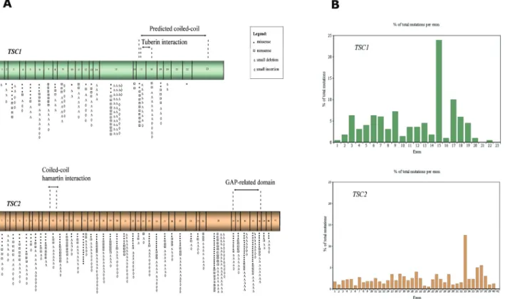
Figure 1A schematically shows the structure ofTSC1
andTSC2genes, their coding exons and the main domains of hamartin and tuberin. These proteins bind each other via their respective coiled-coil domains to form an intracellular complex that integrates signals to control cellular homeo-stasis, oxygen levels, presence of nutrients, energy pool, and stimulation by growth factors. Such signals regulate Rheb (a Ras homologue enriched in brain), responsible for the activation of mTOR (mammalian target of rapamycin) kinase. mTOR, in turn, regulates the translation of a signifi-cant proportion of cellular proteins, including those respon-sible for the control of cell growth and proliferation (Kwiatkowski, 2003).
Figure 2 shows the role of the TSC2:TSC1 complex in the mTOR pathway. Loss of function mutations inTSC1
72 Rossetet al.
Figure 1-TSC1and TSC2gene structure, domains and distribution of point mutations. (A) Schematic representation ofTSC1andTSC2exons and the domains of hamartin and tuberin, respectively, codified by them. The symbols represent the number of different mutations described at each exon. (B) The graph shows the percentage of the total number of described mutations that occur at eachTSC1andTSC2exon.
genes, the inactivation of bothTSC1or bothTSC2alleles is necessary for benign or malignant tumor formation. The first hit is an inherited germline mutation inTSC1orTSC2,
which can be detected in approximately 85% of patients with the clinical features of TSC, and the second hit is so-matic. There are multiple possible mechanisms for somatic inactivation of the wild-type alleles ofTSC1andTSC2, in-cluding loss of heterozygosity, mutation and promoter methylation. It is possible that epigenetic silencing medi-ated by micro-RNAs also occurs. Moreover, binding of TSC1 to TSC2 appears to stabilize intracellular TSC2 lev-els since uncomplexed TSC2 is subject to ubiquitin-mediated degradation (Chong-Koperaet al., 2006). Thus, TSC1 has a role in stabilizing the complex, while TSC2 has the GTPase activity. For this reason, inactivating mutations in either gene give rise to the same clinical disorder. Clearly, both proteins play pivotal roles in several pro-cesses that are crucial for normal brain development. In ad-dition, because they are widely expressed throughout the mature brain, these proteins likely have important homeos-tatic regulatory functions in neurons during adult life.
Although several TSC families exhibit an autosomal dominant pattern of inheritance, 70% of the cases result fromde novogermline mutations. Linkage studies initially suggested that there would be equivalent numbers of fami-lies with mutations in each TSCgene (Benvenuto et al., 2000). However, the frequency of mutations reported in
TSC2is consistently higher than inTSC1; TSC1mutations account for only 10% to 30% of the families identified with TSC. In sporadic TSC, there is an even greater excess of mutations in TSC2. Nonetheless, identification of TSC1
mutations appears to be twice as likely in familial cases as in sporadic cases. The disparity in mutational frequency may reflect an increased rate of germline and somatic muta-tions inTSC2as compared withTSC1,as well as an ascer-tainment bias, since mutations inTSC2are associated with more severe disease (Dabora et al., 2001; Jansen et al., 2008; Kothareet al., 2014). In patients with the TSC phe-notype and no identifiable mutations in either TSC1 or
TSC2(15% to 20%), the disease is usually milder (Dabora
et al., 2001). A milder phenotype has also been described in rare individuals with mosaicism for mutations inTSC1or
TSC2. Caignecet al.(2009) reported a unique family with three independent pathogenic mutations inTSC2mapping to distinct haplotypes. The three mutations were most likely
de novo, as parents of the affected patients did not present any features of TSC. In addition, findings consistent with gonadal mosaicism were seen in one branch of the family.
Molecular diagnosis in TSC
The development of novel techniques and great ad-vances in high-throughput genetic analysis in the last few years made mutation screening of the TSC1 and TSC2
genes feasible. Recent massively parallel sequencing tech-nologies (Next-Generation Sequencing, NGS) and copy
number variation testing (Multiplex Ligation-dependent Probe Amplification - MLPA and array-Comparative Genomic Hybridization - aCGH) have been validated for clinical use in many disorders including TSC, rendering the analysis much faster and more cost-effective.
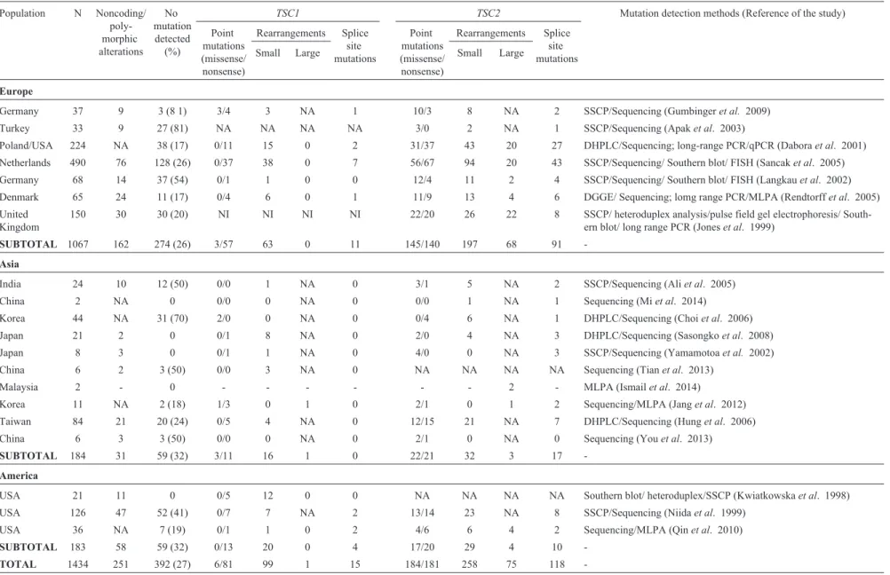
Extensive studies of theTSC1andTSC2genes in pa-tients with TSC have revealed a wide spectrum of muta-tions. We searched the PubMed database to retrieve available published literature in English from 1998 to 2014 that described mutations atTSC1andTSC2genes and es-tablished genotype-phenotype correlations for tuberous sclerosis disease. The following keywords were used:
TSC1mutations;TSC2mutations; tuberous sclerosis com-plex;TSC mutations;TSC molecular analysis; genotype-phenotype correlation on tuberous sclerosis. Twenty-seven studies were included in the final analysis. Table 2 summa-rizes the results obtained in the main studies performed with unrelated TSC patients worldwide; many of the changes listed were found for the first time in the investi-gated population. The most frequent mutation type is point mutations. Large gene rearrangements are less frequently reported, both because of their true prevalence in TSC and also because several studies did not use methodologies that are directed to the identification of such mutations. As ex-pected, the observed mutation detection rate is not always complete. In this group, a mutation could exist in an intronic region distant from the exon-intron boundaries, which could have an effect on the splicing process or gene regulation, causing a reduction of normal mRNA tran-script. Although a third gene for TSC may exist and explain this lack of mutation atTSC1andTSC2genes in some pa-tients, there is currently no concrete evidence for this. Also, somatic and germ line mosaicism is a credible explanation for the failure to detect mutations in some patients, and spe-cialized methods can be used to enhance detection of these specific situations. Most studies in TSC patients were con-ducted in Europe and Asia. The largest cohorts are from the Netherlands and Poland/USA. As expected through ob-served mutation frequencies, in all populations described, the germline mutation rate at theTSC2 locus was higher than that at theTSC1locus. Also, the frequency of small re-arrangements (small insertions/deletions) is higher than missense, nonsense and splice site mutations in all popula-tions.
fre-74
Rosset
et
al.
Table 2- Type and frequency of mutations found inTSCgenes in patients from different studies in the world and diagnostic strategy (1998-2014).
Population N Noncoding/
poly-morphic alterations
No mutation detected
(%)
TSC1 TSC2 Mutation detection methods (Reference of the study)
Point mutations (missense/ nonsense)
Rearrangements Splice site mutations
Point mutations (missense/ nonsense)
Rearrangements Splice site mutations
Small Large Small Large
Europe
Germany 37 9 3 (8 1) 3/4 3 NA 1 10/3 8 NA 2 SSCP/Sequencing (Gumbingeret al. 2009)
Turkey 33 9 27 (81) NA NA NA NA 3/0 2 NA 1 SSCP/Sequencing (Apaket al. 2003)
Poland/USA 224 NA 38 (17) 0/11 15 0 2 31/37 43 20 27 DHPLC/Sequencing; long-range PCR/qPCR (Daboraet al. 2001)
Netherlands 490 76 128 (26) 0/37 38 0 7 56/67 94 20 43 SSCP/Sequencing/ Southern blot/ FISH (Sancaket al. 2005)
Germany 68 14 37 (54) 0/1 1 0 0 12/4 11 2 4 SSCP/Sequencing/ Southern blot/ FISH (Langkauet al. 2002)
Denmark 65 24 11 (17) 0/4 6 0 1 11/9 13 4 6 DGGE/ Sequencing; lomg range PCR/MLPA (Rendtorffet al. 2005)
United Kingdom
150 30 30 (20) NI NI NI NI 22/20 26 22 8 SSCP/ heteroduplex analysis/pulse field gel electrophoresis/
South-ern blot/ long range PCR (Joneset al. 1999)
SUBTOTAL 1067 162 274 (26) 3/57 63 0 11 145/140 197 68 91
-Asia
India 24 10 12 (50) 0/0 1 NA 0 3/1 5 NA 2 SSCP/Sequencing (Aliet al. 2005)
China 2 NA 0 0/0 0 NA 0 0/0 1 NA 1 Sequencing (Miet al. 2014)
Korea 44 NA 31 (70) 2/0 0 NA 0 0/4 6 NA 1 DHPLC/Sequencing (Choiet al. 2006)
Japan 21 2 0 0/1 8 NA 0 2/0 4 NA 3 DHPLC/Sequencing (Sasongkoet al. 2008)
Japan 8 3 0 0/1 1 NA 0 4/0 0 NA 3 SSCP/Sequencing (Yamamotoaet al. 2002)
China 6 2 3 (50) 0/0 3 NA 0 NA NA NA NA Sequencing (Tianet al. 2013)
Malaysia 2 - 0 - - - 2 - MLPA (Ismailet al. 2014)
Korea 11 NA 2 (18) 1/3 0 1 0 2/1 0 1 2 Sequencing/MLPA (Janget al. 2012)
Taiwan 84 21 20 (24) 0/5 4 NA 0 12/15 21 NA 7 DHPLC/Sequencing (Hunget al. 2006)
China 6 3 3 (50) 0/0 0 NA 0 2/1 0 NA 0 Sequencing (Youet al. 2013)
SUBTOTAL 184 31 59 (32) 3/11 16 1 0 22/21 32 3 17
-America
USA 21 11 0 0/5 12 0 0 NA NA NA NA Southern blot/ heteroduplex/SSCP (Kwiatkowskaet al. 1998)
USA 126 47 52 (41) 0/7 7 NA 2 13/14 23 NA 8 SSCP/Sequencing (Niidaet al. 1999)
USA 36 NA 7 (19) 0/1 1 0 2 4/6 6 4 2 Sequencing/MLPA (Qinet al. 2010)
SUBTOTAL 183 58 59 (32) 0/13 20 0 4 17/20 29 4 10
-TOTAL 1434 251 392 (27) 6/81 99 1 15 184/181 258 75 118
quency of 9.5% (determined as the percentage of mutations per base pairs, considering the size of each exon) and the highest mutation frequency is in exon 13 (14.3%). Con-sidering all exons, the average frequency of observed muta-tions is 5.9%. Seven of the 23 exons have higher values (above 9%). Small deletions are responsible for 41% of the disease and small insertions, for 18.5%. Large rearrange-ments are responsible for 7% of mutations in TSC1 at this database. The predicted coiled-coil domain of hamartin corresponds to exons 17-23, where 21.9% of the mutations are localized. Exons 17-18 are responsible for interaction with tuberin, and they account for 15.9% of the point muta-tions described. Another database, the Leiden Open Varia-tion Database (LOVD), reports 690 unique DNA variants for theTSC1gene.
Considering theTSC2gene, 183 unique missense and 125 nonsense mutations gene are described in the HGMD database, as well as 189 small deletions, 99 small inser-tions, 120 splice site mutations and 148 large rearrange-ments. Point mutations correspond to 82.6% of the mutations, with the largest number of these occurring in exon 33, which is the largest in basepairs (488). Propor-tionally, this exon corresponds to a mutation frequency of 15.45%, and the highest mutation frequency is in exon 37 (22.9%) and exon 38 (22.8%). Considering all exons, aver-age frequency of observed mutations is 11.0%, a higher number than mutation frequency at the TSC1 gene. Twenty-seven of the 41 exons have mutation frequency values above 9% and an overall mutation number and mu-tation frequency higher than that at theTSC1gene. Small deletions are responsible for 32% of the disease versus 41% in the TSC1 gene, and small insertions for 17% versus 18.5% in the TSC1 gene. Large rearrangements are not shown in the table, and are responsible for 17.4% of muta-tions inTSC2, a higher number than the frequency of large rearrangements in theTSC1gene.
The predicted coiled-coil domain of tuberin corre-sponds to exon 10 of theTSC2gene, where only 1.8% of small mutations are localized. Exons 34-38 encode the GAP-domain, responsible for the essential GTPase acti-vaty, and they account for 18.1% of the point mutations de-scribed at this gene, with a high mutation frequency (95.1%). Exons 37 and 38 have shown the highest mutation frequency in theTSC2gene, and these mutations can have a damage effect on the protein since the GAP domain can be disrupted. In the Leiden Open Variation Database, 1925 unique DNA variants onTSC2gene have been reported.
Figure 1A illustrates the distribution of point muta-tions among all exons and domains of theTSC1andTSC2
genes described in these different studies, and Figure 1B graphically represents the occurrence of point mutations in each of theTSC1andTSC2exons (percentage of the total number of described mutations in the HGMD database that occurs in each exon). This percentage is not related to exon
size, but larger exons contain more mutations than smaller exons.
Because TSC can be a devastating disease, family members of affected individuals are often eager to know whether they are carriers ofTSCmutations. Currently, with the adventure of next generation sequencing platforms, it became possible to analyze point mutations in bothTSC1
andTSC2genes at the same time for a lower cost; if no mu-tations are detected, the search for large deletions and du-plications should proceed. Prenatal and preimplantation genetic tests are also becoming more widely available. The mutation status of family members has great implications on genetic counseling. Furthermore, for all clinical diag-nostic criteria, patients with subclinical TSC may not be correctly diagnosed, and genetic testing is also very impor-tant for these cases.
The second International Tuberous Sclerosis Com-plex Consensus Conference brought together 79 experts from 14 countries to finalize diagnostic, surveillance, and management recommendations for patients with TSC (Northrupet al., 2013). At this meeting, the most signifi-cant change recommended was the incorporation of genetic testing in the diagnostic criteria. Molecular testing of the
TSC1andTSC2genes yields a positive mutation result for 75-90% of TSC-affected individuals categorized as having definite Clinical Diagnostic Criteria. The recommendation of the Genetics Panel was to make the identification of a pathogenic mutation inTSC1orTSC2an independent diag-nostic criterion, regardless of the clinical findings. This will facilitate the diagnosis of TSC in some, particularly young individuals, allowing earlier implementation of surveil-lance and treatment with a potential for better clinical out-comes.TSC1andTSC2genetic variants whose functional effect is not definitely pathogenic would not be considered a major diagnostic criterion. Finally, a normal result from
TSC1andTSC2testing does not exclude TSC, since a frac-tion of TSC patients has no mutafrac-tion identified by conven-tional genetic testing. Nonetheless, if the mutation in an affected relative is known, testing for that mutation has very high predictive value for family members.
Genotype-phenotype correlations in TSC
The discovery of the underlying genetic defects in
TSChas furthered our understanding of this complex ge-netic disorder and genotype-phenotype correlations are be-coming possible. In a retrospective study, Kothareet al.
muta-76 Rossetet al.
Table 3- Genotype-phenotype correlations established for TSC patients.
Population N Locus of DNA alteration Amino acid change
Type of alteration Main associated symptoms
Reference
EUA 1039 TSC2 - Any type onTSC2 Mutations in theTSC2
gene were more frequent thanTSC1gene in patients with retinal findings
(Aronowet al., 2012)
Poland 170 TSC2c.5238-5255del 18pb
- Frameshift Epilepsy (Roket al., 2005)
USA 220 Contiguous deletion
TSC2-PKD1
- Large rearrange-ment
Arachnoid cysts and polycystic kidney disease
(Boronatet al., 2014)
Poland/USA 224 TSC2 - Any type onTSC2 Seizures, mental retarda-tion, average tuber count, subependymal nodules, re-nal angiomyolipomas, angiofibromas and fibrous forehead plaques were more common and severe inTSC2patients
(Daboraet al., 2001)
Netherlands 490 TSC1 - Any type onTSC1 Shagreen patches are more frequent in patients with
TSC1mutation
(Sancaket al., 2005)
Netherlands 490 TSC2 - Any type onTSC2 Mental retardation is more frequent in patients with
TSC2mutation
(Sancaket al., 2005)
Netherlands 490 TSC2 - Nonsense and frameshift
Shagreen patches, forehead plaques, facial
angiofibromas, ungual fibromas, renal
angiomyolipomas and renal cysts
(Sancaket al., 2005)
Netherlands 490 TSC2 - Mutations in the GAP domain
Mental retardation, seizures and subependymal nodules
(Sancaket al., 2005)
Korea 11 TSC2 - Mutations in exons 33-41
Cardiac rhabdomyomas (Janget al., 2012)
USA 65 TSC2 - Any type onTSC2 Higher number of cysts thanTSC1woman with pulmonary
lymphangioleiomyomatosis
(Muzykewiczet al., 2009)
Canada 19 families TSC2 R905Q Missense Milder disease severity (Jansenet al., 2006) USA 478 TSC2proximal region
(exons 1-22) and distal region (exons 34-41)
- Missense muta-tions and small in-frame deletions
or insertions
Proximal and distalTSC2
mutations showed a signifi-cantly higher risk of infan-tile spasms compared with mutations in the central re-gion of the gene
(van Eeghenaet al., 2013)
USA and Belgium
919 TSC2 - Any type onTSC2 More frequent occurrence of several kinds of sei-zures/epilepsy subtypes: partial epilepsy, complex partial seizures, infantile spasms, SENs, SGCTs and cognitive impairment.
(Kothareet al., 2014)
United Kingdom
One case report
TSC1intron 10 (c.1030-3 C > G)
- Splice site mutation
Mild phenotype (seizures and small number of hypomelanotic macules)
(Blythet al., 2010)
tions. Jansenet al.(2008) also reported a more severe neu-rologic phenotype, including an earlier age of seizure onset, lower cognition index and more tubers in patients with a
TSC2mutation as compared to those with aTSC1mutation. Another important correlation involves a subgroup of large genomic deletions at TSC2 that also affect the adjacent
PKD1gene, causing early-onset polycystic kidney disease (Osborneet al., 1991).
Table 3 shows a compilation of the main genotype-phenotype correlations described to date. As expected, mostTSC2mutations are generally associated with a more severe phenotype. Only oneTSC2mutation, R905Q, was associated with milder disease. This mutation was found in 25 individuals from the same family, with a phenotype characterized by the complete absence of disfiguring skin lesions, intractable epilepsy, mental retardation, and severe organ involvement. So, the type and location of mutations in bothTSC1andTSC2genes also have an influence in the phenotype. Hamartin and tuberin are known to bind to at least 40 additional proteins, and thus there are numerous potential and yet undefined effects ofTSCgene mutations. Futhermore, it is likely that other events such as mosaicism, the nature and frequency of the second event of inactivation of the second allele and the modifying genes, as well as en-vironmental effects may interfere with the phenotype, which makes it more difficult to establish clear genotype-phenotype correlations. Moreover, polymorphic and non-pathogenic variants in theTSC1andTSC2genes can act as phenotype modifiers in tuberous sclerosis, and they need to be further explored. To date, little is known about non-pathogenic variants in these genes, and phenotype modifi-ers in tuberous sclerosis have not been identified so far.
In light of emerging human genetic and molecular knowledge, molecular diagnosis of TSC and determination of genotyope-phenotype correlations might help in the es-tablishment of personalized treatment for TSC patients and improve quality of life among these patients. Continuous studies in this area can guide future directions in this line.
References
Ali M, Girimaji SC, Markandaya M, Shukla AK, Sacchidanand S and Kumar A (2005) Mutation and polymorphism analysis of TSC1 and TSC2 genes in Indian patients with tuberous sclerosis complex.Acta Neurol Scand 111:54-63.
Apak A, Haliloolu G, Kose G, Yilmaz E, Anlar B and Aysun S (2003) Mutation analysis of TSC2 gene in 33 Turkish famil-ial cases with tuberous sclerosis. Turk J Pediatr 45:1-5. Aronow ME, Nakagawa JA, Gupta A, Traboulsi EI and Singh AD
(2012) Tuberous sclerosis complex: Genotype/phenotype correlation of retinal findings. Ophthalmology 119:1917-1923.
Benvenuto G, Li S, Brown SJ, Braverman R, Vass WC, Cheadle JP, Halley DJ, Sampson JR, Wienecke R and DeClue JE (2000) The tuberous sclerosis-1 (TSC1) gene product ha-martin suppresses cell growth and augments the expression
of the TSC2 product tuberin by inhibiting its ubiquitination. Oncogene 19:6306-6316.
Blyth M, Raponi M, Treacy R, Raymond FL, Yates JR and Baralle D (2010) Expanding the tuberous sclerosis phenotype: Mild disease caused by a TSC1 splicing mutation. J Neurol Neurosurg Psychiatry 81:350-352.
Boronat S, Caruso P, Auladell M, Van Eeghen A and Thiele EA (2014) Arachnoid cysts in tuberous sclerosis complex. Brain Dev 36:801-806.
Caignec CL, Kwiatkowski DJ, Kury S, Hardouin JB, Melki J and David A (2009) Three independent mutations in the TSC2 gene in a family with tuberous sclerosis. Eur J Hum Genet 17:1165-1170.
Castro M, Sheperd CW, Gomez MR and Lie JT (1995) Pulmonary tuberous sclerosis. Chest 107:189-195.
Choi JE, Chae JH, Hwang YS and Kim KJ (2006) Mutational analysis ofTSC1andTSC2in Korean patients with tuberous sclerosis complex.Brain Dev 28:440-446.
Chong-Kopera H, Inoki K, Li Y, Zhu T, Garcia-Gonzalo FR, Rosa JL and Guan KL (2006) TSC1 stabilizes TSC2 by inhibiting the interaction between TSC2 and the HERC1 ubiquitin ligase. J Biol Chem 281:8313-8316.
Cuccia V, Zuccaro G, Sosa F, Monges J, Lubienieky F and Taratuto AL (2003) Subependymal giant cell tumor in chil-dren with tuberous sclerosis. Childs Nerv Syst 19:232-243. Dabora SL, Jozwiak S, Franz DN, Roberts PS, Nieto A, Chung J,
Choy YS, Reeve MP, Thiele E, Egelhoff JC,et al.(2001) Mutational analysis in a cohort of 224 tuberous sclerosis pa-tients indicates increased severity ofTSC2,compared with TSC1disease in multiple organs. Am J Hum Genet 68:64-80.
Dixon BP, Hulbert JC and Bissler JJ (2011) Tuberous sclerosis complex renal disease. Nephron Exp Nephrol 118:15-20. Frèrebeau P, Benezech J, Segnarbieux F, Harbi H, Desy A and
Marty-Double CH (1985) Intraventricular tumors in tuber-ous sclerosis. Childs Nerv Syst 1:45-48.
Gumbinger C, Rohsbach CB, Schulze-Bonhage A, Korinthenberg R, Zentner J, Häffner M and Fauser S (2009) Focal cortical dysplasia: A genotype-phenotype analysis of polymor-phisms and mutations in the TSC genes. Epilepsia 50:1396-1408.
Hung CC, Su YN, Chien SC, Liou HH, Chen CC, Chen PC, Hsieh CJ, Chen CP, Lee WT, Lin WL,et al.(2006) Molecular and clinical analyses of 84 patients with tuberous sclerosis com-plex. BMC Med Genet 7:e72.
Ismail NFD, Malik NMANA, Mohseni J, Rani AM, Hayati F, Salmi AR, Narazah MY, Zabidi-Hussin ZA, Silawati AR, Keng WT,et al.(2014) Two novel gross deletions ofTSC2 in Malaysian patients with Tuberous Sclerosis Complex and TSC2/PKD1 Contiguous Deletion Syndrome. Jpn J Clin Oncol 44:506-511.
Jang MA, Hong SB, Lee JH, Lee MH, Chung MP, Shin HJ, Kim JW and Ki CS (2012) Identification ofTSC1and TSC2 mu-tations in Korean patients with Tuberous Sclerosis Com-plex. Pediatr Neurol 46:222-224.
Jansen FE, Braams O, Vincken KL, Algra A, Anbeek P, Jen-nekens-Schinkel A, Halley D, Zonnenberg BA, van den Ouweland A, van Huffelen AC,et al.(2008) Overlapping neurologic and cognitive phenotypes in patients withTSC1 orTSC2mutations. Neurology 70:908-915.
Joinson C, O’Callaghan FJ, Osborne JP, Martyn C, Harris T and Bolton PF (2003) Learning disability and epilepsy in an epi-demiological sample of individuals with tuberous sclerosis complex. Psychol Med 33:335-344.
Jones AC, Shyamsundar MM, Thomas MW, Maynard J, Idziaszczyk S, Tomkins S, Sampson JR and Cheadle JP (1999) Comprehensive mutation analysis of TSC1 and TSC2 - and phenotypic correlations in 150 families with tu-berous sclerosis. Am J Hum Genet 64:1305-1315.
Jozwiak S, Schwartz RA, Janniger CK, Michalowicz R and Chmielik J (1998) Skin lesions in children with tuberous sclerosis complex: Their prevalence, natural course, and di-agnostic significance. Int J Dermatol 37:911-917.
Knudson AG (1971) Mutation and cancer: Statistical study of reti-noblastoma. Proc Natl Acad Sci U S A 68:820-823. Kothare SV, Singh K, Chalifoux JR, Staley BA, Weiner HL,
Menzer K and Devinsky O (2014) Severity of manifesta-tions in tuberous sclerosis complex in relation to genotype. Epilepsia 55:1025-1029.
Kumasaka T, Seyama K, Mitani K, Sato T, Souma S, Kondo T, Hayashi S, Minami M, Uekusa T, Fukuchi Y,et al.(2004) Lymphangiogenesis in lymphangioleiomyomatosis: Its im-plication in the progression of lymphangioleiomyomatosis. Am J Surg Pathol 28:1007-1016.
Kwiatkowska J, Jozwiak S, Hall F, Henske EP, Haines JL, McNa-mara P, Braiser J, Wigowska-Sowinska J, Kasprzyk-Obara J, Short MP,et al.(1998) Comprehensive mutational analy-sis of the TSC1 gene: Observations on frequency of muta-tion, associated features, and nonpenetrance. Ann Hum Genet 62:277-285.
Kwiatkowski DJ (2003) Rhebbing up mTor: New insights on TSC1 and TSC2, and the pathogenesis of tuberous sclerosis. Cancer Biol Ther 2:471-476.
Langkau N, Martin N, Brandt R, Zügge K, Quast S, Wiegele G, Jauch A, Rehm M, Kuhl A, Mack-Vetter M,et al.(2002) TSC1andTSC2mutations in tuberous sclerosis, the associ-ated phenotypes and a model to explain observed TSC1/TSC2 frequency ratios.Eur J Pediatr 161:393-402. Maheshwar M, Cheadle JP, Jones AC, Myring J, Fryer AE, Harris
PC and Sampson JR (1997) The GAP-related domain of tuberin, the product of the TSC2 gene, is a target for missense mutations in tuberous sclerosis. Hum Mol Genet 6:1991-1996.
Mi CR, Wang H, Jiang H, Sun RP and Wang GX (2014) Mutation screening ofTSC1andTSC2genes in Chinese Han children with tuberous sclerosis complex. Genet Mol Res 13:2102-2106.
Muzykewicz DA, Sharma A, Muse V, Numis AL, Rajagopal J and Thiele EA (2009)TSC1andTSC2mutations in patients with lymphangioleiomyomatosis and tuberous sclerosis com-plex. J Med Genet 46:465-468.
Niida Y, Lawrence-Smith N, Banwell A, Hammer E, Lewis J, Beauchamp RL, Sims K, Ramesh V and Ozelius L (1999) Analysis of both TSC1 and TSC2 for germline mutations in 126 unrelated patients with tuberous sclerosis. Hum Mutat 14:412-422.
Northrup H, Wheless JW, Bertin TK and Lewis RA (1993) Vari-ability of expression in tuberous sclerosis. J Med Genet 30:41-43.
Northrup H, Krueger DA and International Tuberous Sclerosis Complex Consensus Group (2013) Tuberous sclerosis com-plex diagnostic criteria update: Recommendations of the 2012 International Tuberous Sclerosis Complex Consensus Conference. Pediatr Neurol 49:243-254.
O’Callaghan F and Osborne J (2010) Endocrine, gastrointestinal, hepatic, and lymphatic manifestations of tuberous sclerosis complex. In: Kwiatkowski D, Whittemore V and Thiele E (eds) Tuberous Sclerosis Complex: Genes, Clinical Fea-tures, and Therapeutics. Wiley-Blackwell, Weinheim, pp 369-385.
Osborne JP, Fryer A and Webb D (1991) Epidemiology of tuber-ous sclerosis. Ann NY Acad Sci 615:25-127.
Qin W, Chan JA, Vinters HV, Mathern GW, Franz DN, Taillon BE, Bouffard P and Kwiatkowski DJ (2010) Analysis of TSC cortical tubers by deep sequencing ofTSC1, TSC2and KRASdemonstrates that small second-hit mutations in these genes are rare events. Brain Pathol 20:1096-1105.
Rendtorff ND, Bjerregaard B, Frodin M, Kjaergaard S, Hove H, Skovby F, Brøndum-Nielsen K, Schwartz M and Danish Tu-berous Sclerosis Group (2005) Analysis of 65 TuTu-berous Sclerosis complex (TSC) patients by TSC2 DGGE, TSC1/TSC2MLPA,andTSC1Long-Range PCR Sequen-cing, and report of 28 novel mutations. Hum Mutat 26:374-383.
Roach ES, Gomez MR and Northrup H (1998) Tuberous sclerosis complex consensus conference: Revised clinical diagnostic criteria. J Child Neurol 13:624-628.
Robertson DM (1999) Ophthalmic findings. In: Gomez MR, Sampson JR and Whittemore VH (eds) Tuberous Sclerosis Complex. 3rded. Oxford University Press, New York, pp 145-159.
Rok P, Kasprzyk-Obara J, Domanska-Pakiela D and Józwiak S (2005) Clinical symptoms of tuberous sclerosis complex in patients with an identicalTSC2mutation. Med Sci Monit 11:230-234.
Rowley SA, O’Callaghan FJO and Osborne JP (2001) Ophthalmic manifestations of tuberous sclerosis: A population based study. Br J Ophthalmol 85:420-423.
Sampson JR, Maheshwar MM, Aspinwall R, Thompson P, Chea-dle JP, Ravine D, Roy S, Haan E, Bernstein J and Harris PC (1997) Renal cystic disease in tuberous sclerosis: Role of the polycystic kidney disease 1 gene. Am J Hum Genet 61:843-851.
Sancak O, Nellist M, Goedbloed M, Elfferich P, Wouters C, Maat-Kievit A, Zonnenberg B, Verhoef S, Halley D and van den Ouweland A (2005) Mutational analysis of the TSC1 and TSC2 genes in a diagnostic setting: Genotype-pheno-type correlations and comparison of diagnostic DNA tech-niques in Tuberous Sclerosis Complex. Eur J Hum Genet 13:731-741.
Sasongko TH, Wataya-Kaneda M, Koterazawa K, Gunadi, Yusoff S, Harahap IS, Lee MJ, Matsuo M and Nishio H (2008) Novel mutations in 21 patients with tuberous sclero-sis complex and variation of tandem splice-acceptor sites in TSC1exon 14. Kobe J Med Sci 54:73-81.
Schwartz RA, Fernandez G, Kotulska K and Jozwiak S (2007) Tu-berous sclerosis complex: Advances in diagnosis, genetics, and management. J Am Acad Dermatol 57:189-202. Serfontein J, Nisbet R, Howe CJ and de Vries PJ (2011)
Conserva-tion of structural and funcConserva-tional elements of TSC1 and TSC2: A bioinformatic comparison across animal models. Behav Genet 41:349-356.
Shepherd CW, Gomez MR, Lie JT and Crowson CS (1991) Causes of death in patients with tuberous sclerosis. Mayo Clin Proc 66:792-796.
Sun XF, Yan CL, Fang L, Shen FM and Liao KH (2005) Cutane-ous lesions and visceral involvement of tuberCutane-ous sclerosis. Chin Med J (Engl) 118:215-219.
Tian H, Wang X, Wu M, Zhou G and Lu X (2013) Three novel mutations ofTSC1gene in Chinese patients with tuberous sclerosis complex. Int J Dermatol 52:1274-1290.
van Eeghena AM, Nellist M, van Eeghena EE and Thiele EA (2013) CentralTSC2missense mutations are associated with a reduced risk of infantile spasms. Epilepsy Res 103:83-87. Webb DW, Fryer AE and Osborne JP (1996) Morbidity associ-ated with tuberous sclerosis: A population study. Dev Med Child Neurol 38:146-155.
Yamamotoa T, Pipob JR, Fengb JH, Takeda H, Nanba E, Nino-miya H and Ohno K (2002) NovelTSC1andTSC2 muta-tions in Japanese patients with tuberous sclerosis complex. Brain Dev 24:227-230.
Yates JR (2006) Tuberous sclerosis. Eur J Hum Genet 14:1065-1073.
You J, Liu H, Fu X, Chen M, Niu G, Tian H and Zhang F (2013) Two novelTSC2mutations in Chinese patients with tuber-ous sclerosis complex. Indian J Dermatol Venereol Leprol 79:104-105.
Internet resources
HGMD - Human Genome Mutation Database,
http://www.hgmd.cf.ac.uk/ac/index.php (accessed in No-vember, 2014).
LOVD - Leiden Open Variation Database
(http://www.lovd.nl/3.0/home), access in November, 2014. Orphanet: Tuberous Sclerosis,
http://www.orpha.net/consor/cgi-bin/Dis-ease_Search.php?lng=EN&data_id=660&MISSING%20C
ONTENT=Tuberous-sclerosis&search=Dis-ease_Search_Simple&title=Tuberous-sclerosis (accessed in May, 2015).
Associate Editor: Carlos F. M. Menck