Microencapsulation of
Bifidobacterium animalis
subsp.
lactis
and
Lactobacillus acidophilus
in cocoa butter using spray chilling technology
D.L. Pedroso, M. Dogenski, M. Thomazini, R.J.B. Heinemann, C.S. Favaro-Trindade
Faculdade de Zootecnia e Engenharia de Alimentos, Universidade de São Paulo, Pirassununga, SP, Brazil.
Submitted: March 23, 2012; Approved: November 13, 2012.
Abstract
In the present study, the cells ofBifidobacterium animalissubsp.lactis(BI-01) andLactobacillus acidophilus(LAC-04) were encapsulated in cocoa butter using spray-chilling technology. Survival assays were conducted to evaluate the resistance of the probiotics to the spray-chilling process, their resistance to the simulated gastric and intestinal fluids (SGF and SIF), and their stability during 90 days of storage. The viability of the cells was not affected by microencapsulation. The free and en-capsulated cells of B. animalis subsp. lactis were resistant to both SGF and SIF. The micro-encapsulated cells of L. acidophilus were more resistant to SGF and SIF than the free cells; the viability of the encapsulated cells was enhanced by 67%, while the free cells reached the detection limit of the method (103CFU/g). The encapsulated probiotics were unstable when they were stored at 20 °C. The population of encapsulatedL. acidophilusdecreased drastically when they were stored at 7 °C; only 20% of cells were viable after 90 days of storage. The percentage of viable cells of the en-capsulatedB. animalissubsp.lactis, however, was 72% after the same period of storage. Promising results were obtained when the microparticles were stored at -18 °C; the freeze granted 90 days of shelf life to the encapsulated cells. These results suggest that the spray-chilling process using cocoa butter as carrier protectsL. acidophilusfrom gastrointestinal fluids. However, the viability of the cells during storage must be improved.
Key words:probiotic, spray cooling, spray congealing, lipid matrix, solid lipid microparticles.
Introduction
There is considerable scientific and commercial inter-est in the use of probiotic bacteria. Probiotics are live mi-croorganisms that confer health benefits to the host when administered at adequate levels (FAO WHO, 2006). How-ever, to exert these benefits, the microorganisms must re-main viable during the processing and storage of food and must be resistant to gastrointestinal fluids (Favaro-Trin-dade et al., 2011). Because probiotics are sensitive to a number of factors, including the presence of oxygen and acidic media, microencapsulation has been studied as a method of increasing the viability of probiotic cells (Fava-ro-Trindade and Grosso, 2002; Funget al., 2011; Kim et al., 2008; Kwoket al.(1992); Oliveiraet al.(2007) a and b; Okuroet al.(2013)a).
Microencapsulation of probiotics is a process that surrounds probiotic microorganisms in a polymeric mem-brane, protecting them and, in certain cases, allowing their release under specific conditions. The techniques com-monly applied to encapsulate probiotics are extrusion, atomization or spray drying, emulsion, coacervation and immobilization in fat or starch granules (Favaro-Trindade et al., 2011). Polysaccharides, such as alginate, gellan,
k-carrageenan, and starch are the most commonly used ma-terials in the microencapsulation of bifidobacteria and lactobacilli (Rokka and Rantamaki 2010). Lahtinenet al. (2007), however, immobilized bifidobacteria in a lipid ma-trix of cocoa butter. This process resulted in the increased viability of the cells during storage in model systems that simulated fermented and unfermented beverages. This re-sult suggests that the lipid matrix protects cells against
stor-Send correspondence to C.S. Favaro-Trindade. Universidade de São Paulo, Faculdade de Zootecnia e Engenharia de Alimentos, Pirassununga-SP, Brazil. E-mail: carmenft@usp.br.
age stress and may shield them from exposure to water and stressors, such as H+ions. Furthermore, due to the composi-tion of lipid materials, they are easily digested in the intes-tine by lipases, which release the encapsulated microorgan-isms (Favaro-Trindade et al., 2011). Therefore, the incorporation of probiotics into lipid microparticles is an interesting approach for food products.
Spray chilling technology (also called spray cooling or spray congealing) uses lipids as wall materials. This pro-cess is similar to spray drying with respect to the production of fine droplets. However, spray chilling is based on the in-jection of cold air, which enables the solidification of the particle; a molten matrix that contains the bioactive com-pound is atomized such that it forms drops that quickly so-lidify when they contact the cold air (Champagne and Fustier 2007; Okuroet al., 2013b). The spray chilling pro-cess is typically referred to as matrix encapsulation in the literature because the microcapsules present a structure in which the core material is homogeneously distributed throughout the carrier material (Rokka and Rantamaki, 2010; Okuroet al., 2013b).
The microparticles that are produced by this process can present a number of disadvantages, which include a low encapsulation capacity and the expulsion of core material during storage due to the crystalline structure and polymor-phic arrangement characteristic of many lipid materials during the solidification and crystallization process (Wes-tesenet al., 1997; Sato and Ueno, 2005). However, spray chilling is considered to be the cheapest encapsulation tech-nology that can be used in industrial-scale manufacture (Westesenet al., 1997; Gouin, 2004; Sato and Ueno, 2005). According to Champagne and Fustier (2007), spray chilling deserves greater consideration as a means of microen-capsulation of probiotics because it may expand the range of matrices that are used. Moreover, this technology could be used to generate smaller beads, which may be desirable in food processing.
To date, spray chilling has been applied for conserv-ing enzymes, flavors, minerals, and proteins (de Vos 2010). In previous works our group used spray chilling to produce microparticles of B.animalis subsp. lactis and L. acidophilususing an interesterified fat with palm and palm kernel as carrier (Pedrosoet al., 2012; Okuroet al., 2013a). In this work, however, we used a natural carrier with large application in food products. Thus, the aim of this study was to microencapsulate B.animalissubsp. lactis andL. acidophilusin cocoa butter using the spray chilling tech-nology.
Materials and Methods
Carrier
The cocoa butter, which was kindly donated by Emfal (Betim, Brazil), was used as meltable carrier. It is com-posed of saturated and unsaturated fatty acids with
predom-inance of acid stearic (C18:0) and acid oleic (C18:1). Its melting point is 36.5 °C.
Culture preparation
B. animalissubsp.lactis(BI-01) andL. acidophilus (LAC-04), which were donated by Danisco (Cotia, Brazil) were activated according to the method described by Liser-reet al.(2007), with some modifications. The freeze-dried cultures were activated in reconstituted skim milk (100 g/L) that contained 1% (w/v) glucose, 1% (w/v) so-dium citrate, 1% (w/v) yeast extract and 0.05% (w/v) cys-teine for 20 hours at 37 °C. The cells were harvested by centrifugation at 9,500 rpm for 8 min and were washed twice in NaCl solution (0.85% w/v). The pellet was resus-pended in the saline solution to obtain a suspension that contained approximately 109-1010cfu/g. The suspension of free cells was maintained into a glass vessel with cover at -18 °C, 7 °C and 20 °C for the future assays of resistance and viability.
Enumeration of cells
The viable B. animalis subsp. lactis and L. acidophiluscells were counted by the pour-plate technique on MRS agar according to the method of Grosso and Fávaro-Trindade (2004) with some modifications. ForB. animalissubsp.lactisMRS agar was supplemented with lithium chloride (0.1%), L-cysteine and aniline blue (0.01%). Serial dilutions were prepared with a 2% sodium citrate solution. The plates were incubated in anaerobiosis with the anaerobic system (Probac, São Paulo, Brazil) at 37 °C for 72 hours. The plating was performed in duplicate. For the enumeration of probiotics in the microparticles, however, a pre-heated sodium citrate solution at 40 °C was used in order to melt the fat matrix and promote the release of the cells during serial dilutions.
Preparation of microparticles
Characterization of microparticles
The morphologies of the microparticles were evalu-ated using scanning electron microscopy (SEM) (JSM, Jeol, Tokyo, Japan) according to Oliveiraet al.(2007a) and optical microscopy (Bel, Monza, Italy) using a digital cam-era and the Global Lab Image program (Bel MicroImage Analyzer, Monza, Italy). The particle-size distribution of the microparticles was measured by laser diffraction with Shimadzu Sald-201V (Tokyo, Japan) equipment with ethylic alcohol as the dispersant agent.
Evaluation of the resistance ofB. animalissubsp.
lactisandL. acidophilusto the microencapsulation process
To determine the effect of the spray chilling process on the viability of the microorganisms, cell counts were performed before the microencapsulation process (at the emulsion step) and in the microparticles.
Evaluation of the resistance ofB. animalissubsp.
lactisandL. acidophilusto simulated gastric and intestinal fluids
The resistance of the probiotics to simulated gastric fluid (SGF) and simulated intestinal fluid (SIF)in vitrowas determined according to the method of Gbassiet al.(2009). Free and encapsulated cells were incubated in SGF for 120 min and in SIF for 180 min at 37 °C. The SGF consisted of 9 g/L of sodium chloride (Synth, Diadema, Brazil) and 3 g/L of pepsin from porcine stomach mucosa (Sigma-Aldrich, St. Louis, USA), and the pH was adjusted to 1.8 with hydrochloric acid (Synth, Diadema, Brazil). The SIF consisted of 9 g/L of sodium chloride that contained pan-creatin and trypsin from bovine pancreas (Sigma-Aldrich, St. Louis, USA) at 10 g/L each and 3 g/L of bile salts (Oxgall, Difco, Hampshire, UK), and the pH was adjusted to 6.5 with sodium hydroxide (Synth, Diadema, Brazil). Survey assays were conducted at 0, 60 and 120 min (SGF) and 0, 90 and 180 min (SIF) of incubation.
Evaluation of the stability of the microencapsulated microorganisms during storage
This analysis was designed to determine the stability of the obtained microparticles during storage. The number of viable cells was counted after 1, 30, 60 and 90 days of storage at -18 °C, 7 °C or 20 °C (Oliveiraet al., 2007 b).
Results and Discussion
Morphological characterization and mean diameter of the microparticles
After atomization, a solidified and cooled free-flowing powder was collected. Optical microscopy of the powder revealed that microparticles or, more specifically, microspheres were produced. The microspheres were clearly multinucleate, and the probiotic suspension was
evenly distributed within the entire volume of the particle, which exhibited a typical matrix structure. Because the pro-cess does not involve the evaporation of solvents, the microparticles produced were dense, non-porous and sphe-rical (Figure 1A, 1B). The sphesphe-rical shape is interesting be-cause this format facilitates the flow of material, although it is not only shape that determines flowability. This shape was also observed by Savolainenet al.(2002) and Chambi et al. (2008) when they encapsulated drugs and water-soluble compounds by spray chilling with stearic acid or a mixture of stearic acid, lauric acid and sorbitan tristearate as the encapsulant agents, respectively.
SEM micrographs (Figure 1C, 1D) also revealed that although they were spherical in shape, the walls of the parti-cles were irregular and quite wrinkled, which was attrib-uted by Rodriguezet al.(1999) and Chambiet al.(2008) to the presence of stearic acid. Another possibility is that melt-ing occurred durmelt-ing the transfer of the microparticles into the microscopy equipment, considering the relatively low melting point (36.5 °C) of the carrier.
The cells were not visible on the surface of the micro-particles using SEM photomicrography, even at high mag-nification (3.00 k x). However, a small portion of the cells probably remained on the surfaces of microparticles, due to the matrix structure of the microparticles.
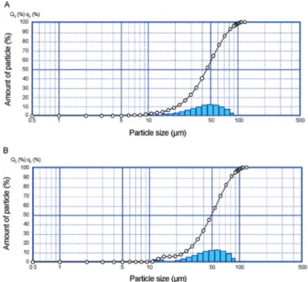
The size of the microcapsules is an important parame-ter that affects the sensory properties of foods; a larger par-ticle size correlates with an increasingly detrimental effect on the food’s texture. Hansenet al.(2002) suggested that a desirable size is approximately 100mm. The laser diffrac-tion confirmed the presence of different particles sizes with a fraction of which were smaller than 100mm, however. The B. animalis subsp. lactisand L. acidophilus micro-particles presented similar diameters, even with the differ-ent portions of reactivated cells:cocoa butter that were applied. The mean diameter of theB. animalissubsp.lactis microparticles was 44.4mm, and the particle size ranged from 1.6 to 126.9mm (Figure 2A); the mean diameter of the L. acidophilusparticles was 49.6mm, and the size ranged from 2.2 to 126.9mm (Figure 2B).
Resistance ofB. animalissubsp.lactisandL. acidophilusto the microencapsulation process
expand the use of probiotics. However, little is known about the effects of microencapsulation on the viability of the cells and the protection of probiotics against external agents. Counts in the emulsion (8.3 log10 cfu/g for B.
animalissubsp. lactis; 6.5 log10cfu/g for L. acidophilus) and in the microcapsules (8.2 log10cfu/g forB. animalis subsp.lactis; 6.6 log10cfu/g forL. acidophilus) suggest that spray chilling did not affect the viability of the probiotics. The conditions that were employed in the process, such as atomization and chilling, were sufficiently mild to maintain the cellular integrity of the majority of the population.
Resistance ofB. animalissubsp.lactisandL. acidophilusto simulated gastric and intestinal fluids
BeforeB. animalissubsp. lactis and L. acidophilus can be used as functional products, they must survive until they reach the gastrointestinal tract, pass through it, and colonise the intestine (Kimet al., 1988). However, one of the major problems in the efficacy of probiotic foods is the low survival rate of the microorganisms at gastric pH and in the high concentrations of bile salts in the intestine (Sabikhi
Figure 1- Optical microscopy images of microparticles prepared by spray chilling containingB. animalissubsp.lactis(A) andL. acidophilus(B) and Scanning electron microscopy images of microparticles containingL. acidophilus.
et al., 2010). Figure 3 shows that both free and encapsulated cells ofB. animalissubsp.lactiswere resistant to SGF and SIF; only a 1.3 logarithmic cycle reduction in the popula-tion was observed during 5 hours of incubapopula-tion.L. acido-philusreached the method detection limit (103 cfu/g) in 210 min in its free form. However, when this bacterium was encapsulated, 67% of the cells remained viable with a 2.7 logarithmic cycle reduction in the population during 5 hours of incubation. So, the solid lipid microparticles pro-duced with cocoa butter offered protection to L. acido-philusinto SGF and SIF. However the 33% of loss could be because of the heterogenic composition and low melting point of the cocoa butter. Some of its triglycerides may have melted at the temperature of this test (37 °C), causing premature release of the probiotics into the SGF. Therefore, considering the loss of viability of the encapsulated B. animalissubsp. lactisand L. acidophilus, microparticles should contain a higher initial number of viable cells in or-der to have an appropriate number of cells released into the intestine or microparticles should be produced with a lipid matrix with higher melted point.
Other studies have confirmed the increase in the resis-tance of the microencapsulated probiotics when they are exposed to simulated gastric and intestinal fluids compared with the survival of free cells.L. acidophiluscells that were encapsulated in sodium alginate were more resistant than the free cells when they were submitted to tests that simu-lated gastric and intestinal conditionsin vitro(Kimet al., 2008; Mokarramet al., 2009; Sabikhiet al., 2010). How-ever, L. acidophilus that were encapsulated in calcium alginate (Favaro-Trindade and Grosso 2000) or in a mix-ture of alginate and starch Sultanaet al.(2006) were not more resistant than the free cells when they were submitted to tests that simulated the stomach acidity and bile condi-tionsin vitro.
Stability of the microencapsulated microorganisms during storage
Cell damage and a loss of activity may occur during the processing and storage of the cells. Therefore, a suitable microencapsulation process should ensure that the microor-ganisms survive processing and remain viable during stor-age (Oliveiraet al., 2007 a and b).
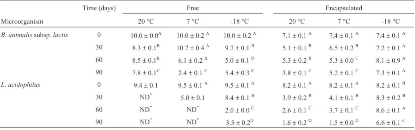
In the present study, according to results that are pre-sented in Table 1, the freeB. animalissubsp.lactis popula-tion was unstable during storage; it displayed logarithmic cycle reductions of 2.2 (20 °C), 7.6 (7 °C) and 4.6 (-18 °C) log cfu/g. Inactivation during storage can be related to a number of factors, such as the formation of free radicals in the presence of oxygen, fatty acid oxidation, and DNA damage (Castroet al., 1997). Microencapsulation did not improve the stability ofB. animalissubsp.lactiswhen the microparticles were stored at 20 °C. However, the encapsu-lated cells were more stable at 7 °C, where 72% of the cells remained viable after 90 days of storage, and at -18 °C, where the microparticle assured a minimum level of 106cfu/g for 90 days (Brasil, 2000).
The population of free and encapsulated cells ofL. acidophilusdecreased drastically during 90 days of storage
Figure 3- Survival of free and encapsulated cells ofB. animalissubsp.
lactisandL. acidophilusafter their exposure to simulated gastric fluid (SGF) and simulated intestinal fluid (SIF).
Table 1- Stability of free and encapsulatedB. animalissubsp. lactis/L. acidophilusduring 90 days of storage (count in Log10cfu/g).
Time (days) Free Encapsulated
Microorganism 20 °C 7 °C -18 °C 20 °C 7 °C -18 °C
B. animalissubsp. lactis 0 10.0±0.0A 10.0±0.2A 10.0±0.2A 7.1±0.1A 7.4±0.1A 7.4±0.1A
30 8.3±0.1B 10.7±0.4A 9.7±0.1B 5.1±0.1B 6.5±0.2B 7.2±0.1A
60 8.5±0.1B 6.1±0.2B 5.0±0.1D 5.3±0.2B 5.3±0.0C 8.1±0.9A
90 7.8±0.1C 2.4±0.1C 5.4±0.3C 3.8±0.1C 5.2±0.1C 7.3±0.1A
L. acidophilus 0 9.4±0.1 9.5±0.1A 9.5±0.1A 8.2±0.1A 8.2±0.1A 8.2±0.1B
30 ND* 5.0±0.1 8.4±0.1B 3.9±0.2B 4.1±0.1B 8.3±0.2B
60 ND* ND* 2.0±0.0C 2.6±0.1C 3.7±0.1C 8.6±0.1A
90 ND* ND*
3.5±0.2D 1.6±0.2D 1.5±0.0D 6.6±0.1C
*
ND = no detected cells.
at 20 °C and 7 °C; the viable free cells were not detected (with limit method detection of 10 cfu/g) after 30 and 60 days of storage, respectively. Moreover only 20% of the encapsulated cells remained viable after 90 days of storage at 7 °C (Table 1). A study by Oliveiraet al.(2007b) showed thatL. acidophilusdisplayed greater viability at a storage temperature of 7 °C; however, in this work, the micro-capsules were dehydrated. The probiotics that were encap-sulated by complex coacervation and were dehydrated by spouted bed showed viability when they were stored at 7 °C for 120 days, but they showed a loss in viability prior to 90 days of storage at 37 °C. In the present study, promising results were observed with freezing conditions, which as-sured 90 days of shelf life; in these conditions, micro-encapsulation increased theL. acidophilus viability from 36.6% (free cells) to 81.1% (microencapsulated cells). These results allow us to conclude that only freezing condi-tions provided a long and satisfactory microparticle shelf life. This result was probably observed because in these conditions, the cells are in a latent state. In freezing temper-atures as -18 °C, the molecular mobility is low, the meta-bolic reactions occur more slowly and thus the microbial activity is very reduced. This result limits the application of these microparticles in food products. Because the mi-croorganisms are metabolically active in the micropar-ticles at 7 °C and 20 °C, the production of such compounds as metabolic acids and bacteriocins or the ab-sence of subtracts may cause inactivation of the viable cells in the microparticles.
The present study shows that spray chilling with co-coa butter as a carrier to produce solid lipid microparticles was efficient in protecting the probiotics against the pas-sage through gastric and intestinal fluids, and they could also be stored at freezing temperatures. In addition, the morphologies and sizes of the microparticles may facilitate the flow of material and would probably cause no harmful effects to food texture. Therefore, this method is an innova-tive matrix for the application of probiotics at low cost with the potential of scaling up. Other advantages are the use of low temperatures, the absence of organic solvents and the possible release of probiotics in the intestine during the di-gestion of the cocoa butter.
A future challenge in further studies is to choose a carrier or a process operation condition that improves the viability of the cells at refrigerated and room temperatures to increase the use of these microparticles in food products and to facilitate their storage.
Acknowledgments
The authors thank FAPESP in Brazil for their finan-cial support (Process 2009/11713-2) and the scholarship that was granted (Process 2009/03140-2). The microorgan-isms and fat were kindly provided by Danisco (Brazil) and Emfal (Brazil), respectively.
References
Brazil, Ministério da Agricultura, Pecuária e Abastecimento (2000) Padrão de identidade e qualidade de leites fermen-tados. Resolução n. 05, de 13 de novembro de 2000. Brasília, 2000. Available at: http://extranet.agricultura.gov.br/ sislegis-consulta/consultarLegislacao.do?operacao=visuali zar&id = 3285.
Castro HP, Teixeira PM Kirby R (1997) Evidence of membrane damage in Lactobacilus bulgaricus following freeze drying. J Appl Microbiol 82:87-94.
Chambi HNM, Alvim ID, Barrera-Arellano D, Grosso CRF (2008) Solid lipid microparticles containing water-soluble compounds of different molecular mass: Production, char-acterization and release profiles. Food Res Int 41:229-236. Champagne CP, Fustier P (2007) Microencapsulation for the
im-proved delivery of bioactive compounds into foods. Food Biotechnol18:184-190.
de Vos P, Faas MM, Spasojevic M, Sikkema J (2010) Encapsula-tion for preservaEncapsula-tion of funcEncapsula-tionality and targeted delivery of bioactive food components. Int Dairy J20:292-302. FAO/WHO (2006) Probiotics in food. Health and nutritional
properties and guidelines for evaluation, FAO Food and Nu-trition Paper No. 85. World Health Organization and Food and Agriculture Organization of the United Nations, Rome. Favaro-Trindade CS, Grosso CRF (2000) The effect of the
immo-bilization of Lactobacillus acidophilus and Bifidobacterium lactis in alginate on their tolerance to gastro-intestinal secre-tions. Milchwissenschaft 55:496-499.
Favaro-Trindade CS, Grosso CRF (2002) Microencapsulation of L. acidophilus (La-05) and B. lactis (Bb-12) and evaluation of their survival at the pH values of the stomach and in bile. J Microencapsul19:485-494.
Favaro-Trindade CS , Heinemann RJB, Pedroso DL (2011) De-velopments in probiotic encapsulation. CAB Reviews: Per-spectives in Agriculture, Veterinary Science, Nutrition and Natural Resources 6:1-8.
Fung WY, Yuen KH, Liong MT (2011) Agrowaste-based nano-fibers as a probiotic encapsulant: fabrication and character-ization. J Agric Food Chem 59:8140-8147.
Gbassi GK, Vandamme T, Ennahar S, Marchioni E (2009) Micro-encapsulation of Lactobacillus plantarum spp in an alginate matrix coated with whey proteins. Int J Food Microbiol 129:103-105.
Gouin S (2004) Microencapsulation: industrial appraisal of exist-ing technologies and trends. Trends Food Sci Technol 15:330-347.
Grosso CRF, Favaro-Trindade CS (2004). Stability of free and immobilized Lactobacillus acidophilus and Bifidobacterium lactis in acidified milk and immobilized B. lactis in yoghurt. Braz J Microbiol 35:151-156.
Hansen LT, Allan-Wojtas PM, Jin LA, Paulson AT (2002) Sur-vival of Ca-alginate microencapsulated Bifidobacterium spp. in milk and simulated gastrointestinal conditions. Food Microbiol 19:35-45.
Kim HS, Kamara BJ, Good IC, Enders Jr GI (1988) Method for the preparation of stabile microencapsulated lactic acid bac-teria. J Ind Microbiol 3:253-257.
Kwok KK,Groves MJ, Burgess DJ (1992) A novel method for the determination of sterility of microparticles and measure-ment of viability of encapsulated organisms. Pharmac Res 13:410-413.
Lahtinen SJ, Ouwehand AC, Salminen SJ, Forssell P, Myllärinen P (2007) Effect of starch and lipid-based encapsulation on the culturability of two Bifidobacterium longum strains. Lett Appl Microbiol 44:500-5.
Liserre AM, Ré MI, Franco BDGM (2007) Microencapsulation of Bifidobacterium animalis subsp. lactis in modified algi-nate-chitosan beads and evaluation of survival in simulated gastrointestinal conditions. Food Biotechnol 21:1-16. Mokarram RR, Mortazavi SA, Habibi Najafi MB, Shahidi F
(2009) The influence of multi stage alginate coating on sur-vivability of potential probiotic bacteria in simulated gastric and intestinal juice. Food Res Int 42:1040-1045.
Okuro PK, Baliero JCC, Liberal RDCO, Favaro-Trindade CS (2013a) Co-encapsulation of Lactobacillus acidophilus with inulin or polydextrose in solid lipid microparticles provides protection and improves stability. Food Res Int 53:96-103. Okuro PK, Matos Jr FE, Favaro-Trindade CS (2013b)
Technolog-ical Challenges for Spray-Chilling Encapsulation of Func-tional Food Ingredients. Food Technol Biotechnol 51:171-182.
Oliveira AC, Moretti TS, Boschini C, Balieiro JCC, Freitas O, Favaro-Trindade CS (2007a) Stability of microencapsulated B. lactis (BI 01) and L. acidophillus (LAC 4) by complex coacervation followed by spray drying dehydration. J Microencapsul 24:685-693.
Oliveira Oliveira AC, Moretti TS, Boschini C, Balieiro JCC, Freitas LAP,Freitas O, Favaro-Trindade CS (2007b) Micro-encapsulation of B. lactis (BI 01) and L. acidophilus (LAC 4) by complex coacervation followed by spouted-bed dry-ing. Dry Technol 25:1687-1693.
Pedroso DL, Thomazini M, Heinemann RJB, Favaro-Trindade CS (2012) Protection of Bifdobacterium lactis and Lacto-bacillus acidophilus by microencapsulation using spray-chilling. Int Dairy J 26:127-132.
Rodriguez L, Passerini N, Cavallari C, Cini M, Sancin P, Fini A (1999) Description and preliminary evaluation of a new ul-trasonic atomizer for pray-73 congealing processes. Int J Pharm 183:133-143.
Rokka S, Rantamaki, P. (2010). Protecting probiotic bacteria by microencapsulation: challenges for industrial applications. Eur Food Res Technol 231, 1-12.
Sabikhi L, Babu R, Thompkinson DK, Kapila S (2010) Resistance of microencapsulated Lactobacillus acidophilus LA1 to pro-cessing treatments and simulated gut conditions. Food Bio-process Technol 3:586-593.
Sato K, Ueno S (2005) Polymorphism in fats and oils. Industrial oil and fat products edible oil and fat products: Chemistry properties and health effects. Inc F. Shahidi Ed., New York, USA.
Savolainen M, Khoo C, Glad H, Dahlqvist C, Juppo AM (2002) Evaluation of controlled-release polar lipid microparticles. Int J Pharm 244:151-161.
Sultana K, Godward G, Reynolds N, Arumugaswamy R Peiris P, Kailasapathy K (2006) Encapsulation of probiotic bacteria with alginate-starch and evaluation of survival in simulated gastrointestinal conditions and in yoghurt. Int J Food Micro-biol 62:47-55.
Westesen K, Bunjes H, Koch MHJ (1997) Physicochemical char-acterization of lipid nanoparticles and evaluation of their drug loading capacity and sustained release potential. J Con-trol Release 48:223-236.