Identities among actin-encoding cDNAs of the Nile tilapia
(
Oreochromis niloticus
) and other eukaryote species revealed
by nucleotide and amino acid sequence analyses
Andréia B. Poletto
1, Adriane P. Wasko
2, Claudio Oliveira
1, Alexandre Azevedo
1, Robson F. Carvalho
1,
Maeli Dal Pai Silva
1, Fausto Foresti
1and Cesar Martins
11
Departamento de Morfologia, Instituto de Biociências, Universidade Estadual Paulista, Campus de
Botucatu, Botucatu, SP, Brazil.
2
Departamento de Genética, Instituto de Biociências, Universidade Estadual Paulista, Campus de Botucatu,
Botucatu, SP, Brazil.
Abstract
Actin-encoding cDNAs of Nile tilapia (Oreochromis niloticus) were isolated by RT-PCR using total RNA samples of different tissues and further characterized by nucleotide sequencing andin silico amino acid (aa) sequence analysis. Comparisons among the actin gene sequences ofO. niloticus and those of other species evidenced that the isolated genes present a high similarity to other fish and other vertebrate actin genes. The highest nucleotide resemblance was observed betweenO. niloticus and O. mossambicusα-actin andβ-actin genes. Analysis of the predicted aa se-quences revealed two distinct types of cytoplasmic actins, one cardiac muscle actin type and one skeletal muscle actin type that were expressed in different tissues of Nile tilapia. The evolutionary relationships between the Nile tilapia actin genes and diverse other organisms is discussed.
Key words:actin, expression pattern, Nile tilapia,Oreochromis niloticus.
Received: August 22, 2006; Accepted: April 18, 2007.
Actin is a ubiquitous protein of eukaryotic cells that has a crucial role in muscle contraction, cell motility, cyto-skeletal structure, cell division, intracellular transport, and cell differentiation (Herman, 1993). In yeast and some pro-tozoans, actin is encoded by a single gene only (Hightower and Meagher, 1986; Reeceet al., 1997). However, in the nuclei of all animals, plants and in many protozoans exam-ined to date, actin proteins are encoded by a multigene fam-ily. In these organisms it seems that actin isoforms are encoded by a set of structurally related genes that resulted from gene duplications followed by functional divergence (Hightower and Meagher, 1986). The number of actin iso-forms varies greatly in different lineages. While mammals posses at least six different isoforms (Vandekerckhove and Weber, 1978), teleost fishes contain at least nine (Ven-kateshet al., 1996) and echinoderm genomes at least eight (Fang and Brandhorst, 1994) distinct actin isoforms. Simi-larly, insects have at least six actin genes (Fyrberget al., 1980). The actin gene family of plants is much larger,
com-prising 8-44 genes, depending on the taxon (Reeceet al., 1992; Drouin and de Sá, 1996).
The actin gene family can be divided into two broad categories: cytoplasmic (β and γ) and muscle (α) type actins. Invertebrate muscle and cytoplasmic actins seem to be more similar to chordate cytoplasmic actins than to chordate muscle actins (Vandekerckhove and Weber, 1984). It has been suggested that the muscle actins of ar-thropods differ from the muscle actins of deuterostomes to such an extent that two independent divergence events of muscle actin genes probably occurred, one within the protostome lineage and one within the deuterostome lin-eage (Mounieret al., 1992).
Although there are data on the evolution of mamma-lian actin genes, the evolutionary origin, pattern of organi-zation, and the diversity of these genes in other vertebrates, especially fishes, remain to be investigated. To date, an in-depth research on the diversity of actin gene types and tissue expression profiles in a fish species was only per-formed onTakifugu rubripes, revealing nine different actin genes: six muscle-type actin genes that include twoα -skel-etal actins, three α-cardiac actins, an α-anomalous tes-tis-type actin, and three cytoplasmic actins that include two Send correspondence to Cesar Martins. Departamento de
Morfolo-gia, Instituto de Biociências, Universidade Estadual Paulista, Cam-pus de Botucatu, 18618-000 Botucatu, SP, Brazil. E-mail: [email protected].
β-cytoplasmic actins and oneβ-cytoplasmic vascular-type actin (Venkateshet al., 1996). The purpose of the present study was the isolation and characterization of distinct actin cDNAs from different tissues of the Nile tilapia (Oreochromis niloticus) - one of the most important food fish species intensively exploited in tropical and subtropi-cal aquaculture (Pullin, 1991) - not only to enhance our knowledge of the species, but also to provide a better under-standing of the organization of the actin multigene family in fish genomes.
Total RNA samples were obtained from different tis-sues (gills, heart, ovaries, skeletal muscle, liver, and brain) of two adult individuals of Oreochromis niloticus using TRIzol reagent (Gibco-Brl Life Technologies), following the manufacturer’s instructions. First-strand cDNA synthe-sis reactions were performed with the SuperScript First-Strand Synthesis System for RT-PCR (Invitrogen Life Technologies) using random hexamer primers. cDNA am-plification was performed using the primer setsαActF (5’-ATGAGACTACCGCCCTTGTG-3’) andαActR (5’-AAT CCACATCTGCTGGAAGG-3’) forα-actin gene amplifi-cation, and βActF (5’-TGTTGACAATGGATCCGGTA-3’) andβActR (5’-CTGCTGGAAGGTGGAGAGAG-3’) forβ-actin gene amplification. Both primer sets were de-signed using the software Primer3 (Rozen and Skaletsky, 2000) based on α-actin and β-actin gene sequences de-scribed previously forT. rubripes(Venkateshet al., 1996). RT-PCR products were electrophoresed and visualized on a 1% agarose gel, purified and ligated into pCR2.1 plasmid (TA Cloning Kit, Invitrogen) used to transform DH5αE. colicompetent cells. Plasmid DNA was purified with the Wizard Plus SV Minipreps DNA Purification System Kit (Promega) and submitted to nucleotide sequencing on an ABI 377 Automated DNA Sequencer (Applied Bio-systems).
Nucleic acid and amino acid sequences ofO. niloticus
were analyzed using BLASTn and BLASTx (Altschulet al., 1990). Additionally, sequences from different organ-isms obtained from GenBank, were aligned with O. niloticus sequence data using the software ClustalW (Thompsonet al., 1994); alignment was checked by eye and adjusted as necessary. Phylogenetic analyses using the putative aa sequences derived fromO. niloticusclones and other aa sequences from previously published actin genes in GenBank were performed using MEGA version 3.1 (Kumaret al., 2004). Phylogenetic trees were constructed using the neighbor-joining method (Saitou and Nei, 1987).
RT-PCR ofOreochromis niloticus cDNA samples, obtained from total RNA extracted from gills, liver, heart, ovary, skeletal muscle and brain, was performed using the two sets of primersαActF/αActR andβActF/βActR in or-der to amplifyα-actin andβ-actin gene sequences, respec-tively. Irrespective of the tissue or the primer set used, all PCR amplifications resulted in one fragment of
approxi-mately 1,100 base pairs (bp) that was cloned and sequenced.
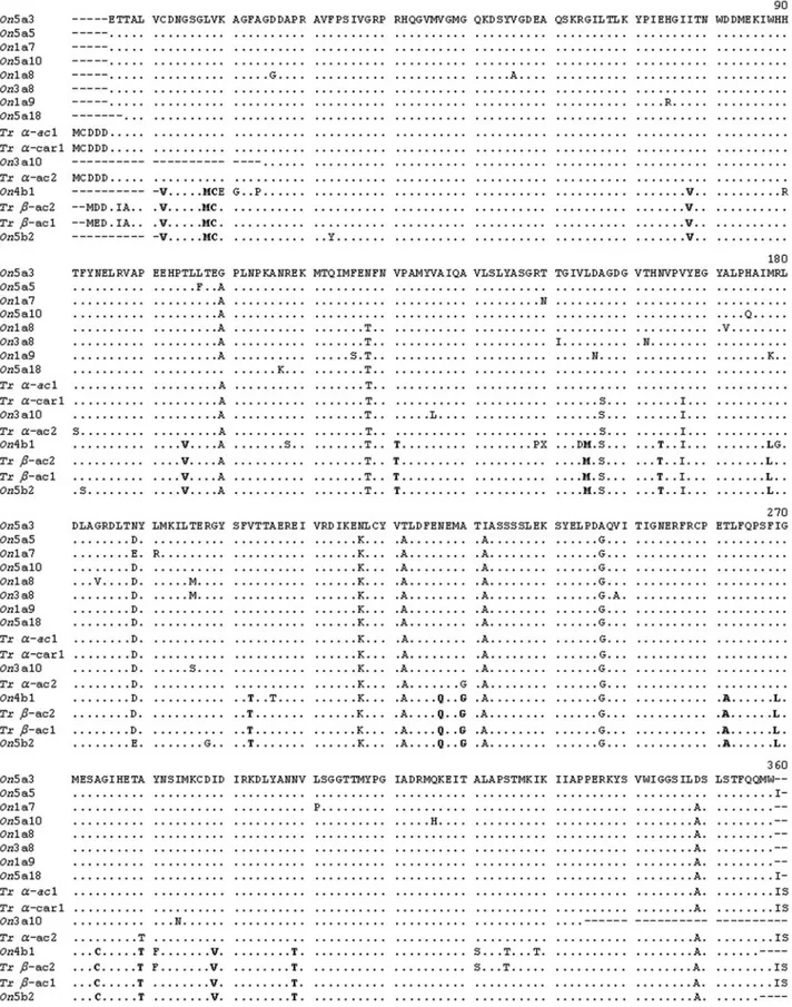
A total of 11 clones were isolated and sequenced in both directions from two individuals ofO. niloticus. These represented three skeletal muscle typeα-actins from gills (On1a7, On1a8, and On1a9); one skeletal muscle type α-actin from heart (On3a8); one cardiac muscle type α -actin from heart (On3a10c); one β-actin from ovary (On4b1); four skeletal muscle type α-actins (On5a18,
On5a10,On5a3 andOn5a5) and oneβ-actin (On5b2) from skeletal muscle tissue (Figure 1). The nucleotide sequences were deposited in the GenBank database under the acces-sion numbers EF206791-EF206801. The obtained se-quences of the amplified cDNAs ofO. niloticus revealed segments ranging in size from 884 to 1,063 bp. The differ-ences in the sequence sizes were due to failures in the se-quencing procedure.
Searches in the NCBI database by means of BLASTn indicated that the isolated cDNA nucleotide sequences from O. niloticuswere very similar to several fish actin gene sequences, especially toα-actins andβ-actins of O. mossambicus(99% mean nucleotide identity between the two species). The putative amino acid sequences of the iso-lated cDNAs fromO. niloticuswere also compared to skel-etal actin1, skelskel-etal actin2, cardiac1 alpha actin, cytoplas-mic actin1 and cytoplascytoplas-mic actin2 genes of T. rubripes
(Venkateshet al., 1996), which resulted in a 98,4% mean identity (Figure 1).
Although the nucleotide sequences of the isolated cDNAs ofO. niloticusand the actin genes ofT. rubripes
differ by several nucleotides, their inferred aa sequences present a high degree of similarity, approximately 98%, since most nucleotide variation corresponds to synony-mous substitutions and, thus, their aa residues are mostly identical (Figure 1). Diagnostic aa positions that distin-guish α-muscle actin from β-cytoplasmic actin of Nile tilapia could be observed (Figure 1). These aa positions were also identified in the isolated cDNAs ofT. rubripes
(Venkateshet al., 1996). Most amino acids that distinguish fish α-striated muscle actins from β-cytoplasmic actins correspond to those that also distinguish mammalianα- and β-actins (Mounier and Sparrow, 1997). Diagnostic amino acids 281, 321 and 325 that distinguishT. rubripesβ -cyto-plasmic actin2 fromβ-cytoplasmic actin1 were identified in the isolated cDNA sequence On4b1 of O. niloticus, which validates the occurrence of a second type of cyto-plasmic actin gene expressed in fish species. This is a novel finding, since the expression ofβ-cytoplasmic actin2 has not yet been reported, only its genomic sequence (Ven-kateshet al., 1996).
myxamoeba speciesDictyostelium discoideumwas used as an outgroup. The animal actins were clearly discriminated from plant actins in 95% of the recovered trees. Not surpris-ingly, a close relationship was observed between Nile tila-pia and other vertebrate α-actins. The clustering of the isolated Nile tilapia α-actins with other vertebrate and ascidian muscle actins in the same clade was strongly ported in 100% of bootstrap replicates. This strongly sup-ported relationship between vertebrate and ascidian muscle actins has been already demonstrated by the comparison of diagnostic aa and phylogenetic analyses, suggesting that the chordate muscle-type actins probably diverged from a nonmuscle-like actin before the divergence of urochordates and vertebrates, but presumably after the divergence of echinoderms and chordates (Kusakabeet al., 1997). The actin genes allowed the discrimination of higher taxa, but
were not informative for lower taxonomic levels because of the high conservation at DNA sequence level.
The clade composed of the isolated Nile tilapia cyto-plasmic actins and vertebrate cytocyto-plasmic actins is sup-ported in 84% of the bootstrap replicates. An interesting point was the presence of invertebrate muscle and cytoplas-mic actins in the clade of vertebrate cytoplascytoplas-mic actins (tree node percentage recovery of 65%). This relationship is in accordance with previous analyses that suggest that
non-Figure 2- Molecular phylogenetic tree inferred by the neighbor-joining method from predicted amino acid (aa) sequences of actin genes. The myxamoeba speciesDictyostelium discoideumwas used as an outgroup. Branch lengths are proportional to evolutionary distances. Scale bar indi-cates an evolutionary distance of 0.05 aa substituition per position in the sequences. The numbers at each node indicate the percentage recovery (> 60%) of the particular node (500 bootstrap replicates) in which the same internal branch was recovered. Sources and accession numbers for the actin sequences are described in Material and Methods.
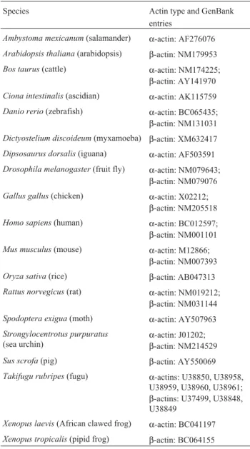
Table 1- Species and accession numbers of actin cDNA sequences ob-tained from GenBank.
Species Actin type and GenBank
entries
Ambystoma mexicanum(salamander) α-actin: AF276076
Arabidopsis thaliana(arabidopsis) β-actin: NM179953
Bos taurus(cattle) α-actin: NM174225; β-actin: AY141970
Ciona intestinalis(ascidian) α-actin: AK115759
Danio rerio(zebrafish) α-actin: BC065435; β-actin: NM131031
Dictyostelium discoideum(myxamoeba) β-actin: XM632417
Dipsosaurus dorsalis(iguana) α-actin: AF503591
Drosophila melanogaster(fruit fly) α-actin: NM079643; β-actin: NM079076
Gallus gallus(chicken) α-actin: X02212; β-actin: NM205518
Homo sapiens(human) α-actin: BC012597; β-actin: NM001101
Mus musculus(mouse) α-actin: M12866; β-actin: NM007393
Oryza sativa(rice) β-actin: AB047313
Rattus norvegicus(rat) α-actin: NM019212; β-actin: NM031144
Spodoptera exigua(moth) α-actin: AY507963
Strongylocentrotus purpuratus
(sea urchin)
α-actin: J01202; β-actin: NM214529
Sus scrofa(pig) β-actin: AY550069
Takifugu rubripes(fugu) α-actins: U38850, U38958, U38959, U38960, U38961; β-actins: U37499, U38848, U38849
Xenopus laevis(African clawed frog) α-actin: BC041197
chordate muscle actin genes are more closely related to ver-tebrate cytoplasmic actins than to verver-tebrate muscle actins (Kusakabe et al., 1997). The actins expressed in muscle cells of non-chordates have traditionally been considered to be cytoplasmic-like (Vandekerckhove and Weber, 1984), and non-muscle actins are likely to represent ancestral actin forms.
As evidenced for other organisms, different fish actin types also seem to be under different evolutionary selection pressures, leading to the conjecture that these isoforms seem to have somewhat different roles. Further analyses comparing the organization of distinct actin isoforms from several species will be useful for understanding the molec-ular evolution and function of these genes in fishes.
Acknowledgments
This work was supported by grants from FAPESP (Fundação de Amparo à Pesquisa do Estado de São Paulo), CNPq (Conselho Nacional de Desenvolvimento Científico e Tecnológico), and CAPES (Coordenação de Aperfeiçoa-mento de Pessoal de Nível Superior).
References
Altschul SF, Gish W, Miller W, Myers EW and Lipman DJ (1990) Basic local alignment search tool. J Mol Biol 215:403-410. Drouin G and de Sá MM (1996). Phylogeny and substitution rates
of angiosperm actin genes. Mol Biol Evol 13:1198-1212. Fang H and Brandhorst BP (1994) Evolution of actin gene
fami-lies of sea urchins. J Mol Evol 39:347-356.
Fyrberg EA, Kindle KL and Davidson N (1980) The actin genes ofDrosophila: A dispersed multigene family. Cell 19:365-378.
Herman IM (1993) Actins isoforms. Curr Opin Cell Biol 5:48-55. Hightower RC and Meagher RB (1986) The molecular evolution
of actin. Genetics 114:315-332.
Kumar S, Tamura K and Nei M (2004) MEGA 3: Integrated soft-ware for molecular evolutionary genetics analysis and se-quence alignment. Brief Bioinform 5:150-163.
Kusakabe T, Araki I, Satoh N and Jeffrey WR (1997) Evolution of chordate actin genes: Evidence from genomic organization and amino acid sequences. J Mol Evol 44:289-298. Mounier N and Sparrow JC (1997) Structural comparisons of
muscle and nonmuscle actins give insights into the evolution of their functional differences. J Mol Evol 44:89-97. Mounier N, Guoy M, Mouchiroud D and Prudhomme C (1992)
Insect muscle actins differ distinctly from invertebrate and vertebrate cytoplasmic actins. J Mol Evol 34:406-415.
Pullin RSV (1991) Cichlids in aquaculture. In: Keenleyside MHA (ed) Cichlid Fishes: Behaviour, Ecology and Evolution. Chapman & Hall, New York, pp 280-309.
Reece KS, McElroy D and Wu R (1992) Function and evolution of actins. Evol Biol 26:1-34.
Reece KS, Siddall ME, Burreson EM and Graves JE (1997) Phylogenetic analysis ofPerkinsusbased on actin gene se-quences. J Parasitol 83:417-423.
Rozen S and Skaletsky HJ (2000) Primer3 on the WWW for gen-eral users and for biologist programmers. In: Krawetz S, Misener S (eds) Bioinformatics Methods and Protocols: Methods in Molecular Biology. Humana Press, Totowa, pp 365-386.
Saitou N and Nei M (1987) The neighbor-joining method: A new method for reconstructing phylogenetic trees. Mol Biol Evol 4:406-425.
Thompson JD, Higgins DG and Gibson TJ (1994) Clustal W: Im-proving the sensitivity of progressive multiple sequence alignment through sequence weighting, position-specific gap penalties and weight matrix choice. Nucleic Acids Res 22:4673-4680.
Vandekerchkove J and Weber K (1978) At least six different actins are expressed in a higher mammal: An analysis based on the amino acid sequences of the amino-terminal tryptic peptide. J Mol Biol 126:783-802.
Vandekerckhove J and Weber K (1984) Chordate muscle actin differ distinctly from invertebrate muscle actins. The evolu-tion of the different vertebrate muscle actins. J Mol Biol 179:391-413.
Venkatesh B, Tay BH, Elgar G and Brenner S (1996) Isolation, characterization and evolution of nine pufferfish (Fugu rubripes) actin genes. J Mol Biol 259:655-665.
Internet Resources
BLAST: The Basic Local Alignment Search Tool (BLAST) is a WWW service of National Center for Biotechnology Infor-mation (NCBI). Available from http://ncbi.nml.nih.gov/ blast/ (May 20, 2006).
ClustalW: WWW Service at the European Bioinformatics Insti-tute. Available from http://www.ebi.ac.uk/clustalw (May 20, 2006).
GenBank: GenBank® is the National Institutes of Health (NIH) genetic sequence database, an annotated collection of all publicly available DNA sequences. Available from http://www.ncbi.nlm.nih.gov/GenBank/ (January 03, 2007). Primer3: Primer3 is a widely used program for designing PCR primers. Available from http://fokker.wi.mit.edu/primer3/ (May 20, 2006).
Associate Editor: Pedro Manoel Galetti Junior