Microtubule-Mediated Inositol Lipid Signaling
Plays Critical Roles in Regulation of Blebbing
Tatsuroh Sugiyama1, Md. Kamruzzaman Pramanik1,2, Shigehiko Yumura1*
1Department of Functional Molecular Biology, Graduate School of Medicine, Yamaguchi University, Yamaguchi, Japan,2Microbiology & Industrial Irradiation Division, IFRB, AERE, Bangladesh Atomic Energy Commission, Dhaka, Bangladesh
*yumura@yamaguchi-u.ac.jp
Abstract
Cells migrate by extending pseudopods such as lamellipodia and blebs. Although the sig-nals leading to lamellipodia extension have been extensively investigated, those for bleb extension remain unclear. Here, we investigated signals for blebbing inDictyosteliumcells using a newly developed assay to induce blebbing. When cells were cut into two pieces with a microneedle, the anucleate fragments vigorously extended blebs. This assay enabled us to induce blebbing reproducibly, and analyses of knockout mutants and specific inhibitors identified candidate molecules that regulate blebbing. Blebs were also induced in anucleate fragments of leukocytes, indicating that this assay is generally applicable to ani-mal cells. After cutting, microtubules in the anucleate fragments promptly depolymerized, followed by the extension of blebs. Furthermore, when intact cells were treated with a micro-tubule inhibitor, they frequently extended blebs. The depolymerization of micromicro-tubules induced the delocalization of inositol lipid phosphatidylinositol 3,4,5-trisphosphate from the cell membrane. PI3 kinase-null cells frequently extended blebs, whereas PTEN-null cells extended fewer blebs. From these observations, we propose a model in which microtubules play a critical role in bleb regulation via inositol lipid metabolism.
Introduction
Various locomotive cells such as neutrophils, fibroblasts, keratocytes, andDictyosteliumcells extend lamellipodia via actin polymerization. Actin polymerizes at the leading edge and pushes against the anterior cell membrane, resulting in the extension of lamellipodia [1]. However, certain cells migrate by extending blebs via a process that is independent of the force of actin polymerization [2,3]. Blebs are extended when the cell membrane is locally decoupled and sep-arated from the underlying actin cortex, which induces outward cytoplasmic flow via intracel-lular pressure. The intracelintracel-lular pressure (hydrostatic pressure) is generated by the contraction of cortical actin and myosin II [2,4]. The power generated by myosin II appears to be crucial for blebbing, which is mediated by signaling via the small G protein Rho and Rho-associated protein kinase (ROCK) in mammalian cells [3,5].
a11111
OPEN ACCESS
Citation:Sugiyama T, Pramanik M.K, Yumura S (2015) Microtubule-Mediated Inositol Lipid Signaling Plays Critical Roles in Regulation of Blebbing. PLoS ONE 10(8): e0137032. doi:10.1371/journal. pone.0137032
Editor:Yulia Komarova, University of Illinois at Chicago, UNITED STATES
Received:May 7, 2015
Accepted:August 11, 2015
Published:August 28, 2015
Copyright:© 2015 Sugiyama et al. This is an open access article distributed under the terms of the
Creative Commons Attribution License, which permits unrestricted use, distribution, and reproduction in any medium, provided the original author and source are credited.
Data Availability Statement:All relevant data are within the paper and its Supporting Information files.
Funding:The authors have no support or funding to report.
Bleb-driven migration is especially prominent in three-dimensional environments, such as in collagen gel, whereas lamellipodia predominate during migration on flat surfaces, such as on a coverslip [6,7]. Furthermore, the experimental induction of blebbing enables cells to invade into three-dimensional environments [8,9]. Germ cells move to their correct locations in zebra-fish embryos simply by repeated directional blebbing [10]. Some cancer cells can migrate by switching between lamellipodia extension and blebbing, and the extension mechanisms leading lamellipodia and blebs are mutually exclusive [11]. For example, upon knocking down Brick 1, which is a subunit of the WAVE complex that is involved in actin polymerization to drive lamellipodia, HeLa cells extend blebs rather than lamellipodia [12]. A balance between the activities of Rho and Rac is implicated as a signal for the switch [13,14]; however, a comprehen-sive picture of the signaling scheme for blebbing has not yet been obtained.
Although an abundance of literature exists regarding the physiological role of blebbing, blebs are occasionally considered to be by-products of apoptotic and necrotic processes or as pathological phenomena that occur under physical or chemical stress. However, blebs are not essential for these processes [15] and have recently been recognized as protrusions representing a distinct mode of cell migration. Bleb-mediated cell migration toward chemotactic signals has been reported in fish embryos [10,16] andDictyosteliumcells [17].
The cellular slime moldDictyosteliumhas been studied as a model organism for cell migra-tion, chemotaxis, and cytokinesis [18–22].Dictyosteliumcells can extend both lamellipodia and blebs [23]. When these cells are uniformly stimulated with a chemoattractant, they extend blebs [24]. A recent study has revealed that cells extend blebs toward a chemoattractant gradi-ent, indicating that blebs can be integrated into chemotactic cell migration [17]. However, the frequency of bleb extension is too low to be analyzed experimentally in a quantitative manner.
In the present study, we developed a new assay to investigate blebbing inDictyosteliumcells. When aDictyosteliumcell was cut into two pieces with a microneedle, the anucleate fragment vigorously extended blebs. This assay enabled us to induce blebbing and to identify candidates involved in blebbing regulation in many knockout mutants. After cutting, microtubules in the anucleate fragments immediately depolymerized, followed by bleb extension. The depolymeri-zation of microtubules resulted in delocalidepolymeri-zation of the inositol lipid PIP3 from the cell mem-brane. Furthermore, PI3 kinase-null cells extended blebs more frequently, whereas PTEN-null cells extended fewer blebs. From these observations, we proposed a model in which microtu-bules play a role in blebbing via regulating inositol lipid metabolism.
Results
Lamellipodia and blebs in
Dictyostelium
cells
Dictyosteliumcells extend both lamellipodia and blebs.Fig 1Ashows live images of a typical cell expressing GFP-ABD, a probe for actin filaments. A representative kymograph shows that actin constantly assembled in the forward direction as the lamellipodia extended (Fig 1C). In contrast, blebs suddenly extended and presented a rounder and smoother shape than lamellipodia (Fig 1B). Actin filaments were not detected along the edges of blebs immediately following extension, and the actin cortex remained at the basal region of the blebs (Fig 1D), suggesting that the cell membrane was detached from the actin cortex and that unlike lamellipodia extension, bleb exten-sion is not driven by actin polymerization. Approximately 1.5 seconds after the blebs extended, actin filaments began to appear along their edges, and the old actin cortex gradually disappeared from the basal region (Fig 1B and 1D). The velocity of bleb extension (Fig 1E, 3.09 ± 0.87μm /
sec, n = 21) was 4 times faster than that of lamellipodia extension (0.73 ± 0.17μm / sec, n = 21).
Induction of blebbing by microsurgery
Swanson and Taylor [25] showed that when aDictyosteliumcell is cut in half, the nucleate frag-ment resumes normal migration within seconds, while the anucleate fragfrag-ment does not migrate and instead repeats bleb-like extensions. We first attempted to confirm this observation.Fig 2Ashows a typical microsurgery experiment in which a migrating cell expressing GFP-ABD was cut by a microneedle under confocal microscopy. The anucleate fragments frequently extended bleb-like extensions (arrows).Fig 2Bshows enlarged sequential images of a typical Fig 1. Lamellipodia and blebs inDictyosteliumcells.(A and B) Phase contrast and fluorescence images of a typical cell expressing GFP-ABD, an actin filament probe. The cell extended lamellipodia (arrow) followed by blebs (large arrowheads). (C) Kymographs generated from the rectangles in panel A. Note that actin assembly advanced as the lamellipodium extended. Here, the white lines indicate the leading edge of the lamellipodium. (D) Kymographs generated from the rectangles in panel B. A bleb showing a sudden extension (asterisks). Note that actin was not detected along the edge of the initial extension of the bleb and that the actin cortex remained at the basal region of the bleb (small arrowheads in panel B). Approximately 2 seconds later, actin gradually appeared along the edge (arrow). (E) The velocity of lamellipodia and bleb extension (n = 21). (F) The frequency of the appearance of lamellipodia and blebs. Note that lamellipodia were frequently observed (14.4±7.9 times / 5 min), whereas blebs were rarely observed (0.7±1.8 times / 5 min). Bars, 2.5μm.
Fig 2. Induction of blebs.(A) A typical microsurgery experiment. A cell expressing GFP-ABD was cut using a microneedle under confocal microscopy. The nucleate fragment continued to migrate, extending a lamellipodium even after cutting. Here, the asterisks indicate the nucleus. The anucleate fragment did not migrate and instead repeatedly extended blebs (arrows). Bar, 10μm. (B) Time course of bleb-like extension
bleb-like extension of an anucleate fragment.Fig 2Cshows a kymograph generated from the rectangles inFig 2B. The average velocity of their extension (3.33 ± 1.02μm / sec, n = 24) was
approximately comparable to that of bleb extensions in uncut cells. Actin filaments were not detected along the leading edges of the blebs at the initial time of extension, indicating that the extensions in anucleate fragments are‘blebs,’as judged from the above criteria. The nucleate fragments did not extend blebs and normally migrated by extending lamellipodia, suggesting that the wound caused by microsurgery does not induce blebbing; rather, the loss of the nucleus or a nucleus-related factor induces blebbing.
Reversible induction of blebbing
Interestingly, upon disconnecting the cytoplasm in the middle of a cell by placing a micronee-dle through the cell, the half without a nucleus vigorously extended blebs (Fig 2D), whereas the half with a nucleus did not extend blebs. In this experiment, the cell was not actually cut because both halves of the cytoplasm rejoined, the blebbing ceased, and the cell resumed nor-mal migration once the microneedle was removed. These experiments could be repeated in the same cells to reversibly induce blebbing, supporting the idea that the nucleus or a nucleus-related factor suppresses blebbing.
Genetic analysis of blebbing
There is little information available regarding the intracellular signals that induce bleb forma-tion. Many knockout mutants defective in chemotaxis-, cytoskeleton-, and membrane traffic-related genes are available in theDictyosteliumresearch community. The microsurgery assay enabled us to survey what signals and proteins are necessary for bleb formation. The number of blebs was examined upon cutting mutant cells by microsurgery.Fig 3Asummarizes the results (seeS1 Tablefor detailed information regarding the mutants). The mutant cells are cate-gorized into the following three groups: signal proteins, membrane trafficking proteins, and cytoskeleton-related proteins. They extended blebs with varying frequencies. Regarding signal proteins, cells deficient ingbpC-/gbpD(guanylate cyclases),pkgB(serine/threonine-protein kinase),iplA(Ca2+channel), orpi3k(phosphatidylinositol 3-kinase) extended blebs more fre-quently than wild type cells. In contrast, cells deficient inpkbA(AKT/PKB),pten(phosphatase and tensin homolog), orpakC(homolog of mammalian p21-activated kinase) extended blebs infrequently. Regarding membrane trafficking proteins, cells deficient inLvsA(contractile vac-uole protein) extended blebs more frequently than wild-type cells.
Chemotactic signaling inDictyosteliumcells has been extensively investigated; there are 4 main routes of signal transduction from membrane receptor to cytoskeleton [26]. Mutant anal-ysis revealed that the transduction route involving phosphoinositol was clearly involved in the bleb formation, although other routes, such as those involving PLA2 and IplA, may also have roles in this process (Fig 3B). Thus, we focused on phosphoinositol signaling in the latter part of this study.
that the bleb-like structure extended very quickly and that actin did not localize to its leading edge
immediately after extension, suggesting that these extensions from the anucleate fragments are‘blebs.’Bar, 2.5μm. (D) The cytoplasm was disconnected in the middle of the cell by placing a microneedle into the cell
under DIC microscopy. After disconnecting the cytoplasm, the half without a nucleus (arrowhead) vigorously extended blebs (arrows), and the other half with a nucleus did not extend them. Both sides of the cytoplasm rejoined when the microneedle was removed, showing that the cytoplasm was not actually cut. After the microneedle was removed, the blebbing ceased. These disconnection experiments could be repeated in the same cells and reversibly induced blebbing. Bar, 10μm.
Regarding cytoskeleton-related proteins,ctxA/ctxB(cortexillin)-null cells andlimB(LIM domain-containing protein)-null cells extended blebs more frequently than wild-type cells, suggesting that these actin-binding proteins contribute to the regulation of bleb formation. In contrast, myosin II-null cells extended virtually no blebs.
Fig 3. Genetic analysis of blebbing.(A) The frequency of blebbing in various knockout mutants. Knockout mutants in signal proteins, membrane trafficking proteins, and cytoskeleton-related proteins were subjected to the microsurgery-based blebbing assay. Detailed information regarding these mutants is provided inS1 Table. The blebs were counted for 5 min after the mutant cells were cut by microsurgery. (B) Mapping of the assayed genes onto the chemotactic signal network. The binding of cAMP to the cAMP receptor on the membrane activates multiple intracellular signaling cascades, transmits signals to cytoskeletal proteins, and finally drives cell migration toward the chemoattractant. Red, mutants that showed increased blebbing; blue, mutants that showed reduced blebbing; green, no detectable difference compared to wild type cells (as determined by the t-test statics).
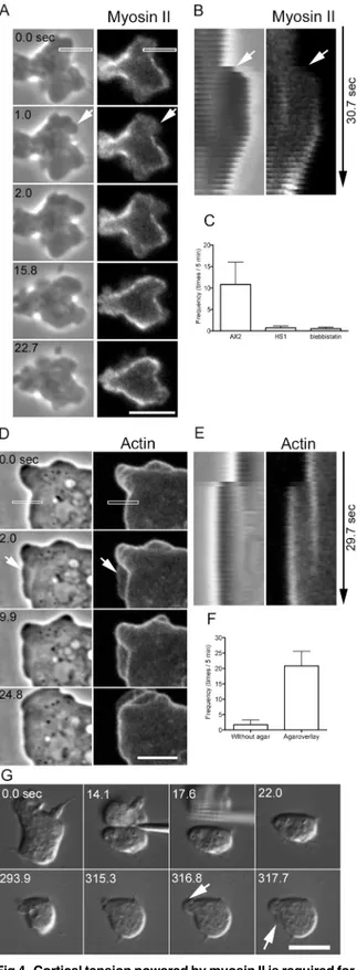
Cortical tension powered by myosin II is required for blebbing
It has been reported that myosin II contributes to bleb formation in various types of cells. We next investigated the role and dynamics of myosin II in the blebbing of cell fragments. Cells expressing GFP-myosin II were cut, and the dynamics of myosin II in the anucleate fragments were observed by confocal microscopy (Fig 4A). In many cases, blebs extended from cortexes that possessed little myosin II. Similar to actin, myosin II was not found along the leading edges of the blebs immediately after extension, and the preexisting cortical myosin II remained in the basal region. Myosin II subsequently appeared along the bleb edge (Fig 4B), followed by bleb retraction, suggesting that the recruitment of myosin II powers the retraction. However, the blebs did not always retract; in some cases, new blebs extended from pre-existing blebs (data not shown).
Next, we investigated whether myosin II contributes to bleb extension. Uncut myosin II-null cells (HS1) did not extend blebs; this observation is consistent with previous works [24]. When myosin II-null cells were cut, virtually no blebs were observed in the anucleate fragments (Fig 4C), although very small and thin extensions were sometimes observed (data not shown). When wild type cells were cut in the presence of blebbistatin, which is an inhibitor of myosin ATPase, virtually no blebs were observed (Fig 4C). Therefore, the activity of myosin II is required for the extension of blebs in anucleate fragments as well.
Dictyosteliumcells do not have large actomyosin bundles such as stress fibers. Myosin II mainly localizes at the cortex [27,28]. The extension of blebs may be caused by the intracellular pressure generated by the cortical actomyosin. To assess this possibility, pressure was applied to cells expressing GFP-ABD using an agar block. Under this pressure, the cells frequently extended bleb-like extensions (arrows inFig 4D). Under agar pressure, the morphology of the extensions appeared different from that of blebs not subjected to pressure (Fig 1B).Fig 4E shows a kymograph generated from the rectangle inFig 4D, indicating that these extensions are‘blebs,’according to the criteria mentioned in the previous section.Fig 4Fshows the fre-quency of blebbing in uncut cells with and without pressure, showing that cells under pressure extended blebs more frequently. Myosin II-null cells also extended blebs after being pressed with agar (data not shown). In addition, anucleate fragments in myosin II-null cells also extended blebs upon being pressed with agar (arrows inFig 4G).
Taken together, these results indicate that myosin II contributes to the extension of blebs by increasing intracellular pressure.
Microtubule disassembly induces blebbing
After cells expressing GFP-tubulin were cut, microtubules disassembled in the anucleate frag-ments, whereas the nucleate fragments retained a microtubule network (Fig 5A). Most likely, the microtubule-organizing center (MTOC), which is firmly associated with the nucleus [29], thereby follows the nucleus to the nucleate fragment after cutting. In the anucleate fragments, the physically cut microtubules must disassemble because they have free minus ends.
Blebbing always began after the microtubules disappeared, suggesting that the disassembly of microtubules induces blebbing (arrows inFig 5A). To examine this potential role of microtu-bules, uncut cells were treated with thiabendazole, a depolymerizer ofDictyostelium microtu-bules. In the presence of thiabendazole, a substantial number of microtubules disappeared, except at MTOC (Fig 5B). These cells extended blebs at a high frequency (13.5 ± 3.2 times/5 min; n = 20,Fig 5C).
the half that did not have a nucleus. After the microtubules depolymerized, blebbing began in that half (S1 Fig).
Next, cells were cut in the presence of paclitaxel, a stabilizer of microtubules. This drug did not inhibit the blebbing and disassembly of microtubules in the anucleate fragments (data not shown). Paclitaxel was probably ineffective onDictyosteliumcell microtubules due to its cell membrane impermeability; indeed, neither cell morphology nor microtubule networks changed in the presence of this drug.
Collectively, these results suggest that microtubules play a role in the suppression of blebbing.
fragments of wild-type (AX2), myosin II-null (HS1), and blebbistatin-treated wild-type cells. (D) A wild-type cell expressing GFP-ABD was observed under pressure applied via an agar block overlay. The cell frequently extended blebs under pressure (arrow). (E) Kymographs of the dynamics of phase contrast and fluorescence images in the rectangles shown in D. (F) Frequency (times per 5-min period) of blebbing with and without an agar overlay (n = 20). (G) After a myosin II-null cell was cut (14.1 sec), the anucleate fragment was pressed with an agar block (293.9–317.7 sec). DIC microscopy shows that the blebs extended under the pressure of the agar (arrows). Bars, 5μm.
doi:10.1371/journal.pone.0137032.g004
Fig 5. Disassembly of microtubules induced blebbing.(A) When cells expressing GFP-tubulin were cut, the microtubules disassembled within 6.6±4.9 sec (n = 19) in the anucleate fragments, whereas the nucleate
fragments retained their microtubule networks (arrowheads). (B) When uncut cells were treated with thiabendazole, most of the microtubules disappeared except at MTOC (arrowheads). These cells frequently extended blebs (arrows). (C) The frequency of blebbing in untreated cells and cells treated with
thiabendazole. Bar, 10μm.
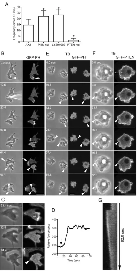
PIP2 is a key regulator of blebbing
The intracellular signals for lamellipodia extension have been intensively investigated. In par-ticular, the inositol lipid phosphatidylinositol 3,4,5-trisphosphate (PIP3) plays an important role in actin assembly in lamellipodia [30]. The production of PIP3 is regulated by two enzymes. PI3 kinase (PI3K) phosphorylates phosphatidylinositol 4,5-bisphosphate (PIP2) to generate PIP3. Conversely, PTEN (a phosphatase and tensin homolog) is a phosphatase that dephosphorylates PIP3 to generate PIP2. To investigate the roles of these inositol lipids in bleb-bing, PI3K-null cells and PTEN-null cells were cut, and the frequency of blebbing was exam-ined. The PI3K-null cells used in this study had mutations in all five PI3K genes. The level of PIP3 in this quintuple PI3K mutant is only approximately 10% of the wild type level [31]. However, the PTEN-null mutant has a higher level of PIP3 than wild-type cells [32]. In the cut-ting experiments, the anucleate fragments of PI3K-null cells exhibited more frequent blebbing than those of wild type cells, and those of PTEN-null cells showed significantly reduced bleb-bing (Fig 6A). When wild type cells were cut in the presence of the PI3K inhibitor LY294002, the anucleate fragments frequently extended blebs (Fig 6A).
Next, the localization of PIP3 was observed in live cells expressing the GFP-labeled pleck-strin homology (PH) domain of Akt/PKB, which is an indicator of the location of PIP3. GFP-PH was localized along the leading edge of lamellipodia (arrows inFig 6B). When a cell was cut into two fragments, the nucleate fragments continued to migrate, and GFP-PH contin-ued to localize along the leading edge. In contrast, in the anucleate fragments, GFP-PH disap-peared from the lamellipodia after retraction.Fig 6Cshows enlarged images of an anucleate fragment.Fig 6Dshows a time course of fluorescence intensity in the cytoplasm (box inFig 6C), indicating that GFP-PH became evenly diffused throughout the cytoplasm and did not localize to the blebs (arrowheads inFig 6B and 6C). Incidentally, GFP-PH also did not localize to the blebs in uncut cells (data not shown).
When cells expressing GFP-PH were observed in the presence of thiabendazole, they fre-quently extended blebs, but GFP-PH did not localize to the blebs (Fig 6E).
We next examined the location of PIP2 in blebbing cells expressing GFP-PTEN (G129E) in the presence of thiabendazole. GFP-PTEN (G129E) is an indicator of PIP2 and has negligible phosphatase activity [33,34]. PTEN was localized along the margins of resting cells and did not localize along the leading edges of blebs immediately after extension; rather, it gradually accu-mulated there in a similar manner to myosin II (Fig 6F and 6G).
Together, PIP3 localizes to the cortex depending on the presence of microtubules. Most likely, microtubules stabilize PIP3 in the cell membrane, which in turn suppresses bleb forma-tion. PIP2 may positively regulate myosin II in cortex outside of blebs, as discussed later.
Blebs can be induced in leukocytes by microsurgery
Discussion
In the present study, we developed a new quantitative assay using microsurgery to investigate the signals involved inDictyosteliumcell blebbing. Using this assay, blebs could be experimen-tally induced in a reproducible manner; this was also the case in human leukocytes. The anucle-ate fragments vigorously extended blebs. The extension of blebs was dependent on the
contraction force generated by myosin II because myosin-null cells did not extend blebs. Most likely, the intracellular pressure induced by cortical actomyosin powers the extension of blebs;
microscopy. Time course of phase contrast and fluorescence images before and after cutting. PIP3 was localized along the edges of the lamellipodia (arrow). When the cell was cut into two fragments, the nucleate fragments continued to migrate, and PIP3 was localized along the leading edge (arrow). However, in the anucleate fragment, PIP3 delocalized and became evenly distributed in the cytoplasm. The anucleate fragment frequently extended blebs, but PIP3 did not localize to the blebs (arrowheads). The fluorescence in the blebs appears slightly higher than that in the cytoplasm, which was caused by the exclusion volume [58]. (C) Enlarged images of the fragment shown in panel B. (D) The time course of fluorescence intensity in the fragment (rectangle in panel C), demonstrating that the fluorescence signal increases in the cytoplasm after cutting (arrow). (E) When microtubules were disassembled in cells expressing the GFP-PH domain in the presence of thiabendazole (TB), the cells frequently extended blebs (arrowheads). Note that GFP-PH diffused in the cytoplasm and did not localize to the bleb cortex. (F) Uncut cells expressing GFP-PTEN (G129E), a marker of PIP2, were observed in the presence of thiabendazole (TB). (G) Kymograph of the rectangle in panel F. Note that PIP2 did not localize along the leading edge of the bleb (arrowheads) immediately after extension but rather gradually accumulated there, followed by the retraction of the bleb. Bars, 10μm.
doi:10.1371/journal.pone.0137032.g006
Fig 7. Blebs can be induced in leukocytes by microsurgery.(A) Human leukocytes were individually cut into two fragments under DIC microscopy. The arrow shows the nucleus. After cutting, the nucleate fragment continued normal migration by extending lamellipodia. However, the anucleate fragment frequently extended blebs (arrowheads). (B) Sequential images of lamellipodium extension (arrowhead) and a kymograph generated from the rectangle. (C) Sequential images of bleb extension (arrowhead) and a kymograph generated from the rectangle. (D) Velocities of lamellipodia and blebs. Note that blebs extend much faster than lamellipodia. (E) Bleb frequencies in untreated and nocodazole-treated uncut cells. Note that leukocytes extend more blebs in the presence of nocodazole. Bars, 10μm.
this pressure could be mimicked by applying pressure to the cells with an agar block. In the anucleate fragments, the microtubules depolymerized after cutting, and the blebs then began to extend. The microtubule inhibitor also induced blebbing in uncut cells, suggesting that the depolymerization of microtubules induces blebbing. The depolymerization of microtubules delocalized the inositol lipid PIP3 from the cell membrane. In addition, PI3K-null cells extended blebs more frequently, whereas PTEN-null cells extended them less frequently than wild-type cells. Therefore, microtubules play a critical role in blebbing-related signaling via inositol lipid metabolism.
Blebs extended by detachment of the cell membrane from the underlying actin cortex. It was previously proposed that this detachment is caused by a local decrease of adhesion between the membrane and the cortex or by local disassembly of the actin cortex [2]. In the present study, no actin cortex disassembly was observed in either the anucleate fragments or in uncut
Dictyosteliumcells, supporting the former scenario.
There are three distinct, successive steps of bleb extension: (1) specification of a bleb site, (2) local detachment of the cell membrane from the actin cortex, and (3) expansion of the bleb. At present, we do not know how the location of bleb extension is specified; microtubules may be the best potential candidate, as is discussed later. Regarding local detachment, a reduction of the attachment energy between the cell membrane and actin cortex must occur locally to initi-ate blebbing. Indeed, interfering with the activities of linking molecules such as ezrin / radixin / moesin (ERM) and class I myosins frequently induced blebbing in zebrafish prechordal plate mesendoderm progenitor cells [35]. Local pulsed-laser-induced ablation of the cell membrane can induce blebbing [36,37], which also supports the above idea. In the present study, virtually no blebs were observed in the anucleate fragments of myosin II-null cells; instead, very small extensions were often observed. Most likely, blebbing locations are still specified in myosin II-null cells, but without myosin II intracellular pressure is insufficient to induce blebbing, thereby resulting only in small extensions.
Experiments in which membrane tethers were pulled using optical tweezers have shown that decreased PIP2 in fibroblast cell membranes reduces the adhesion between the actin cortex and cell membrane [38]. InDictyosteliumcells, live imaging using GFP-PTEN (G129E), an indicator of PIP2, has shown that PIP2 localizes to the rear cortexes in migrating cells and at the furrow regions of dividing cells [33,39,40]. These localization patterns are similar to those of myosin II [27,34]. PTEN-null cells show aberrant myosin II localization, resulting in cytoki-nesis failure and reduced cell migration. This finding suggests that PTEN is an upstream regu-lator of proper myosin II localization in cells [34]. In the present study, both PTEN and myosin II mainly localized along the cortex, except in blebs of the anucleate fragments. The observation that blebs extend at cortical regions that possess less PIP2 is consistent with the results of the membrane tethering experiments, which indicated that PIP2 increases the strength of adhesion between the actin cortex and cell membrane [38]. Recent observations of blebs inXenopusprimordial germ cells and of fixation-induced blebs in human leukemia cells also indicate the important role of PIP2 in bleb formation [41,42].
Furthermore, because PIP2 localizes to the cortex rather than to bleb sites, it can induce the localization of myosin II to the same region and enhance actomyosin contraction, resulting in increased intracellular pressure and bleb expansion.
is a strong candidate regulator of this switching. Switching regulators in other organisms have been reported. In the course of zebrafish gastrulation, noncanonical Wnt signaling regulates the balance between lamellipodia-driven (mesenchymal mode) and blebs-driven cell migration by regulating myosin activity via the Rho pathway [44,45]. In melanoma tumor cells, a balance between the activities of the small GTPases Rac and Rho is important in switching between lamellipodia-driven and blebs-driven cell migration [14]. Inhibiting activities of either of these small GTPases has been shown to induce switching between the modes of migration
[13,14,46].
In the present study, we demonstrated that microtubules stabilize PIP3 in the cell mem-brane, likely by attaching to the cell cortex via their distal ends. Although the molecular mecha-nism for PIP3 stabilization is not clear at present, some signals are probably carried along microtubules to activate PI3K or inactivate PTEN at the cell cortex. It has been reported that microtubules play an important role in the polarization of cells for chemotactic migration and proper cell division. Tsunami, a Hedgehog signaling protein kinase, localizes to microtubule networks inDictyosteliumcells and is related to PIP3 production and actin assembly. Mutant cells deficient intsunamishow a defect in chemotaxis due to their inability to become polarized and to correctly orient lamellipodia in chemoattractant gradients [47]. The 14-3-3 protein coordinates microtubules and the actin cytoskeleton to control cell mechanics and cytokinesis [48].DictyosteliumLis1, originally identified as a target for the sporadic mutations that cause lissencephaly in humans, localizes to the microtubule network and modulates actin dynamics by binding to Rac1A [49]. EB1, a microtubule end-binding protein, is required for the intracel-lular localization of SCAR/WAVE, an actin nucleation factor [50].
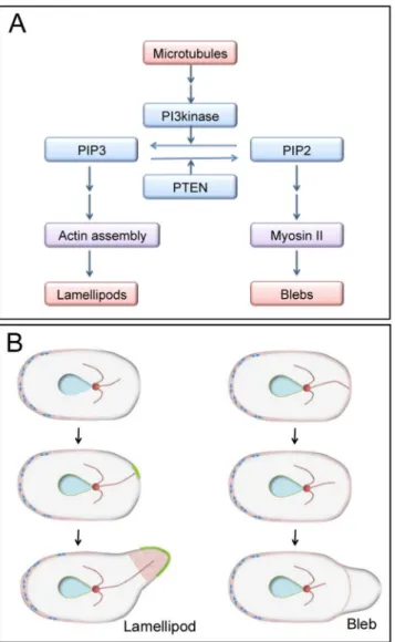
Taken together, our data lead us to propose a model in which microtubules play a critical role in bleb extension-related signaling via inositol lipid metabolism (Fig 8A). In uncut cells, when microtubules detach from the cell cortex by physical displacement or via shortening due to dynamic instability, PIP3 locally delocalizes from the cell cortex. This delocalization occurs due to PI3K inactivation or PTEN activation, which may specify the location of blebbing (right inFig 8B). PIP2 localizes to the cortex, except in nascent blebs, and recruits and activates myo-sin II, resulting in bleb expansion by increamyo-sing the intracellular pressure. However, when microtubules attach to the cortex, PIP3 is locally enriched in the cell membrane, resulting in polymerization of actin filaments and extension of lamellipodia (Fig 8B, left). At present, there is no direct evidence of such a role for microtubules in migratingDictyosteliumcells, but it is known that astral microtubules can induce actin polymerization that results in development of mitosis-specific dynamic actin structures (MiDAS), a substratum adhesion, in dividing Dic-tyosteliumcells [51]. It is also known that dynamics of microtubules affects blebbing during fibroblast spreading [52]. In epithelial cells, the depolymerization of microtubules by colcemid activates RhoA/ROCK, thus leading to blebbing [53].
Blebs are prominent in cells in 3D environments, such as cancer cells and primordial germ cells. In such constricted environments, cells receive outside pressure, which increases their intracellular hydrostatic pressure, and may stimulate bleb extension, as shown by agar overlay experiments in the present study. Alternatively, the pressure of the agar overlay may separate the distal ends of the microtubules from the cortex, thereby inducing blebbing.
In conclusion, our findings demonstrate that microtubules play a critical role in regulating blebbing via inositol lipid metabolism. Many intracellular signals, actin-related proteins, and membrane trafficking proteins participate in blebbing. The complete picture of the signaling cascade, from microtubule changes to bleb extension, remains to be clarified in the future. Fig 8. Current model for blebbing signaling.(A) The results of our present study indicate that microtubules can be positioned upstream of bleb extension. In the presence of microtubules, PIP3 localizes to the anterior membrane and induces lamellipodia formation by enhancing the assembly of actin at that location. Without microtubules, PIP3 delocalizes from the cell membrane. PIP2 recruits myosin II along the cortex, generating more internal pressure inside of the cell. A bleb will extend at the weakest part of the cortex (where the levels of PIP2 and myosin II proteins are low) because myosin II contributes to the rigidity of the cortex. (B) A model of lamellipodium and bleb extension in uncut cells. When microtubules extend to the cortex, unknown signals are conveyed along the microtubules that locally induce PIP3 accumulation and actin assembly in the cortex, resulting in the extension of lamellipodium (left). However, when microtubules retract or detach from the cortex, PIP3 delocalizes from the cortex. In the presence of intracellular pressure, based on the power of myosin II, the cell membrane detaches from the weakest part of the actin cortex, resulting in bleb formation. Blue double arrows, myosin II; red, actin filaments; green, PIP3.
Materials and Methods
Cell Preparation
Dictyostelium discoideumwild type (AX2) cells and mutant cells were cultured in plastic dishes at 22°C in HL5 medium (1.3% bacteriological peptone, 0.75% yeast extract, 85.5 mM D-
glucose
, 3.5 mM Na2HPO412H2O, and 3.5 mM KH2PO4, pH 6.3). The sources of thesecells are listed inS1 Table. GFP-ABD, GFP-myosin II, GFP-@-tubulin, and GFP-PH domain
expression vectors were transformed by electroporation, as previously described [54]. Trans-formed cells were selected in HL5 medium supplemented with 10μg/ml G418 (Sigma). The
medium was exchanged with BSS (3 mM CaCl2, 10 mM KCl, 10 mM NaCl, and 3 mM MES,
pH 6.3) and cells were incubated in the solution for 6–8 h. Details regarding the mutant strains are presented inS1 Table.
Neutrophils were isolated using an established method [55]. Briefly, blood was sampled by finger pricking with Medisafe Finetouch (Terumo). The authors were the source of blood. Approximately 50μl of blood was placed in a glass-bottom chamber at 37°C. After 45 min, the
clot was removed and washed repeatedly with HBS (1 mM MgCl2, 1 mM CaCl2, 5 mM KCl,
150 mM NaCl, and 20 mM HEPES, pH 7.3) supplemented with 10 mMD-
glucose
, 0.2% (w/ v) bovine serum albumin (BSA), and 20 nM formyl-Met-Leu-Phe.Microscopy
Cells were placed in a glass-bottomed chamber and observed under a differential interference contrast (DIC) or phase contrast microscope (IX71, Olympus). Cells expressing GFP-ABD, GFP-myosin, GFP-tubulin, or GFP-PH were observed under a confocal microscope (LSM 510 META, Carl Zeiss) equipped with a 100x objective lens (Plan-Neofluar NA x 1.3) as previously described [56]. A 488-nm argon laser beam was used for excitation of GFP, and fluorescence was detected using a 505-530-nm band-pass filter.
Microsurgery
Glass microneedles were made from glass rods (GD-1.5, Narishige) using a puller (PG-1, Nar-ishige). Each was bent at a 25-degree angle with a micro-forge (MF-830, NarNar-ishige). The tips of the microneedles were dipped in Sigmacote (Sigma) to make them hydrophobic, thereby pre-venting the cells from sticking to the microneedle. The microneedle was mounted on a manip-ulator (3Man, S Company) that was attached to an inverted DIC microscope or a confocal microscope. To cut the cells, the microneedle was placed onto a cell and quickly pulled.
For the cell pressing experiments, an agar block (0.17-mm thick, 1.5% agarose) was pre-pared as described previously [27]. The cells were cut using a microneedle under the agar block, and the external solution was then carefully removed with a piece of filter paper to apply pressure to the cells or cell fragments.
Inhibitors
Blebbistatin, thiabendazole, nocodazole, and LY294002 were dissolved in dimethyl sulfoxide (DMSO) to create stock solutions of 20 mM, 100 mM, 10 mM, and 5 mM, respectively. The final concentrations of these inhibitors for the experiments were 150μM, 0.1 mM, 17μM, and
20μM in BSS. The experiments with blebbistatin, nocodazole and LY294002 were conducted
To completely depolymerize their microtubules,Dictyosteliumcells were incubated on ice for 30 min in the presence of thiabendazole [57]. These cells were observed by confocal micros-copy in the presence of thiabendazole at 22°C. Under these conditions, all of the microtubules except at the microtubule organizing center (MTOC) were completely depolymerized.
Nocodazole (17μM) in HBS (supplemented with D-glucose plus BSA and 20 nM
formyl-Met-Leu-Phe) was used to depolymerize the microtubules in neutrophils.
Image analysis
Time courses of the extension and retraction of lamellipodia and blebs were analyzed by creat-ing kymographs in Image J (http://rsbweb.nih.gov/ij/).
Ethics
This study was conducted in accordance with the ethical standards of the Helsinki Declaration.
Supporting Information
S1 Fig. Microtubules reversibly depolymerize in anucleate halves of cells in disconnection experiments.When the cytoplasm of a cell expressing GFP-tubulin was disconnected by press-ing with a microneedle under confocal microscopy, the microtubules quickly depolymerized in the anucleate half. After the microtubules depolymerized, blebbing began (arrows). Both sides of the cytoplasm rejoined after removing the microneedle, and the blebbing then ceased. After joining, the cell again had an intact network of microtubules. Bars, 10μm.
(TIF)
S1 Table. Blebs in mutant cells.The frequency of blebs was examined in mutant cells for 5 min after cutting. The frequency is plotted in the graph inFig 4A. Most of the mutants were provided by the DictyBase stock center (www.Dictybase.org).‘n’represents the number of examined cells.
(DOCX)
Acknowledgments
We are grateful to members of theDictyosteliumresearch community, the Dicty Stock Center, and the NBRP Nenkin for the many mutants. We thank Dr. T. Q. P. Uyeda for providing con-structs of GFP-tubulin, GFP-ABD, GFP-myosin II, and GFP-PH. We also thank Drs. T. Q. P. Uyeda and T. Kitanishi-Yumura for their critical reading of the manuscript and their helpful comments.
Author Contributions
Conceived and designed the experiments: TS MKP SY. Performed the experiments: TS MKP SY. Analyzed the data: TS MKP SY. Contributed reagents/materials/analysis tools: TS MKP SY. Wrote the paper: TS MKP SY.
References
1. Pollard TD, Borisy GG. Cellular motility driven by assembly and disassembly of actin filaments. Cell. 2003; 112: 453–465. PMID:12600310
2. Charras G, Paluch E. Blebs lead the way: how to migrate without lamellipodia. Nat Rev Mol Cell Biol. 2008; 9: 730–736. doi:10.1038/nrm2453PMID:18628785
3. Fackler OT, Grosse R. Cell motility through plasma membrane blebbing. J Cell Biol. 2008; 181: 879–
4. Paluch EK, Raz E. The role and regulation of blebs in cell migration. Curr Opin Cell Biol. 2013; 25: 582–590. doi:10.1016/j.ceb.2013.05.005PMID:23786923
5. Pinner S, Sahai E. PDK1 regulates cancer cell motility by antagonising inhibition of ROCK1 by RhoE. Nat Cell Biol. 2008; 10: 127–137. doi:10.1038/ncb1675PMID:18204440
6. Petrie RJ, Gavara N, Chadwick RS, Yamada KM. Nonpolarized signaling reveals two distinct modes of 3D cell migration. J Cell Biol. 2012; 197: 439–455. doi:10.1083/jcb.201201124PMID:22547408
7. Sixt M. Cell migration: fibroblasts find a new way to get ahead. J Cell Biol. 2012; 197: 347–349. doi:10. 1083/jcb.201204039PMID:22547405
8. Gadea G, de Toledo M, Anguille C, Roux P. Loss of p53 promotes RhoA-ROCK-dependent cell migra-tion and invasion in 3D matrices. J Cell Biol. 2007; 178: 23–30. PMID:17606864
9. Tournaviti S, Hannemann S, Terjung S, Kitzing TM, Stegmayer C, Ritzerfeld J, et al. SH4-domain-induced plasma membrane dynamization promotes bleb-associated cell motility. J Cell Sci. 2007; 120: 3820–3829. PMID:17959630
10. Blaser H, Reichman-Fried M, Castanon I, Dumstrei K, Marlow FL, Kawakami K, et al. Migration of zeb-rafish primordial germ cells: a role for myosin contraction and cytoplasmic flow. Dev Cell. 2006; 11: 613–627. PMID:17084355
11. Bergert M, Chandradoss SD, Desai RA, Paluch E. Cell mechanics control rapid transitions between blebs and lamellipodia during migration. Proc Natl Acad Sci U S A. 2012; 109: 14434–14439. doi:10. 1073/pnas.1207968109PMID:22786929
12. Derivery E, Fink J, Martin D, Houdusse A, Piel M, Stradal TE, et al. Free Brick1 is a trimeric precursor in the assembly of a functional wave complex. PLoS One. 2008; 3: e2462. doi:10.1371/journal.pone. 0002462PMID:18560548
13. Sahai E, Marshall CJ. Differing modes of tumour cell invasion have distinct requirements for Rho/ ROCK signalling and extracellular proteolysis. Nat Cell Biol. 2003; 5: 711–719. PMID:12844144
14. Sanz-Moreno V, Gadea G, Ahn J, Paterson H, Marra P, Pinner S, et al. Rac activation and inactivation control plasticity of tumor cell movement. Cell. 2008; 135: 510–523. doi:10.1016/j.cell.2008.09.043 PMID:18984162
15. Shiratsuchi A, Mori T, Nakanishi Y. Independence of plasma membrane blebbing from other biochemi-cal and biologibiochemi-cal characteristics of apoptotic cells. J Biochem. 2002; 132: 381–386. PMID:12204106
16. Fink RD, Trinkaus JP. Fundulus deep cells: directional migration in response to epithelial wounding. Dev Biol. 1988; 129: 179–190. PMID:3410158
17. Zatulovskiy E, Tyson R, Bretschneider T, Kay RR. Bleb-driven chemotaxis of Dictyostelium cells. J Cell Biol. 2014; 204: 1027–1044. doi:10.1083/jcb.201306147PMID:24616222
18. Insall R. The interaction between pseudopods and extracellular signalling during chemotaxis and directed migration. Curr Opin Cell Biol. 2013; 25: 526–531. doi:10.1016/j.ceb.2013.04.009PMID: 23747069
19. Loomis WF. Cell signaling during development of Dictyostelium. Dev Biol. 2014; 391: 1–16. doi:10. 1016/j.ydbio.2014.04.001PMID:24726820
20. Swaney KF, Huang CH, Devreotes PN. Eukaryotic chemotaxis: a network of signaling pathways con-trols motility, directional sensing, and polarity. Annu Rev Biophys. 2010; 39: 265–289. doi:10.1146/ annurev.biophys.093008.131228PMID:20192768
21. Van Haastert PJ. Chemotaxis: insights from the extending pseudopod. J Cell Sci. 2010; 123: 3031–
3037. doi:10.1242/jcs.071118PMID:20810783
22. Yumura S, Uyeda TQ. Myosins and cell dynamics in cellular slime molds. Int Rev Cytol. 2003; 224: 173–225. PMID:12722951
23. Yoshida K, Soldati T. Dissection of amoeboid movement into two mechanically distinct modes. J Cell Sci. 2006; 119: 3833–3844. PMID:16926192
24. Langridge PD, Kay RR. Blebbing of Dictyostelium cells in response to chemoattractant. Exp Cell Res. 2006; 312: 2009–2017. PMID:16624291
25. Swanson JA, Taylor DL. Local and spatially coordinated movements in Dictyostelium discoideum amoebae during chemotaxis. Cell. 1982; 28: 225–232. PMID:6277507
26. Wang Y, Chen CL, Iijima M. Signaling mechanisms for chemotaxis. Dev Growth Differ. 2011; 53: 495–
502. doi:10.1111/j.1440-169X.2011.01265.xPMID:21585354
27. Yumura S, Mori H, Fukui Y. Localization of actin and myosin for the study of ameboid movement in Dic-tyostelium using improved immunofluorescence. J Cell Biol. 1984; 99: 894–899. PMID:6381508
29. Kuriyama R, Sato C, Fukui Y, Nishibayashi S. In vitro nucleation of microtubules from microtubule-orga-nizing center prepared from cellular slime mold. Cell Motil. 1982; 2: 257–272. PMID:7172222
30. Funamoto S, Milan K, Meili R, Firtel RA. Role of phosphatidylinositol 3' kinase and a downstream pleck-strin homology domain-containing protein in controlling chemotaxis in dictyostelium. J Cell Biol. 2001; 153: 795–810. PMID:11352940
31. Hoeller O, Kay RR. Chemotaxis in the absence of PIP3 gradients. Curr Biol. 2007; 17: 813–817. PMID: 17462897
32. Iijima M, Devreotes P. Tumor suppressor PTEN mediates sensing of chemoattractant gradients. Cell. 2002; 109: 599–610. PMID:12062103
33. Iijima M, Huang YE, Luo HR, Vazquez F, Devreotes PN. Novel mechanism of PTEN regulation by its phosphatidylinositol 4,5-bisphosphate binding motif is critical for chemotaxis. J Biol Chem. 2004; 279: 16606–16613. PMID:14764604
34. Pramanik MK, Iijima M, Iwadate Y, Yumura S. PTEN is a mechanosensing signal transducer for myosin II localization in Dictyostelium cells. Genes Cells. 2009; 14: 821–834. doi:10.1111/j.1365-2443.2009. 01312.xPMID:19515202
35. Diz-Munoz A, Krieg M, Bergert M, Ibarlucea-Benitez I, Muller DJ, Paluch E, et al. Control of directed cell migration in vivo by membrane-to-cortex attachment. PLoS Biol. 2010; 8: e1000544. doi:10.1371/ journal.pbio.1000544PMID:21151339
36. Kelly CV, Kober MM, Kinnunen P, Reis DA, Orr BG, Banaszak Holl MM. Pulsed-laser creation and characterization of giant plasma membrane vesicles from cells. J Biol Phys. 2009; 35: 279–295. doi: 10.1007/s10867-009-9167-7PMID:19669579
37. Tinevez JY, Schulze U, Salbreux G, Roensch J, Joanny JF, Paluch E. Role of cortical tension in bleb growth. Proc Natl Acad Sci U S A. 2009; 106: 18581–18586. doi:10.1073/pnas.0903353106PMID: 19846787
38. Raucher D, Stauffer T, Chen W, Shen K, Guo S, York JD, et al. Phosphatidylinositol 4,5-bisphosphate functions as a second messenger that regulates cytoskeleton-plasma membrane adhesion. Cell. 2000; 100: 221–228. PMID:10660045
39. Funamoto S, Meili R, Lee S, Parry L, Firtel RA. Spatial and temporal regulation of 3-phosphoinositides by PI 3-kinase and PTEN mediates chemotaxis. Cell. 2002; 109: 611–623. PMID:12062104
40. Janetopoulos C, Borleis J, Vazquez F, Iijima M, Devreotes P. Temporal and spatial regulation of phos-phoinositide signaling mediates cytokinesis. Dev Cell. 2005; 8: 467–477. PMID:15809030
41. Tarbashevich K, Dzementsei A, Pieler T. A novel function for KIF13B in germ cell migration. Dev Biol. 2011; 349: 169–178. doi:10.1016/j.ydbio.2010.10.016PMID:21029731
42. Zhao S, Liao H, Ao M, Wu L, Zhang X, Chen Y. Fixation-induced cell blebbing on spread cells inversely correlates with phosphatidylinositol 4,5-bisphosphate level in the plasma membrane. FEBS Open Bio. 2014; 4: 190–199. doi:10.1016/j.fob.2014.02.003PMID:24649401
43. Devreotes P, Janetopoulos C. Eukaryotic chemotaxis: distinctions between directional sensing and polarization. J Biol Chem. 2003; 278: 20445–20448. PMID:12672811
44. Weiser DC, Pyati UJ, Kimelman D. Gravin regulates mesodermal cell behavior changes required for axis elongation during zebrafish gastrulation. Genes Dev. 2007; 21: 1559–1571. PMID:17575056
45. Weiser DC, Row RH, Kimelman D. Rho-regulated myosin phosphatase establishes the level of protru-sive activity required for cell movements during zebrafish gastrulation. Development. 2009; 136: 2375–
2384. doi:10.1242/dev.034892PMID:19515695
46. Vidali L, Chen F, Cicchetti G, Ohta Y, Kwiatkowski DJ. Rac1-null mouse embryonic fibroblasts are motile and respond to platelet-derived growth factor. Mol Biol Cell. 2006; 17: 2377–2390. PMID: 16525021
47. Tang L, Franca-Koh J, Xiong Y, Chen MY, Long Y, Bickford RM, et al. tsunami, the Dictyostelium homo-log of the Fused kinase, is required for polarization and chemotaxis. Genes Dev. 2008; 22: 2278–
2290. doi:10.1101/gad.1694508PMID:18708585
48. Zhou Q, Kee YS, Poirier CC, Jelinek C, Osborne J, Divi S, et al. 14-3-3 coordinates microtubules, Rac, and myosin II to control cell mechanics and cytokinesis. Curr Biol. 2010; 20: 1881–1889. doi:10.1016/ j.cub.2010.09.048PMID:20951045
49. Rehberg M, Kleylein-Sohn J, Faix J, Ho TH, Schulz I, Graf R. Dictyostelium LIS1 is a centrosomal pro-tein required for microtubule/cell cortex interactions, nucleus/centrosome linkage, and actin dynamics. Mol Biol Cell. 2005; 16: 2759–2771. PMID:15800059
51. Itoh G, Yumura S. A novel mitosis-specific dynamic actin structure in Dictyostelium cells. J Cell Sci. 2007; 120: 4302–4309. PMID:18029399
52. Tvorogova AV, Vorob'ev IA. Microtubules suppress blebbing and stimulate lamellae extension in spreading fibroblasts. Tsitologiia. 2012; 54: 742–753. PMID:23285727
53. Pletjushkina OJ, Rajfur Z, Pomorski P, Oliver TN, Vasiliev JM, Jacobson KA. Induction of cortical oscil-lations in spreading cells by depolymerization of microtubules. Cell Motil Cytoskeleton. 2001; 48: 235–
244. PMID:11276073
54. Yumura S, Matsuzaki R, Kitanishi-Yumura T. Introduction of macromolecules into living Dictyostelium cells by electroporation. Cell Struct Funct. 1995; 20: 185–190. PMID:7586008
55. Pierini LM, Maxfield FR. Optical microscopy-based migration assay for human neutrophils. Curr Protoc Cell Biol. 2003; Chapter 12: Unit 12.6.
56. Yumura S. Myosin II dynamics and cortical flow during contractile ring formation in Dictyostelium cells. J Cell Biol. 2001; 154: 137–146. PMID:11448996
57. Kitanishi T, Shibaoka H, Fukui Y. Disruption of microtubules and retardation of development of Dictyos-teliumwith ethyl N-phenylcarbamate and thiabendazole. Protoplasma. 1984; 120: 185–196.