BIOMEDICAL SCIENCES
AND
CLINICAL INVESTIGATION
www.bjournal.com.br
www.bjournal.com.br
Braz J Med Biol Res, January 2010, Volume 43(1) 57-67
Immune cells and oxidative stress in the endotoxin tolerance
mouse model
E.S. Melo, H.V. Barbeiro, S. Ariga, T. Goloubkova, R. Curi, I.T. Velasco, D. Vasconcelos and
F.G. Soriano
Institutional Sponsors
Immune cells and oxidative stress in
the endotoxin tolerance mouse model
E.S. Melo
1, H.V. Barbeiro
1, S. Ariga
1, T. Goloubkova
1, R. Curi
2, I.T. Velasco
1,
D. Vasconcelos
3and F.G. Soriano
11Disciplina de Emergências Clínicas, Faculdade de Medicina, 2Departamento de Fisiologia e Biofísica, Instituto de Ciências Biomédicas, and 3Laboratório de Investigação em Dermatologia e Imunodeficiência,
Departamento deDermatologia, Instituto de Medicina Tropical, Universidade de São Paulo, São Paulo, SP, Brasil
Abstract
Sepsis is a systemic inflammatory response that can lead to tissue damage and death. In order to increase our understanding of sepsis, experimental models are needed that produce relevant immune and inflammatory responses during a septic event. We
describe a lipopolysaccharide tolerance mouse model to characterize the cellular and molecular alterations of immune cells during sepsis. The model presents a typical lipopolysaccharide tolerance pattern in which tolerance is related to decreased production and secretion of cytokines after a subsequent exposure to a lethal dose of lipopolysaccharide. The initial lipopolysaccharide exposure also altered the expression patterns of cytokines and was followed by an 8- and a 1.5-fold increase in the T helper 1 and 2 cell subpopulations. Behavioral data indicate a decrease in spontaneous activity and an increase in body temperature following exposure to lipopolysaccharide. In contrast, tolerant animals maintained production of reactive oxygen species and nitric oxide when terminally challenged by cecal ligation and puncture (CLP). Survival study after CLP showed protection in tolerant compared to naive animals. Spleen mass increased in tolerant animals followed by increases of B lymphocytes and subpopulation Th1 cells. An increase in the number of stem cells was found in spleen and bone marrow. We also showed that administration of spleen or bone marrow cells from tolerant to naive animals transfers the acquired resistance status. In con-clusion, lipopolysaccharide tolerance is a natural reprogramming of the immune system that increases the number of immune cells, particularly T helper 1 cells, and does not reduce oxidative stress.
Key words: Sepsis; Cytokines; Immune tolerance; Lipopolysaccharide; Murine sepsis model; T helper cells
Introduction
Lipopolysaccharide (LPS) is one of the most potent known inducers of inflammation and exerts its effects via the Toll-like receptor (TLR) 4, which is essential for the recognition of distinct bacterial cell wall components (1-3). Inhibition of TLR signaling by inducible negative regulators, anti-inflammatory cytokines and alterations in the TLR sig -naling complex has been attributed to the phenomenon of “LPS tolerance” (1). Endotoxin tolerance has been defined as reduced responsiveness to a LPS challenge following a first encounter with endotoxin (4,5). Tolerance to LPS involves the participation of macrophages and mediators such as glucocorticoids, prostaglandins, interleukin-10 (IL-10), and transforming growth factor beta (4). However, the assumption that LPS tolerance is caused by a “generalized” down-regulation of cellular responses and mediator release
is not accurate (4,6). A similar loss of LPS reactivity has been repeatedly reported in circulating leukocytes of septic patients (4,7). Studies of cellular signaling within leukocytes from septic patients have revealed numerous alterations similar to those observed in endotoxin-tolerant cells (4,7). However, the altered responsiveness to LPS of leukocytes from septic patients is not synonymous with LPS tolerance. The term “cellular reprogramming” is helpful in describing the immune status of circulating leukocytes during the es-tablishment of endotoxin tolerance (4,8). Tolerance to LPS has been shown to protect against bacterial infections; it is a model of an adequate immune response in contrast to the deadly dysregulation of immune function in sepsis (4). In other studies, monophosphoryl lipid A has been used to produce tolerance and has been shown to protect animals
Correspondence: F.G. Soriano, Laboratório da Disciplina de Emergências Clínicas, FM, USP, Av. Dr. Arnaldo, 455, Sala 3187, 01246-913 São Paulo, SP, Brasil. Fax: +55-11-3061-7170. E-mail: [email protected]
from subsequent infections (4,9).
Animal models represent a critical research bridge between experimental results from the laboratory and findings at the clinical level. Any disease, or intoxication, of humans that is established experimentally in animals should ideally mimic the natural sequelae seen in humans. The establishment of simple, economical, and robust animal models that can be used in different laboratories should facilitate the discovery of vaccines or therapies. One goal of the proposed murine model is to accelerate the develop-ment of vaccines or therapies that target a select family of bacterial protein toxins.
The aim of the present study was to evaluate a murine model of LPS tolerance. We characterized the complex cellular alterations associated with tolerance, including increased lymphocyte, monocyte and neutrophil prolifera-tion.
Material and Methods
The present study was approved by the Research Ethics Committee of the University of São Paulo School of Medicine (protocol #331/06). All animals were treated according to institutional rules for laboratory animal care.
Unless otherwise indicated, reagents were purchased from Sigma-Aldrich (USA).
Induction of LPS tolerance
Six-week-old male BALB/c mice weighing 20 to 25 g were randomized into two groups. The naive group (N = 47) received daily subcutaneous injections of 1 mL saline, and the tolerant group (N = 47) received daily subcutane-ous injections of either 0.5 or 1 mg/kg LPS [Sigma-Aldrich (LPS Escherichia coli serotype 026:B6) catalogue number L3755] for 5 days and was allowed to rest for 2 days.
Lethal LPS challenge
Following induction of LPS tolerance and a 2-day rest, on day 8 some animals received either the lethal dose of LPS (20 mg/kg; 80% mortality in naive mice) or a supramaximal LPS dose (40 mg/kg; 100% mortality in naive mice) ip. Some animals from the tolerant and naive groups were sacrificed at 0 (zero), 2, 4, 6, and 8 h after the lethal challenges for biochemical measurements and cytokine analysis.
Cecal ligation and puncture (CLP) model
Mice were anesthetized with a mixture of ketamine (80 mg/kg) and xylazine (10 mg/kg) given intraperitoneally. Under aseptic conditions, a 2-cm midline laparotomy was performed to allow exposure of the cecum with adjoining intestine. The cecum was tightly ligated with a 3.0 silk suture (Ethicon; Johnson & Johnson, USA) at its base, below the ileocecal valve, and was perforated twice (top and bottom) with an 18-gauge needle (Becton Dickinson, USA). The cecum was returned to the peritoneal cavity
and the laparotomy was closed with 4.0 silk sutures. The animals were returned to their cages with free access to food and water.
Study of animal survival
Survival was assessed after CLP challenge or the ad-ministration of a lethal dose of LPS. Each group comprised 30 animals, and survival was assessed every 8 h up to 72 h after LPS or CLP. After 72 h, there was no change in survival.
Measurement of cytokines
Blood was collected from 6 mice per group using tubes containing ethylenediamine tetraacetic acid (1 mg/mL). Blood was centrifuged, and the supernatant was harvested to identify secreted cytokines. Production of IL-6, IL-10, macrophage inflammatory protein 2 (MIP-2), tumor necrosis factor alpha (TNF-α) and IL-1β was evaluated by enzyme-linked immunosorbent assay (ELISA) using a DuoSet kit (R&D Systems, USA). We performed all ELISA assays at the same time to avoid any kind of variation.
Reverse transcriptase-polymerase chain reaction A reverse transcriptase-polymerase chain reaction (RT-PCR) assay was performed to measure the differential mRNA expression of TNF-α, IL-10 and IL-1β in cells from naive and tolerant mice. Primer sequences were designed using information accessed from GenBank of the National Center for Biotechnology Information (NCBI, www.ncbi. nlm.nih.gov). For semi-quantitative PCR analysis, the housekeeping glyceraldehyde-3-phosphate dehydrogenase (GAPDH) gene sequence was used as a reference.
PCR analysis
The PCR products were analyzed on 1.5% agarose gels containing 0.5 µg/mL ethidium bromide and electrophoresed for 1 h at 100 V. Gels were photographed using a Kodak DC120 Zoom Digital Camera System (Eastman Kodak, USA). Images were processed and analyzed with the Kodak Digital Science 1D Image Analysis software (Eastman Kodak). Band intensities were absorbance normalized with respect to GAPDH expression. Samples from naive mice (N = 6) received an arbitrary value of 1, and the values for samples from tolerant mice (N = 6) were in relation to those for the respective naive animals.
Thiobarbituric acid-reactive substances
cooling to room temperature, the samples were cleared by centrifugation at 10,000 g (10 min) and their absorbance was measured at 532 nm, using 1,1,3,3-tetramethoxypropane (Sigma-Aldrich) as an external standard. The level of lipid peroxides was reported as µmol TBARS/mg protein.
Nitrite and nitrate
Plasma nitrite and nitrate (NOx) levels were measured by the classic Griess method. After being passed through 50-kDa ultrafilters, 40 μL plasma was diluted with 240 μL assay buffer and mixed with 10 μL cofactor and 10 μL nitrate reductase (NOx colorimetric assay kit; Cayman Chemical, USA). After the plasma had been maintained at room tem-perature for 3 h to convert nitrate to nitrite, total nitrite was measured at 540 nm absorbance by reaction with Griess reagent (sulfanilamide and naphthalene-ethylene diamine dihydrochloride). Amounts of plasma nitrite were estimated by a standard curve obtained from enzymatic conversion of NaNO3 to nitrite.
Spleen tissue preparation
Spleen tissue (50 mg) was pulverized in liquid nitro-gen and homonitro-genized in RIPA buffer (10 mM Tris-HCl, pH 7.5), 1% tergitol, 0.1% sodium dodecyl sulfate, 1% Na-deoxycholate, 150 mM NaCl, 40 µg/mL phenylmeth-ylsulfonyl fluoride, and 10 µg/mL pepstatin (Sigma, USA). Samples were centrifuged at 14,000 g for 45 min and protein concentrations from supernatants were determined by the Bradford method (10,11).
Quantification of leukocytes
For flow cytometry analysis, spleen tissue was gently ground to dissociate cells. After hypotonic lysis of red blood cells, and following cell membrane permeabilization with saponin, leukocytes were incubated for 30 min at room temperature with fluorescein isothiocyanate-labeled anti-mouse monoclonal antibodies specific for murine CD3+ and CD4+ T-lymphocytes, and phycoerythrin-labeled antibodies to TNF-α, interferon-γ, IL-10, and IL-4. The analysis was performed using the Coulter EPICS XL-MCL flow cytometer (Beckman Coulter, UK) with gating on lymphocytes using characteristic forward and side scatter.
Pluripotent cells
The anti-apoptotic transcription factor Octamer-4 (Oct-4) is a member of the Pit-Oct-Unc family and a marker of pluri-potent cells. The undifferentiated state of embryonic stem cells is characterized by a high level of Oct-4 expression. To determine levels of Oct-4, we used an embryonic stem cell recognition kit that includes antibodies to Oct-4 and SSEA-1 (ES Cell Marker Sample Kit; Chemicon, USA). In brief, spleen cells were allowed to adhere to glass slides, after which they were washed twice with rinse buffer (TBST, 20 mM Tris-HCl, pH 7.4, 150 mM NaCl, 0.05% Tween 20). Cells were then fixed with paraformaldehyde and permeabilized
with 0.1% Triton X-100. Nonspecific markers were blocked with 5% bovine serum albumin, and the cells were incubated with primary antibody (anti-Oct-1 or anti-SSEA-1) at room temperature for 1 h. After three washes in TBST, the cells were incubated with fluorescein isothiocyanate-conjugated secondary antibody for 30 min. Cells were mounted on glass slides with antifade solution.
The slides were viewed under epifluorescence micros -copy using a Zeiss Axioplan II microscope (Carl Zeiss, Germany) with a reticulated lens (100 grids of 1 x 1 mm). In each of 20 random fields, we counted the number of Oct-4- or SSEA-1-labeled cells among 200 nucleated cells, and the result is reported as the percent of the total. Three independent examiners performed the counts, and the final value represents the mean ± SEM of those three measure-ments. The digital images were recorded using a cooled charge-coupled device camera (PCO Computer Optics, Germany), and analyzed using the ISIS program (MetaSys-tems, Germany). The final magnification was 100X.
Transfer of tolerance
Spleen tissue from tolerant mice was ground gently to dissociate cells. After hypotonic lysis of red blood cells and separation with Lymphoprep (Axis-Shield, Norway), leukocytes were resuspended in PBS buffer. Bone mar-row cells were collected by flushing PBS into the marmar-row space. The collected mixture was resuspended in PBS for injection. During the first post-CLP hour, suspensions of cells from the spleen or bone marrow cells from tolerant or naive mice were injected intravenously into naive animals (40,000 cells/animal).
Behavior and clinical parameters
Mice were placed under anesthesia (3% halothane for induction and 1.5% for maintenance in 30% O2 and 70%
N2O). Using aseptic techniques, a temperature
transmit-ter (E-mittransmit-ter; Mini Mitter, USA) was placed in the perito-neal cavity via a midline incision. After surgery, mice were housed in individual cages, each with a running wheel and a telemetry system (Vitalview 4000 series; Mini Mitter). Temperature was monitored telemetrically. Signals were received by an antenna below the cage (ER-4000 Receiver; Mini Mitter) and relayed to a signal processor connected to a computer. Wheel revolutions were monitored magnetically and relayed to a signal processor connected to a computer. Measurements of mean temperature and accumulated wheel revolutions were collected every 1 min.
Statistical analysis
statistically significant at P < 0.05.
Results
Survival study
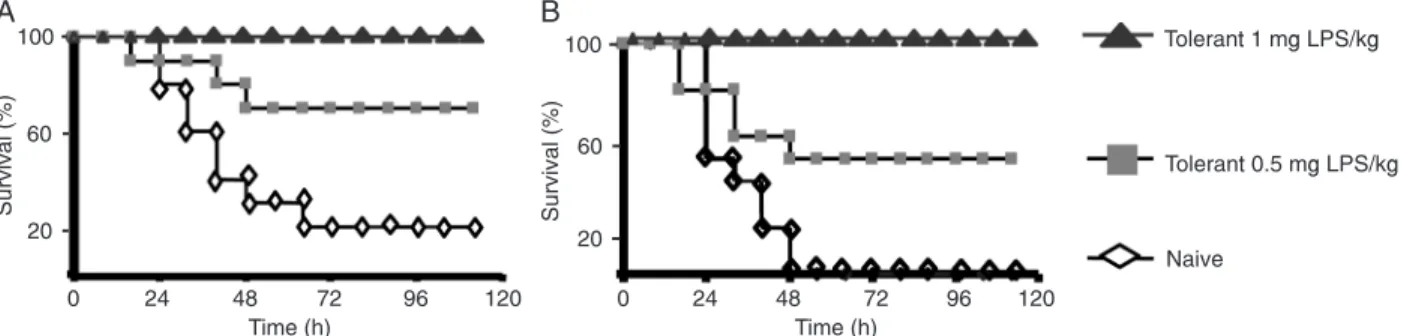
The rate of survival following lethal LPS challenges was higher in animals that received prior treatment with LPS (tolerant) than in those not so treated (naive), as can be seen in Figure 1A,B. All animals received the lethal dose of LPS (either 20 or 40 mg/kg). Overall survival for naive animals after 72 h was 10%. Tolerant mice previously treated with 0.5 mg/kg LPS had a survival rate of 60%, whereas animals that received treatment with 1 mg/kg LPS showed 100% survival compared to controls.
Characterization of LPS tolerance
In BALB/c mice, LPS induced tolerance in a dose-dependent manner, as shown by the reduced mortality after a subsequent lethal dose of LPS (Figure 1A,B). In tolerant mice, production of proinflammatory cytokines (TNF-α, IL-1β and IL-6; Figure 2A-C, respectively) and of the chemokine MIP-2 (Figure 2D) decreased in comparison to naive mice. In contrast, there was no difference in plasma levels of the anti-inflammatory cytokine IL-10 (Figure 2E). Levels of plasma NO and ROS (as measured via their NOx and TBARS products) remained unaffected in tolerant animals following LPS challenge (Figure 3A,B).
Leukocyte analysis
The spleen is the largest peripheral lymphatic organ and plays an important role in the inflammatory response. Following lethal LPS challenge (20 mg/kg), control (N = 10) and tolerant (N = 10) animals were submitted to splenectomy and the spleens were weighed. Tolerant animals presented higher spleen mass and a reduction in body weight relative to controls (Figure 4A,B).
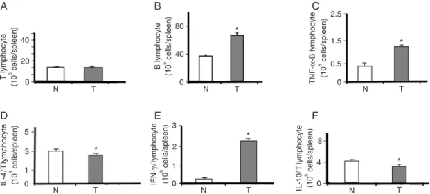
Compared with those of controls, the spleens of toler-ant animals contained larger numbers of B cells and T lymphocytes (CD4+ and CD8+), as shown in Figure 5A-F. The concentration of neutrophils in the spleen and blood was also higher in tolerant animals (data not shown). To determine whether a specific cell population was stimulated by LPS tolerance, T cells were analyzed by flow cytometry. Expression of TNF-α, IFN-γ, IL-4, and IL-10 was measured from cytoplasm. An elevated number of Th1 (TNF-α and
IFN-γ) lymphocytes (P < 0.05) was observed in tolerant compared to naive mice (Figure 5C-F). When animals were subjected to CLP, the spleens from the naive group exhibited a reduction in the number of cells containing TNF-α (Figure 5C) compared with those from the LPS-tolerant group. In addition, tolerant animals presented higher numbers of CD4+ T cells expressing IFN-γ (Figure 5E), IL-4 and IL-10 (Figure 5D,F) than did the controls. Compared with those of the controls, the spleens of tolerant animals also exhibited an increase in pluripotent cells that was maintained even after CLP challenge (Figure 6A). A significant reduction in mortality was observed when cells extracted from the spleens and bone marrow of tolerant mice were injected intravenously into naive animals previously subjected to CLP (Figure 6B). In contrast, the injection of spleen or bone marrow cells from naive animals did not confer any protection (Figure 6C).
Clinical and behavioral change
The body weight of the experimental animals was mea-sured daily during induction of tolerance and after lethal LPS challenge. There was a continuous reduction in the weight of the animals after each LPS dose, as shown in Figure 4. Control animals showed no weight change during the first five days. After the lethal dose of LPS, both groups showed some reduction in body weight, which was greater in the control group. Temperature and spontaneous activity were
Figure 2. Cytokine production. BALB/c mice were treated with 1 mg/kg lipopolysaccharide (LPS; tolerant group, N = 6) or saline pla-cebo (naive group, N = 6) daily for 5 days. Following a lethal dose of LPS (20 mg/kg) on day 8, LPS-tolerant (T) and naive (N) animals
were sacrificed at 0 (zero), 2, 4, 6, and 8 h to obtain cytokine data. Blood serum concentrations of cytokines: A, Tumor necrosis factor
alpha, TNF-α; B, interleukin, IL-1β; C, IL-6; D, macrophage inflammatory protein 2, MIP-2, and E, IL-10. F, Absorbanceof
PCR-am-plified products (TNF-α, IL-10 and IL-1β) are presented as the ratio cytokine/glyceraldehyde-3-phosphate dehydrogenase (GAPDH).
Data are representative of 10 animals per group. *P < 0.05 compared to naive mice (analysis of variance with the Tukey-Kramer post hoc test).
Figure 3. Thiobarbituric acid-reactive substances (TBARS) and nitrite/nitrate production. Oxidative stress response and nitric oxide production in lipopolysaccharide-tolerant animals (tolerant group) and controls (naive group). A, TBARS per mg of protein was used as an index of oxidative stress. B, Blood plasma concentration of nitrite and nitrate (NOx). Data are representative of N = 10 animals per
Figure 4. Body and spleen weight. Body and spleen weight of BALB/c mice treated with 1 mg/kg lipopolysaccharide (LPS; tolerant group) or saline placebo (naive group) daily for 5 days. Animals received a lethal dose of LPS (20 mg/kg) on day 8, following a two-day rest. A, Change in daily body weight among tolerant and naive animals. Data are reported as means ± SEM. *P < 0.05 compared to naive mice (analysis of variance with the Tukey-Kramer post hoc test). B, Spleen weight of tolerant and naive animals. *P < 0.05 compared to naive mice (unpaired t-test).
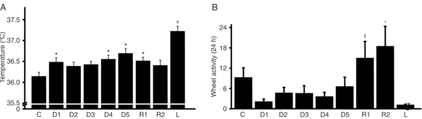
measured throughout the period of LPS tolerance induction (Figure 7A,B). Temperature data showed a progressive increase in mean body temperature following each LPS dose. The temperature response when the lethal LPS dose (20 mg/kg) was applied was substantially higher than that observed for previous stimuli (Figure 7A). Spontaneous movement was measured by wheel activity. Animals showed a reduction in spontaneous activity during LPS tolerance induction and a large reduction after administration of the lethal dose of LPS (Figure 7B).
Discussion
A murine model of endotoxin tolerance offers an in vivo
system for analyzing the regulatory feedback mechanisms involved in cytokine synthesis (5). Endotoxin tolerance has been divided into an early, non-immunological phase and a later immunological phase in which circulating antibod-ies might play a significant role in endotoxin detoxification
(12,13). Based on literature data, we hypothesized that LPS tolerance produces an “adaptation” of the innate immune response (1). Previous studies have presented conflicting findings regarding cytokine production during lethal LPS challenges, as well as regarding the extent to which tolerance provides resistance to infection (4). Most of these differences are due to differences in the model used, doses used to establish tolerance, injection times, administration routes, and the time of delay before restimula-tion with LPS or infecrestimula-tion (4-6,14,15). In the present study, we established and characterized a reliable murine model of endotoxin tolerance in which we applied subcutaneous injection of LPS for inducing tolerance. While intraperitoneal injections are commonly reported in the literature, they cannot exclude the chance that protection against CLP is a local effect, perhaps activating local macrophages and the large number of neutrophils located in the peritoneal cavity (4-6,14,15). Because stress produces glucocorticoids that down-regulate proinflammatory cytokines, we waited for
Figure 6. Pluripotent cells and transfer of tolerance. Suspensions of spleen or bone marrow cells from lipopolysaccharide-tolerant BALB/c mice were injected intravenously in naive animals submitted to cecal ligation and puncture (CLP) or a sham procedure. A, Intensity of Oct-4 in a sample of pooled spleen and bone marrow cells from tolerant and naive mice under sham and CLP conditions
(magnification, 100X). Mortality following CLP (B) was 100% in naive mice after 48 h, 50% in naive mice injected with spleen cells after
two days after establishing tolerance before challenging animals with LPS or CLP (4,16). Our aim was to identify changes in cell populations and gene expression produced by tolerance and to discover novel protection mechanisms in addition to the known effects of glucocorticoids. Models that do not allow animals to rest prior to a re-exposure to LPS, and others in which CLP is performed during the cytokine storm and inflammatory response, can result in investiga -tors analyzing immunosuppression or immunoparalysis rather than a true tolerance phenomenon (4,5,10,11,15). In addition, we established tolerance using two doses of LPS and found that protection is dose-dependent.
LPS tolerance model
We report that LPS tolerance is associated with sub -stantial and selective alterations in animal behavior and immune cells. In animals, LPS tolerance is a highly effective mechanism for protection against the lethal syndrome of severe sepsis and other diseases (4,5). This phenomenon, which seemingly prepares the organism for a subsequent infection, was originally described as a form of “blunted endotoxin-associated mortality” not necessarily related to cytokine production (5). The first aim of our study was to confirm that our procedure produced a reliable model of LPS tolerance. This was a critical step, due to the variety of tolerance models in the literature using different LPS doses, frequencies and periods (4,5). We established tolerance using two different doses of LPS. The mortality data demonstrate that the protective effect of LPS-induced tolerance is dose-dependent. A dose of 1 mg/kg LPS per day over 5 days provided complete protection in experimental animals, whereas a dose of 0.5 mg/kg per day provided only partial protection. We then measured the production of selected cytokines after animals had been challenged with a lethal dose of LPS. Because of economic and ethical constraints, cytokine analysis was performed using only the animals that achieved complete protection (i.e., the 1 mg/
Figure 7. Effect of lipopolysaccharide (LPS) tolerance on body temperature and exercise. BALB/c mice (N = 8) were injected daily (D1, D2, D3, D4, and D5) with 1 mg/kg LPS, followed by 2 days of rest (R1 and R2) and lethal (L) injection of LPS (20 mg/kg). Effect of treatment on (A) mean 24 h body temperature and (B) mean 24 h exercise wheel activity. Data are representative of 8 animals. *P < 0.05 vs control group (C); #P < 0.05 vs all other groups; §P < 0.05 vs D1 and D4; +P < 0.05 vs D1-D5 (analysis of variance with the
Tukey-Kramer post hoc test).
kg group). We found that injection of lethal doses of LPS into mice results in transient increases in serum levels of TNF-α, IL-1β, IL-10, IL-6, and MIP-2. The data show that induction of tolerance produced a significant reduction in the plasma levels of TNF-α, IL-1β, IL-6, and MIP-2, but not of IL-10. There are conflicting data in the literature regarding IL-1β; some investigators have reported that production of IL-1β decreases in tolerant animals, whereas others found no difference (4,5). Our experiments showed that, in animals tolerized with a high LPS induction dose, IL-1β production decreased following administration of the lethal LPS dose. In addition, using our model, we found that tolerance induces a clear anti-inflammatory response, since IL-10 production was unchanged in tolerant animals and pro-inflammatory cytokines were reduced. Although a reduction in IL-10 production in tolerant animals during terminal LPS chal-lenge has been described previously, some investigators have found either increases or no change (4,5). These discrepancies require us to carefully characterize our LPS tolerance model. The anti-inflammatory cytokine IL-10 not only deactivates macrophages and neutrophils but also has the ability to alter the type and phase of the immune response (17). The diversity of biologic responses medi-ated by cytokines requires a fuller understanding of their regulation during homeostasis and in various pathological conditions.
cytokines are necessary (11). Sepsis progresses via an exacerbated inflammatory response, including tissue ne -crosis and leukocyte apoptosis (11,22). Therefore, sepsis is an inappropriate strategy to eliminate invading microor-ganisms (11,22). In our study, spleen volume increased in tolerant animals, which, as we showed using cell counts and cytometry, reflects an increased number of immune cells. Patients and animals deficient in specific immune cells are more susceptible to infection and death (23,24). In addition, Hotchkiss and Karl (22) showed that apoptosis of lymphocytes and dendritic cells are associated with death from sepsis in human patients. This observation was con-firmed in experimental studies in which apoptosis inhibition protected against polymicrobial infection (22). The increased number of lymphocytes and neutrophils in the spleens of tolerant animals in our study might signal a greater capacity on the part of the immune system to fight bacteria. In our LPS tolerance model, NOx and TBARS levels were not reduced in response to a lethal LPS challenge. Although production of NO in LPS tolerance has been controversial, Hirohashi and Morrison (25) showed that the release of NO depends on the tolerogenic LPS dose. We highlight here that protection against lethal LPS and CLP challenges found in LPS-tolerant animals is not caused by a reduction in NO. Despite some beneficial effects of NO inhibition, NO is a superoxide that the body uses to kill bacteria (11,26). These data raise the question of whether treating septic patients with antioxidant therapy is appropriate, or whether it is better to block the terminal effects of sepsis, i.e., reducing apoptosis and necrosis (11,22).
In order to demonstrate the clinical relevance of LPS tolerance, we performed mortality studies using CLP, an accepted polymicrobial model of sepsis. Eichacker et al. (27) showed that anti-inflammatory interventions in sepsis were successful when the control group presented mortal-ity rates greater than 60%. Lower mortalmortal-ity rates (40%) in controls were associated with no detectable protection by the various anti-inflammatory strategies proposed as experimental therapeutic approaches for the treatment of sepsis (27). In clinical trials, mortality from sepsis is gener-ally 30-40% (27). This might explain why approaches that produce positive results in experimental studies routinely fail in clinical trials (27). The protection against death con-ferred by LPS tolerance in our study was apparent not only in severe sepsis but also when mortality rates were lower in the control group. This suggests that LPS tolerance is effective in preventing death from sepsis in a wide range of disease severity. Some studies in the literature report that LPS tolerance provides protection against infections (4,14,15). However, that finding has not been confirmed, and some investigators have detected an increase in mortality (4,5). Comparing these studies is difficult because of the different models of tolerance, bacterial infection and fungal infection involved (4,5,14,15). However, as in our study, a preponderance of more recent reports have shown that LPS
tolerance offers a protective effect in sepsis (4,5,14,15). We believe that the LPS tolerance model is characterized by changes in the immune system, and that these changes help it remain correctly regulated during sepsis. As a result, such models are not only useful for studying sepsis, but can point the way to new strategies for protecting patients. Although most studies in the literature have focused on ways to protect animals and patients by halting the clinical progression of sepsis, these strategies have failed to lower the mortality of sepsis (22).
Spleen cells in LPS tolerance
Differences in the qualitative and quantitative immune response to microbial stimulation might be critical for the understanding of the pathophysiology of sepsis (5,18). Although T-cell dysfunction is known to occur in sepsis, the increased release of cytokines has received far more attention than have changes in T-cell populations (28). Here, we observed that the spleens of tolerant mice contained more lymphocytes, monocytes, neutrophils, and pluripotent cells than did those of controls. It is known that T cells express Toll-like receptors, and that CD4+ T cells produce key cytokines and chemokines that can activate and attract macrophages and neutrophils to infection sites (28). Defec-tive T-cell proliferation and cytokine secretion correlates with increased mortality in sepsis (22). In our study, LPS tolerance was not accompanied by similar defects in T-cell activity. Our results show that tolerance induction leads to changes in macrophages and lymphocytes, particularly T cells. In tolerant animals, the Th1 population of T
lympho-cytes was elevated, as evidenced by the cells containing the markers TNF-α and IFN-γ, indicating that the lympho -cytes were activated. These results might seem to contrast with our findings vis à vis cytokine levels in blood plasma. However, it is important to note that baseline cytokine levels were higher in tolerant animals than in controls. However, when the animals were submitted to CLP, the tolerant mice produced lower amounts of cytokines. These results sug-gest that the pre-activated lymphocytes were tempering the inflammatory response, as has been reported in the literature (29).
References
1. Foster SL, Hargreaves DC, Medzhitov R. Gene-specific control of inflammation by TLR-induced chromatin modifica -tions. Nature 2007; 447: 972-978.
2. Poltorak A, He X, Smirnova I, Liu MY, Van HC, Du X, et al. Defective LPS signaling in C3H/HeJ and C57BL/10ScCr mice: mutations in Tlr4 gene. Science 1998; 282: 2085-2088.
3. Takeuchi O, Akira S. Toll-like receptors; their physiological role and signal transduction system. Int Immunopharmacol 2001; 1: 625-635.
4. Cavaillon JM, Adib-Conquy M. Bench-to-bedside review: endotoxin tolerance as a model of leukocyte reprogramming in sepsis. Crit Care 2006; 10: 233.
5. West MA, Heagy W. Endotoxin tolerance: A review. Crit Care Med 2002; 30: S64-S73.
6. Fan H, Cook JA. Molecular mechanisms of endotoxin toler-ance. J Endotoxin Res 2004; 10: 71-84.
7. Rigato O, Salomao R. Impaired production of interferon-gamma and tumor necrosis factor-alpha but not of interleu-kin 10 in whole blood of patients with sepsis. Shock 2003; 19: 113-116.
8. Cavaillon JM, Adrie C, Fitting C, Adib-Conquy M. Repro-gramming of circulatory cells in sepsis and SIRS. J Endo-toxin Res 2005; 11: 311-320.
9. Mata-Haro V, Cekic C, Martin M, Chilton PM, Casella CR, Mitchell TC. The vaccine adjuvant monophosphoryl lipid A as a TRIF-biased agonist of TLR4. Science 2007; 316: 1628-1632.
10. Liaudet L, Murthy KG, Mabley JG, Pacher P, Soriano FG,
Salzman AL, et al. Comparison of inflammation, organ dam -age, and oxidant stress induced by Salmonella enterica serovar Muenchen flagellin and serovar Enteritidis lipopoly-saccharide. Infect Immun 2002; 70: 192-198.
11. Soriano FG, Liaudet L, Szabo E, Virag L, Mabley JG, Pacher P, et al. Resistance to acute septic peritonitis in
poly(ADP-ribose) polymerase-1-deficient mice. Shock 2002; 17: 286-292.
12. Greisman SE, Hornick RB. The nature of endotoxin toler-ance. Trans Am Clin Climatol Assoc 1975; 86: 43-50.
13. Greisman SE, Young EJ, Workman JB, Ollodart RM, Hornick
RB. Mechanisms of endotoxin tolerance. The role of the spleen. J Clin Invest 1975; 56: 1597-1607.
14. Rayhane N, Fitting C, Lortholary O, Dromer F, Cavaillon JM. Administration of endotoxin associated with lipopolysaccha-ride tolerance protects mice against fungal infection. Infect Immun 2000; 68: 3748-3753.
15. Wheeler DS, Lahni PM, Denenberg AG, Poynter SE, Wong
HR, Cook JA, et al. Induction of endotoxin tolerance enhances bacterial clearance and survival in murine polymicrobial sep-sis. Shock 2008; 30: 267-273.
16. Flohe S, Dominguez FE, Ackermann M, Hirsch T, Borger-mann J, Schade FU. Endotoxin tolerance in rats: expression of TNF-alpha, IL-6, IL-10, VCAM-1 and HSP 70 in lung and liver during endotoxin shock. Cytokine 1999; 11: 796-804. 17. Bonig H, Packeisen J, Rohne B, Hempel L, Hannen M,
Klein-Vehne A, et al. Interaction between interleukin 10 and interleukin 6 in human B-cell differentiation. Immunol Invest 1998; 27: 267-280.
18. Deng JC, Cheng G, Newstead MW, Zeng X, Kobayashi K,
Flavell RA, et al. Sepsis-induced suppression of lung innate immunity is mediated by IRAK-M. J Clin Invest 2006; 116: 2532-2542.
19. Kobayashi K, Hernandez LD, Galan JE, Janeway CA Jr, Medzhitov R, Flavell RA. IRAK-M is a negative regulator of Toll-like receptor signaling. Cell 2002; 110: 191-202. 20. Cerutti A, Zan H, Schaffer A, Bergsagel L, Harindranath N, (30,31). Our findings show that LPS tolerance induced an
increase in the number of pluripotent cells in the spleen. Other investigators recently reported that administering bone marrow stem cells to mice reduced mortality in a CLP model of sepsis (32). Stem cells are known to modulate the immune response and to decrease apoptotic events in neighboring cells, a critical and dangerous phenomenon seen in septic patients (32). Our data are insufficient to say whether the protective effect observed was related to the transfer of lymphocytes, monocytes or stem cells. Previously, Freudenberg and Galanos (33) and others (2,4) showed that macrophage transfer protected animals from a lethal LPS dose. It is our view that no single cell type is able to transfer acquired protection to sepsis. In our study, cells were tolerized by exposure to LPS, and the physiol-ogy of naive animals was altered to protect them from CLP. These data show that tolerance alters the innate immune response of cells and that protection does not require the production of antibodies. The cells maintain a memory of exposure to LPS and when they are transferred to a naive animal the protective effect is maintained.
Chronic infection model
Chronic infections frequently cause reduction in body weight and an increase in spleen size, as well as slight increases in body temperature and reduced spontaneous physical activity. Most of these signs have been linked to persistently elevated levels of cytokine release (34). A similar pattern was observed in animals in our study during the period of tolerance induction. Given these results, we hypothesize that the period during which LPS tolerance is induced can be used as a model to study chronic infection and related metabolic and immunological alterations.
The present study sheds new light on the model of LPS tolerance and the immunological alterations that accompany tolerance, as well as showing that LPS tolerance can be used in the study of chronic infections.
Acknowledgments
This study was supported by FAPESP (#02/07430-6)
Max EE, et al. CD40 ligand and appropriate cytokines induce switching to IgG, IgA, and IgE and coordinated germinal center and plasmacytoid phenotypic differentiation in a hu-man monoclonal IgM+IgD+ B cell line. J Immunol 1998; 160: 2145-2157.
21. Duddy ME, Alter A, Bar-Or A. Distinct profiles of human B cell
effector cytokines: a role in immune regulation? J Immunol 2004; 172: 3422-3427.
22. Hotchkiss RS, Karl IE. The pathophysiology and treatment of sepsis. N Engl J Med 2003; 348: 138-150.
23. Hayakawa K, Hardy RR, Herzenberg LA. Peritoneal Ly-1 B cells: genetic control, autoantibody production, increased lambda light chain expression. Eur J Immunol 1986; 16: 450-456.
24. Popi AF, Lopes JD, Mariano M. Interleukin-10 secreted by B-1 cells modulates the phagocytic activity of murine mac-rophages in vitro. Immunology 2004; 113: 348-354. 25. Hirohashi N, Morrison DC. Low-dose lipopolysaccharide
(LPS) pretreatment of mouse macrophages modulates LPS-dependent interleukin-6 production in vitro. Infect Immun 1996; 64: 1011-1015.
26. Alves-Filho JC, de Freitas A, Spiller F, Souto FO, Cunha FQ. The role of neutrophils in severe sepsis. Shock 2008; 30 (Suppl 1): 3-9.
27. Eichacker PQ, Parent C, Kalil A, Esposito C, Cui X, Banks
SM, et al. Risk and the efficacy of antiinflammatory agents: retrospective and confirmatory studies of sepsis. Am J Re-spir Crit Care Med 2002; 166: 1197-1205.
28. Scumpia PO, Delano MJ, Kelly-Scumpia KM, Weinstein JS,
Wynn JL, Winfield RD, et al. Treatment with GITR agonistic
antibody corrects adaptive immune dysfunction in sepsis. Blood 2007; 110: 3673-3681.
29. Kim KD, Zhao J, Auh S, Yang X, Du P, Tang H, et al. Adaptive immune cells temper initial innate responses. Nat Med 2007; 13: 1248-1252.
30. Takahashi K, Tanabe K, Ohnuki M, Narita M, Ichisaka T, Tomoda K, et al. Induction of pluripotent stem cells from
adult human fibroblasts by defined factors. Cell 2007; 131: 861-872.
31. Wernig M, Meissner A, Foreman R, Brambrink T, Ku M,
Hochedlinger K, et al. In vitro reprogramming of fibroblasts into a pluripotent ES-cell-like state. Nature 2007; 448: 318-324.
32. Nemeth K, Leelahavanichkul A, Yuen PS, Mayer B, Par-melee A, Doi K, et al. Bone marrow stromal cells attenuate sepsis via prostaglandin E(2)-dependent reprogramming of host macrophages to increase their interleukin-10 produc-tion. Nat Med 2009; 15: 42-49.
33. Freudenberg MA, Galanos C. Induction of tolerance to lipopolysaccharide (LPS)-D-galactosamine lethality by pretreatment with LPS is mediated by macrophages. Infect Immun 1988; 56: 1352-1357.
34. DeBoer MD, Scarlett JM, Levasseur PR, Grant WF, Marks