ISSN 0100-879X
BIOMEDICAL SCIENCES
AND
CLINICAL INVESTIGATION
www.bjournal.com.br
www.bjournal.com.br
Volume 42 (11) 993-1118 November 2009
Faculdade de Medicina de Ribeirão Preto Campus
Ribeirão Preto
Institutional Sponsors
The Brazilian Journal of Medical and Biological Research is partially financed by
Braz J Med Biol Res, November 2009, Volume 42(11) 1020-1026
Dengue virus-induced regulation of the host cell translational
machinery
Dengue virus-induced regulation of the
host cell translational machinery
C.S.A. Villas-Bôas
1, T.M. Conceição
1, J. Ramírez
2, A.B.M. Santoro
1,
A.T. Da Poian
1and M. Montero-Lomelí
11Programa de Biologia Molecular e Biotecnologia, Instituto de Bioquímica Médica,
Universidade Federal do Rio de Janeiro, Rio de Janeiro, RJ, Brasil
2Departamento de Genética Molecular, Instituto de Fisiologia Celular,
Universidad Nacional Autónoma de México, Mexico City, Mexico
Abstract
Dengue virus (DV)-induced changes in the host cell protein synthesis machinery are not well understood. We investigated the transcriptional changes related to initiation of protein synthesis. The humanhepatoma cell line, HepG2, was infected with DV serotype 2 for 1 h at a multiplicity of infection of one. RNA was extracted after 6, 24 and 48 h. Microarray results showed
that 36.5% of the translation factors related to initiation of protein synthesis had significant differential expression (Z-score ≥ ±2.0). Confirmation was obtained by quantitative real-timereverse transcription-PCR. Of the genes involved in the activation of mRNA for cap-dependent translation (eIF4 factors), eIF4A, eIF4G1 and eIF4B were up-regulated while the negative regulator of translation eIF4E-BP3 was down-regulated. This activation was transient since at 24 h post-infection levels were not
sig-nificantly different from control cells. However, at 48 h post-infection, eIF4A, eIF4E, eIF4G1, eIF4G3, eIF4B, and eIF4E-BP3 were down-regulated, suggesting that cap-dependent translation could be inhibited during the progression of infection. To test
this hypothesis, phosphorylation of p70S6K and 4E-BP1, which induce cap-dependent protein synthesis, was assayed. Both proteins remained phosphorylated when assayed at 6 h after infection, while infection induced dephosphorylation of p70S6K
and 4E-BP1 at 24 and 48 h of infection, respectively. Taken together, these results provide biological evidence suggesting that in HepG2 cells DV sustains activation of the cap-dependent machinery at early stages of infection, but progression of infection
switches protein synthesis to a cap-independent process. Key words: Dengue virus; Flavivirus; Protein synthesis
Introduction
Correspondence: M. Montero-Lomelí, Programa de Biologia Molecular e Biotecnologia, Instituto de Bioquímica Médica, UFRJ, Caixa
Postal 68041, 21941-590 Rio de Janeiro, RJ, Brasil. Fax: +55-21-2270-8647. E-mail: [email protected]
Research supported by ICGEB-OPS-RELAB Collaborative Research Programme; Rede Genoprot-Dengue MCT/FINEP; FAPERJ -
Pensa Rio e Cientistas do Nosso Estado; CNPq to M. Montero-Lomelí and to A.T. Da Poian. C.S.A. Villas-Bôas was the recipient of an undergraduate fellowship and presently of a Master’s degree from CNPq, and T.M. Conceição and A.B.M. Santoroare recipients of doctoral fellowships from CNPq.
This study is dedicated to the memory of our student TaianeMonteiro de Lima and to her colleagues at the Universidade Federal do
Rio de Janeiro who will remember her always and will accomplish a fruitful career in Nursing.
Received January 19, 2009. Accepted September 1, 2009. Available online October 26, 2009.
Dengue virus (DV), an RNA virus member of the Fla-viviridae family, is one of the major human arboviruses in the world, whose clinical manifestations range from a mild self-limited fever to severe and potentially life-threatening diseases such as dengue hemorrhagic fever and dengue shock syndrome (1). The virus is transmitted to humans through the bite of mosquitoes of the genus Aedes infected by one of the four serotypes of the virus and it is estimated that 2.5 billion people live in risk areas and 50-100 million
infections occur annually, leading to about 20 thousand deaths (2,3).
Cellular protein synthesis during dengue virus infection 1021
www.bjournal.com.br Braz J Med Biol Res 42(11) 2009
they have evolved several strategies for expressing their own proteins inside the host cell by modulating cellular transcription and translation. DV-RNA is a single-stranded positive sense molecule that contains an m7G (5’)pppG(5’) N-cap structure (cap structure) at the 5’end and a 3’stem loop of 100 nucleotides, instead of the polyadenylation tail of cellular mRNA (4). Its translation results in a large polyprotein that is processed co- and post-translationally by proteases into at least 10 viral proteins, 3 structural (C, prM and E) and 7 non-structural proteins (NS1, NS2A, NS2B, NS3, NS4A, NS4B, and NS5). Despite the progress in the understanding of flavivirus translation strategies, the complete understanding of how DV interacts with the host cell translational machinery is still lacking.
The initiation of translation is the most crucial step for regulation of protein synthesis in eukaryotic cells. Dur-ing this step it is essential to activate the mRNA by eIF4 factors. The 5’-cap structure of the mRNA is recognized by eIF4E, which is part of the complex known as eIF4F, formed by eIF4E, an RNA helicase, eIF4A and an adaptor protein, eIF4G (5). The mRNA is thought to be circularized by interaction of eIF4G with the poly(A) binding protein, which in turn interacts with the poly(A) tail (6). The ribo-somal 40S subunit carrying tRNA-Met/eIF2-GTP then binds to this structure recognizing the initiation codon AUG. Afterwards the 60S subunit binds to this complex forming monosomes 80S (5). Positive regulation of the formation of the initiation complex occurs mainly by regulation of eIF4E at the transcription level (7) and by inhibition of eIF4E interaction with 4E binding proteins (4E-BPs) by hyper-phosphorylation of the latter (8,9). Ribosomal pro-tein S6 (RPS6) in its phosphorylated form has also been linked to stimulation of translation. S6 is phosphorylated by the p70S6K, which is in turn regulated positively by phosphorylation. Both 4E-BP1 and p70S6K are regulated by two pathways that sense nutrients activating them (10,11). These pathways are phosphoinositide 3’ kinase, protein kinase B (PKB or AKT) and the mammalian target of rapamycin, mTOR (12). Eukaryotic cells also rely on a cap-independent mechanism of translation, or internal ribosome entry site (IRES)-mediated translation initiation, which was discovered when the interaction of poliovirus with the cellular translation machinery was studied (13). Poliovirus inhibits translation by selectively inhibiting the cap-dependent translation by degrading eIF4G and sequestering eIF4E protein that leads to the important switch from expression of cellular proteins to expression of viral proteins (14). However, each virus induces differ-ent changes in the cell translational machinery to promote IRES-mediated translation initiation.
DV does not inhibit host protein translation but interacts with the cap-dependent machinery through its 5’cap struc-ture (15). The flavivirus RNA also binds viral non-structural proteins NS5, NS3 and NS2A and the cellular proteins eEF1A, La and PTB (16). It has been recently shown
that inhibition of the cap-dependent machinery either by suppression of EIF4E expression or by the addition of the inhibitors LY294002 and wortmanin does not inhibit rep-lication and translation of DV, suggesting that translation occurs via a cap-independent mechanism. However, the DEN-5’UTR region does not contain an IRES that could recruit the protein synthesis machinery. The authors sug-gest that probably interactions between the 5’ and 3’UTR sites promote this interaction (17).
The main objective of the present study was to char-acterize how DV interacts with the host cell translational machinery. We used HepG2, a human hepatoma cell line, to follow post-infection changes in the expression of genes involved in the protein synthesis machinery assayed by global gene expression and quantitative real-time PCR (qRT-PCR). The choice of using HepG2 cells was based on the fact that liver dysfunction is a characteristic of severe dengue infection (18). Analyses of liver autopsies obtained from individuals with dengue hemorrhagic fever have revealed extensive areas of tissue damage, with foci of necrosis, steatosis and apoptosis, characterized by the presence of apoptotic bodies (19,20). Viral antigens were detected near the lesioned areas, suggesting an as-sociation between virus replication and hepatic damage. Hepatomegaly (21), liver enzyme abnormality (21-24) and occasional fulminant hepatic failure (25,26) also illustrate the influence of DV infection on liver function. During the early phase of infection, DV activates the cap-dependent protein synthesis machinery in which transcription of genes involved in mRNA activation are transiently up-regulated and the regulators of translation 4E-BP1 and p70S6K are in the phosphorylated state. However, the progression of infection leads to the inhibition of the transcription of essen-tial genes related to the cap-dependent machinery and of ribosomal genes. Accordingly, 4E-BP1 and p70S6K are de-phosphorylated. The results suggest that DV maintains the cap-dependent translation machinery active at early times of infection and progression promotes its inhibition.
Material and Methods
Cell culture and virus propagation
HepG2 cells were acquired from the American Type Cell Collection (USA). Cells were cultured on appropriate medium (minimum essential medium with 5 mM glucose) supplemented with 10% fetal bovine serum (Invitrogen Corporation, USA), 100 U/mL penicillin, 100 μg/mL strep -tomycin, 0.22% sodium bicarbonate, and 0.2% HEPES, pH 7.4, in a CO2 humid incubation chamber at 37°C. HepG2 is
a differentiated human hepatocellular carcinoma cell line, which preserves many of the morphological and functional characteristics of hepatocytes (27-29). Cells were seeded at a density of 4 x 10⁵ cells/mL, and grown on plastic Petri
(mul-tiplicity of infection of 1 plaque forming unit per cell). After 1 h, cells were washed to remove non-adsorbed viruses and incubated with 10 mL of growth medium for the desired periods of time.
DV was propagated in C6/36 Aedes albopictus cells grown in L-15 medium supplemented with 5% fetal bovine serum. The supernatant from infected cells was collected on the 4th day of infection, aliquoted and stored at -70°C.
Global transcription analyses
Samples for RNA isolation were taken from HepG2 cells at 6, 24 and 48 h after infection with DV or after mock infection. Total RNA was extracted with Trizol (Sigma, USA). Ten micrograms of total RNA was used for cDNA synthesis by incorporating dUTP-Cy3 into the mock-infected cDNA and dUTP-Cy5 into the DV-mock-infected cDNA employing the CyScribe First-Strand cDNA labeling kit (Amersham, USA). A dye-swap experiment was performed for each time point sample. Fluorophore incorporation was analyzed using absorbance at 555 nm for Cy3 and 655 nm for Cy5. Equal quantities of labeled cDNA were hybridized using hybridization solution HybIT2 (TeleChem International, Inc., USA) to the collection of a 50-mer oligo human library from MWGBiotech Oligo Sets (Germany). The complete set description of a unique experiment can be found at http://microarrays.ifc.unam.mx/principal.html. Array images were acquired and quantified with a ScanAr -ray 4000 with its accompanying software ScanAr-ray 4000 from Packard BioChips (USA). Data were analyzed with the GenArise software and genes related to the protein synthesis machinery with altered expression (Z score ≥2.0 or ≤-2.0) were selected. The complete list of genes covered by microarray and the results of the microarray experiment are available upon request ([email protected]).
Quantitative real-time PCR
The relative copy number from selected transcripts of three independent biological samples, infected versus mock-infected, was determined by qRT-PCR analysis to confirm differences of gene expression. Total RNA was ex-tracted and reverse-transcribed using the Taq-Man master mix (Applied Biosystems, USA) following the manufacturer protocol. The cDNA samples were used as templates for amplification of the desired genes by PCR employing the Sybr-Green PCR master mix gene expression assay (Applied Biosystems). The conditions for PCR were: 92°C for 2 min, followed by 40 cycles at 92°C for 15 s, 55°C for 30 s, and 72°C for 90 s, followed by elongation at 72°C for 5 min and final dissociation at 95°C for 15 s. The amount of fluorescence was detected using a 7500 real-time PCR machine (Applied Biosystems). The number of PCR cycles (Ct) required to reach a fluorescence intensity above threshold was calculated using the Sequence detection software version 1.3 (Applied Biosystems) by the standard curve method. Relative expression levels for the studied
genes were calculated as described by Ginzinger et al. (30). The measurement of an unrelated control mRNA, GAPDH, was used to normalize the samples. The mean Ct value for three replicates of each gene was subtracted from the mean Ct value for three replicates of the reference gene GAPDH in each sample to obtain ΔCt, [(ΔCt = Ct (gene) - Ct (GAPDH)]. Relative copy number (RCN) in the infected HepG2 to uninfected cells was calculated using the following formula: RCN = 2−ΔΔCt, where ΔΔCt = ΔCt
(infected) - ΔCt (mock). The primers designed for each gene were: eIF4EBP3-f (5’-tggagtgcaagaactcacccattg-3’);
eIF4EBP3-r (5’-tctgtaactccacatgccaggtca-3’); eIF2B2-f (5’-accattggcttctcccgaacagta-3’); eIF2B2-r (5’-tggtctt cgtgccaatgatcacct-3’); eIF4A1-f (5’-agcagcgagccattc taccttgta-3’); eIF4A1-r (5’-tgtagtctcctagtgccatgacca-3’);
Denhel-f (5’-cagctgaaatggaggaagcc-3’), Denhel-r (5’-gct atacttgctgggtctgtg-3’); RPs6-f (5-ggcattcctgttacagacc aagga-3’); RPs6-r (5’-acacgtggagtaacaagacgctga-3’);
EIF4E-f(5’-tggctagagacacttctgtgcctt-3’), EIF4E-r (5’-tcgatt gcttgacgcagtctccta-3’); EIF4G3-f (5’-accagaagcaagaggag aagccaa-3’), EIF4G3-r (5’-tgtgcaagatgtgaggctgggtat-3’);
EIF4G1-f (5’-gaaccacgcaagatcattgccaca-3’), EIF4G1-r (5’-aagttgggctctgaaatggccttc-3’); EIF4B-f (5’-attgaccgtt ccatccttcccact-3’), EIF4B-r (5’-agcaacgtccactcgaatt ctcct-3’); GAPDH-f (5’-tcggagtcaacggatttggtcgta-3’);
GAPDH-r (5’-tgatgacaagcttcccgttctcag-3’).
Immunoblotting
HepG2 cells were infected or mock-infected and after 6, 24 or 48 h cells were harvested by centrifugation, re-suspended in 0.4 mL of extraction buffer (50 mM Tris-HCl, pH 7.5, 50 mM NaF, 1 mM NaVO4, 0.2% Triton X-100, 1
mM PMSF, 1 μg/mL leupeptin, 1 μg/mL pepstatin, and 1 μg/mL aprotinin) and homogenized. Extracts were centri-fuged at 4oC for 10 min at 800 g and supernatants were
collected. Protein concentration was determined by the method of Lowry et al. (31) using bovine serum albumin as standard. Protein samples (30 µg) were separated by electrophoresis on a 10% (for p70S6K) or 12% (for 4E-BP1) SDS-acrylamide gel using the Mini-Protean II system (BioRad, USA). Electrotransfer of protein from the gel to Immobilon-P was performed for 1 h and 45 min at 250 mA per gel in 25 mM Tris, 192 mM glycine and 10% methanol using a Trans-Blot apparatus (The W.E.P. Company, USA). Membranes were blocked with 5% non-fat dry milk in Tris-buffered saline for 1 h at room temperature. Membranes were then incubated overnight at 4oC with antibodies against phospho-p70S6K (Thr389),
Cellular protein synthesis during dengue virus infection 1023
www.bjournal.com.br Braz J Med Biol Res 42(11) 2009
Results
Transcription analysis of HepG2 cells infected with dengue virus
We studied the changes with time of gene transcrip-tion of HepG2 cells 6, 24, and 48 h after infectranscrip-tion with DV by microarray analysis. After cell collection, the cDNAs relative to transcripts were hybridized against a 50-mer oligonucleotide human library containing 10,000 genes of known function or with homology to them. From this list a set of 41 genes was identified as eukaryotic translation factors and 114 genes corresponded to ribosomal proteins. Genes related to protein translation that were differentially expressed (Z-score ≥ ±2.0) at each time relative to con-trol cells were selected. Results showed that 36.5% of
the genes involved in translation initiation and related to activation of mRNA were up-regulated (Figure 1). eIF4A1, eIF4E, eIF4G3, eIF3S2, and RPS3A were transiently up-regulated at 6 h post-infection. Also, mTOR, RPS6 and eIF4E, genes related to the TOR pathway, which is a positive regulator of translation, were up-regulated at this time. At 24 h post-infection eIF3S2, eIF1A, eIF3k, and eIF4B were up-regulated while RPS6, eIF2B1, eIF3S3, eIF5B, and eIF4E-BP2 were down-regulated at 48 h post-infection. Few genes had their transcription inhibited. eIF4E-BP1 was down-regulated during the first 24 h while eIF4G1 was down-regulated only at 6 h after infection.
In order to confirm the microarray results, we studied differential expression by qRT-PCR under the same condi-tions as tested in the microarray experiment (Table 1). These genes are essential for the progression of cap-dependent protein synthesis (32). For this purpose we chose six genes that code for proteins that form the eIF4F complex, eIF4A, eIF4E, eIF4G1, eIF4G3, eIF4B, a negative regulator of the formation of the eIF4F complex eIF4E-BP, and the guanine nucleotide-exchange factor for eukaryotic initiation factor eIF2B. The expression of eIF4A and eIF4G showed a transient up-regulation of transcription at 6 h post-infection and eIF4E-BP3 was down-regulated at this time. At 24 h post-infection their expression level was equivalent to that of non-infected cells, while interestingly at 48 h post-infection the genes assayed were down-regulated except for eIF4E (Table 1). It should also be noted that the eIF4B gene was highly expressed (6 times of induction) at 6 h of infection. eIF4B protein is particularly important for modulating protein synthesis and has been shown to be positively regulated by phosphorylation by its interaction with eIF3 (33). Transcrip -tion of eIF4E-BP3, which is a negative regulator of protein synthesis (34,35), was inhibited at 6 h post-infection, when measured by qRT-PCR, as well as EIF4E-BP1 shown in the microarray experiment. This indicates that DV is favoring cap-dependent translation. However, at 48 h of infection eIF4E-BP3 was also down-regulated, a fact that is not con-sistent with our hypothesis that cap-dependent translation is inhibited at 48 h of infection. Until now eIF4E-BP has not been shown to be regulated at the transcription level, but the role of its phosphorylation state in the regulation of general protein translation has been extensively studied (35). Thus, the transcription levels of RPS6, a positive regulator of cap-dependent translation, was measured as also was the phosphorylation of eIF4E-BP and of the positive regulator of cap-dependent translation, p70S6K.
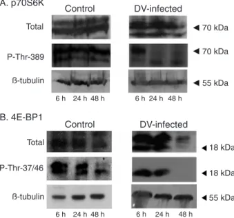
DV infection leads to regulation of p70S6K and 4E-BP1 phosphorylation
RPS6 is phosphorylated by p70S6K and this event is correlated with activation of protein synthesis and cell growth (13). qRT-PCR results showed that the correspond-ing gene for RPS6 is up-regulated (1.85 ± 0.30-fold) after 6 h of infection in relation to the mock-infected control, and
Figure 1. Modification of the expression of genes related to
translation and to the target of rapamycin pathway after infec-tion with dengue virus. Alterainfec-tions in the transcripinfec-tion of genes related to translation are represented. Genes are colored if their
down-regulated, 0.52 ± 0.18 and 0.71 ± 0.09, at 24 and 48 h, respectively (Table 1). This result confirms the fact that protein synthesis is activated during initial times of infection. To further evaluate this result, we have studied the changes induced in the phosphorylation state of p70S6K after 6, 24 and 48 h of DV infection and compared them to control mock-infected cells. The antibody to total p70S6K showed that the expression of p70S6K was not influenced by DV infection (Figure 2A, upper panel) while the antibody against p70S6K phosphorylated at T389 (P-Thr-389), showed that p70S6K continued to be phosphorylated in the 6 h of infection like control cells (Figure 2, middle panel), while dephosphorylation was induced as infection progressed (24 and 48 h).
Another protein that has been involved in the regula-tion of cap-dependent translaregula-tion via the TOR and AKT pathways is the regulatory factor, 4E-BP1. Treatment of cells with insulin or growth factors induces 4E-BP1 phos-phorylation, while its dephosphorylation by stress results in protein synthesis inhibition (10,11). The data for qRT-PCR indicated repression of transcription of the subunit eIF4E-BP3 at 6 and 48 h post-infection, while no alteration was detected at 24 h (Table 1). In this experiment, we expected to observe down-regulation also at 24 h. We decided to directly assay the state of 4E-BP1 phosphorylation during infection by Western blot using a polyclonal anti-4E-BP1 antibody or a phosphospecific antibody (P-Thr-37/46). We observed both differential expression and phosphorylation patterns induced by DV infection. In control cells, two main proteins of 20 and 18 kDa were visualized with the total
antibody and did not change expression throughout the experiment, except for the 18-kDa protein whose expres-sion was down-regulated. On the other hand, DV induced reduced expression of both proteins at 48 h of infection. Regarding phosphorylation, phospho-specific antibodies recognized the two proteins (18 and 20 kDa) in control cells (6 and 24 h) while at 48 h of infection only the 18-kDa protein was observed. In extracts derived from DV-infected cells, only an 18-kDa phosphorylated protein was observed at 6 and 24 h of infection. As infection progressed no phosphorylated 4E-BP1 was observed, most certainly due to inhibition of its expression. In order to test the integrity of proteins in the extract, we probed the same blots with antibodies against β-tubulin and the results showed the same intensity of expression throughout the experiment. Taken together, these results show that both p70S6K and 4E-BP1 are phosphorylated at early times of infection, sug-gesting a cap-dependent translation at this time, which is then inhibited after 24 h of infection, probably changing to a cap-independent translation.
Discussion
A strategy used by several virus classes to replicate inside the cell is to halt the host protein synthesis and to
Figure 2. p70S6K and 4E-BP1 phosphorylation is modified by
dengue virus (DV) infection. Whole cell protein extracts from HepG2 cells infected with DV, or mock-infected for 6, 24 or 48
h, were prepared and 30 µg protein was analyzed by Western blot. Panel A, Western blot analysis of total p70S6K and its phos
-phorylated form (P-Thr-389) using specific antibodies. Panel B,
Western blot analysis of total 4E-BP1 and its phosphorylated form (P-Thr-37/46). β-tubulin was used to determine protein in
-tegrity in both panels. One representative result is shown of three
independent experiments.
Table 1. 4E-BP1 phosphorylation is modified by dengue virus
infection.
Time post-infection 6 h 24 h 48 h eIF4A 1.39 ± 0.22 0.74 ± 0.10 0.71 ± 0.11 eIF4E 1.17 ± 0.42 0.75 ± 0.13 1.15 ± 0.35 eIF4G1 1.66 ± 0.30 0.93 ± 0.26 0.61 ± 0.25 eIF4G3 0.99 ± 0.23 0.98 ± 0.30 0.61 ± 0.22 eIF4B 1.43 ± 0.20 1.21 ± 0.40 0.34 ± 0.17 eIF4E-BP3 0.77 ± 0.20 1.30 ± 0.21 0.66 ± 0.10 eIF2B2 1.01 ± 0.19 1.07 ± 0.33 0.64 ± 0.10 RPS6 1.85 ± 0.35 0.52 ± 0.18 0.71 ± 0.09 Quantitative real-time PCR (qRT-PCR) experiments were carried out to measure mRNA expression of the indicated genes. Specif-ic primers were designed for each gene and cDNAs synthesized using RNA isolated from HepG2 cells mock-infected or infected with dengue virus after 6, 24, and 48 h were used as templates. GAPDH expression was used to normalize the results. The
Cellular protein synthesis during dengue virus infection 1025
www.bjournal.com.br Braz J Med Biol Res 42(11) 2009
sequester the protein synthesis machinery for translating their own proteins. However, the flaviviruses are RNA viruses that do not inhibit cellular protein synthesis. The structure of DV RNA resembles typical mRNA with a 5’cap structure. However, DV RNA is differentiated because it contains a 3’UTR, which forms a stalk structure resembling the poly(A)tail (36). This structure suggests that DV utilizes the cap-dependent translation machinery for protein syn-thesis. However, it has been shown that DV can utilize an alternative mechanism independent of an internal ribosome entry site that might depend on the interaction between the 5’ and 3’UTR for binding to the initiation factors (37). Until now, no study has been done on the interaction of DV with the cap-dependent machinery of the host cell. As a first approach, we analyzed differentially expressed genes post-infection using an oligo-library containing 10,000 of approximately 25,000 genes coded by the human genome. This library was chosen because it contains only genes with known or predicted function. We focused our analyses on the genes related to translation. It is noteworthy that of the 10,000 genes analyzed, 41 represent translation factors, and of these genes with changed expression, a cluster related to protein synthesis was up-regulated in response to DV at an early time of infection (6 h). In contrast to our results, another study that monitored the transcriptional response to DV infection in HepG2 cells did not detect significant differential expression of genes related to protein synthesis from 3 to 72 h of infection (38). Using qRT-PCR methodology, we tested eight genes related to the initiation of translation. It should be noted that all of them are positive factors needed for protein synthesis except for eIF4E-BP3 that is part of eIF4E-BP, which in its dephosphorylated form inhibits eIF4E assembly into the eIF4F complex. At 24 and 48 h of infection all of them were down-regulated. Inhibition of crucial translation factors by DV infection and the studied independence of DV on cap-dependent translation led us to examine whether infection would modulate 4EBP-1 and p70S6K phosphorylation. A key aspect of regulation of protein translation is the phosphorylation state of 4EBP-1
and p70S6K. Both phosphorylated proteins induce cell proliferation and protein synthesis and their phosphorylation is inhibited by rapamycin and wortmanin, which inhibit the mTOR and AKT pathways. Our results showed that at an early time of infection with DV, p70S6K and 4E-BP1 are phosphorylated. However, the progression of infection in HepG2 cells leads to dephosphorylation of both proteins, which may be related to an inhibition of cap-dependent protein synthesis.
In studies using picornavirus models, it has been shown that poliovirus and encephalomyocarditis virus shut off protein synthesis and a primary event is the dephospho-rylation of 4E-BP1 leading to inhibition of cap-dependent translation (39). Furthermore, DV does not rely on cap-dependent protein synthesis, since the addition of wort-manin or LY294002 does not inhibit virus translation (17). On the basis of the results obtained, we propose a model whereby DV enters HepG2 cells and replicates through a cap-dependent translation during the first hours of infection and then switches to a cap-independent translation as seen by inhibition of transcription of translation initiation factors and dephosphorylation of 4E-BP1 and p70S6K. It would be interesting now to determine which initiation factors are re-quired for translation of DV proteins by the cap-independent mechanism, since a potential selective advantage for the DV involving a switch from cap-dependent to cap-independent protein translation mechanisms would be to have a lower requirement for initiation factors that might be rate limiting in the cell. Another selective advantage would be to bypass apoptosis induced by the virus (40), which can promote inhibition of host cap-dependent protein synthesis.
Acknowledgments
We thank José Luis Santillán Torres and Lorena Chávez González, Microarray Unit, Instituto de Fisiologia Celular, Universidad Nacional Autónoma de México, for technical support.
References
1. Guzman MG, Kouri G. Dengue: an update. Lancet Infect Dis
2002; 2: 33-42.
2. Mackenzie JS, Gubler DJ, Petersen LR. Emerging flavivi -ruses: the spread and resurgence of Japanese encephalitis, West Nile and dengue viruses. Nat Med 2004; 10: S98-S109.
3. Whitehead SS, Blaney JE, Durbin AP, Murphy BR. Prospects
for a dengue virus vaccine. Nat Rev Microbiol 2007; 5: 518-528.
4. Clyde K, Kyle JL, Harris E. Recent advances in deciphering viral and host determinants of dengue virus replication and pathogenesis. J Virol 2006; 80: 11418-11431.
5. Hershey JWB, Merrick WC. The pathway and mechanism
of initiation of protein synthesis. In: Sonenberg N, Hershey
JWB, Mathews MB (Editors), Translational control of gene expression. Cold Spring Harbor: Cold Spring Harbor Press;
2000. p 33-88.
6. Sachs AB, Varani G. Eukaryotic translation initiation: there are (at least) two sides to every story. Nat Struct Biol 2000; 7: 356-361.
7. Jones RM, Branda J, Johnston KA, Polymenis M, Gadd M,
Rustgi A, et al. An essential E box in the promoter of the gene encoding the mRNA cap-binding protein (eukaryotic initiation factor 4E) is a target for activation by c-myc. Mol Cell Biol
1996; 16: 4754-4764.
eIF4E and control of cell growth. Curr Opin Cell Biol 1998; 10: 268-275.
9. Burnett PE, Barrow RK, Cohen NA, Snyder SH, Sabatini
DM. RAFT1 phosphorylation of the translational regulators p70 S6 kinase and 4E-BP1. Proc Natl Acad Sci U S A 1998; 95: 1432-1437.
10. Martin-Perez J, Thomas G. Ordered phosphorylation of 40S
ribosomal protein S6 after serum stimulation of quiescent
3T3 cells. Proc Natl Acad Sci U S A 1983; 80: 926-930. 11. Fingar DC, Richardson CJ, Tee AR, Cheatham L, Tsou C,
Blenis J. mTOR controls cell cycle progression through its
cell growth effectors S6K1 and 4E-BP1/eukaryotic transla -tion initia-tion factor 4E. Mol Cell Biol 2004; 24: 200-216. 12. Chung J, Grammer TC, Lemon KP, Kazlauskas A, Blenis J.
PDGF- and insulin-dependent pp70S6k activation mediated
by phosphatidylinositol-3-OH kinase. Nature 1994; 370: 71-75.
13. Sonenberg N, Pelletier J. Poliovirus translation: a paradigm
for a novel initiation mechanism. Bioessays 1989; 11: 128-132.
14. Gradi A, Svitkin YV, Imataka H, Sonenberg N. Proteolysis of human eukaryotic translation initiation factor eIF4GII, but not
eIF4GI, coincides with the shutoff of host protein synthesis after poliovirus infection. Proc Natl Acad Sci U S A 1998; 95: 11089-11094.
15. Holden KL, Harris E. Enhancement of dengue virus transla-tion: role of the 3’ untranslated region and the terminal 3’ stem-loop domain. Virology 2004; 329: 119-133.
16. De Nova-Ocampo M, Villegas-Sepulveda N, del Angel RM. Translation elongation factor-1alpha, La, and PTB interact with the 3’ untranslated region of dengue 4 virus RNA. Virol-ogy 2002; 295: 337-347.
17. Edgil D, Polacek C, Harris E. Dengue virus utilizes a novel strategy for translation initiation when cap-dependent
trans-lation is inhibited. J Virol 2006; 80: 2976-2986.
18. Seneviratne SL, Malavige GN, de Silva HJ. Pathogenesis of liver involvement during dengue viral infections. Trans R Soc Trop Med Hyg 2006; 100: 608-614.
19. Couvelard A, Marianneau P, Bedel C, Drouet MT, Vachon F, Henin D, et al. Report of a fatal case of dengue infection with hepatitis: demonstration of dengue antigens in hepatocytes and liver apoptosis. Hum Pathol 1999; 30: 1106-1110. 20. Bhamarapravati N. Hemostatic defects in dengue
hemor-rhagic fever. Rev Infect Dis 1989; 11 (Suppl 4): S826-S829.
21. Huerre MR, Lan NT, Marianneau P, Hue NB, Khun H, Hung
NT, et al. Liver histopathology and biological correlates in five cases of fatal dengue fever in Vietnamese children.
Virchows Arch 2001; 438: 107-115.
22. Mohan B, Patwari AK, Anand VK. Hepatic dysfunction in childhood dengue infection. J Trop Pediatr 2000; 46: 40-43.
23. Kuo CH, Tai DI, Chang-Chien CS, Lan CK, Chiou SS, Liaw
YF. Liver biochemical tests and dengue fever. Am J Trop Med Hyg 1992; 47: 265-270.
24. Nguyen TL, Nguyen TH, Tieu NT. The impact of dengue
haemorrhagic fever on liver function. Res Virol 1997; 148: 273-277.
25. Wahid SF, Sanusi S, Zawawi MM, Ali RA. A comparison of
the pattern of liver involvement in dengue hemorrhagic fe-ver with classic dengue fefe-ver. Southeast Asian J Trop Med Public Health 2000; 31: 259-263.
26. Subramanian V, Shenoy S, Joseph AJ. Dengue hemorrhagic
fever and fulminant hepatic failure. Dig Dis Sci 2005; 50: 1146-1147.
27. Knowles BB, Howe CC, Aden DP. Human hepatocellular
carcinoma cell lines secrete the major plasma proteins and
hepatitis B surface antigen. Science 1980; 209: 497-499.
28. Kosaki A, Webster NJ. Effect of dexamethasone on the
alternative splicing of the insulin receptor mRNA and insulin action in HepG2 hepatoma cells. J Biol Chem 1993; 268: 21990-21996.
29. Chen Q, Xia Y, Qiu Z. Effect of ecdysterone on glucose metabolism in vitro. Life Sci 2006; 78: 1108-1113.
30. Ginzinger DG, Godfrey TE, Nigro J, Moore DH, Suzuki S,
Pallavicini MG, et al. Measurement of DNA copy number at
microsatellite loci using quantitative PCR analysis. Cancer Res 2000; 60: 5405-5409.
31. Lowry OH, Rosebrough NJ, Farr AL, Randall RJ. Protein
measurement with the Folin phenol reagent. J Biol Chem
1951; 193: 265-275.
32. Pestova TV, Kolupaeva VG. The roles of individual
eu-karyotic translation initiation factors in ribosomal scanning
and initiation codon selection. Genes Dev 2002; 16: 2906-2922.
33. Shahbazian D, Roux PP, Mieulet V, Cohen MS, Raught B, Taunton J, et al. The mTOR/PI3K and MAPK pathways con -verge on eIF4B to control its phosphorylation and activity.
EMBO J 2006; 25: 2781-2791.
34. Pause A, Belsham GJ, Gingras AC, Donze O, Lin TA, Law-rence JC Jr, et al. Insulin-dependent stimulation of protein
synthesis by phosphorylation of a regulator of 5’-cap func -tion. Nature 1994; 371: 762-767.
35. Richter JD, Sonenberg N. Regulation of cap-dependent translation by eIF4E inhibitory proteins. Nature 2005; 433: 477-480.
36. Chambers TJ, Hahn CS, Galler R, Rice CM. Flavivirus ge -nome organization, expression, and replication. Annu Rev Microbiol 1990; 44: 649-688.
37. Edgil D, Harris E. End-to-end communication in the
modu-lation of transmodu-lation by mammalian RNA viruses. Virus Res
2006; 119: 43-51.
38. Fink J, Gu F, Ling L, Tolfvenstam T, Olfat F, Chin KC, et al.
Host gene expression profiling of dengue virus infection in
cell lines and patients. PLoS Negl Trop Dis 2007; 1: e86.
39. Gingras AC, Svitkin Y, Belsham GJ, Pause A, Sonenberg N.
Activation of the translational suppressor 4E-BP1 following infection with encephalomyocarditis virus and poliovirus.
Proc Natl Acad Sci U S A 1996; 93: 5578-5583.