ISSN 0100-879X
BIOMEDICAL SCIENCES
AND
CLINICAL INVESTIGATION
www.bjournal.com.br
www.bjournal.com.br
Volume 43 (3) 182-267 March 2011
Braz J Med Biol Res, March 2011, Volume 44(3) 200-205
doi: 10.1590/S0100-879X2011007500019
CCN2/CTGF silencing blocks cell aggregation in embryonal carcinoma
P19 cell
D.P. Aguiar, J.M. Coelho-Aguiar and J.G. Abreu
Faculdade de Medicina de Ribeirão Preto Campus
Ribeirão Preto
Institutional Sponsors
The Brazilian Journal of Medical and Biological Research is partially financed by
analiticaweb.com.br S C I E N T I F I C Hotsite of proteomics metabolomics
CCN2/CTGF silencing blocks cell aggregation
in embryonal carcinoma P19 cell
D.P. Aguiar, J.M. Coelho-Aguiar and J.G. Abreu
Programa de Biologia Celular e do Desenvolvimento, Instituto de Ciências Biomédicas, Universidade Federal do Rio de Janeiro, Rio de Janeiro, RJ, Brasil
Abstract
Connective tissue growth factor (CCN2/CTGF) is a matricellular-secreted protein involved in extracellular matrix remodeling. The P19 cell line is an embryonic carcinoma line widely used as a cellular model for differentiation and migration studies. In the present study, we employed an exogenous source of CCN2 and small interference RNA to address the role of CCN2 in the P19 cell aggregation phenomenon. Our data showed that increasing CCN2 protein concentrations from 0.1 to 20 nM decreased the number of cell clusters and dramatically increased cluster size without changing proliferation or cell survival, suggesting that CCN2 induced aggregation. In addition, CCN2 specific silencing inhibited typical P19 cell aggregation, which could be partially rescued by 20 nM CCN2. The present study demonstrates that CCN2 is a key molecule for cell aggregation of embryonic P19 cells.
Key words: Teratocarcinoma; Adhesion; Condensation; CCN family; CCN2/CTGF; Extracellular matrix
Introduction
Correspondence: J.G. Abreu, Programa de Biologia Celular e do Desenvolvimento, Instituto de Ciências Biomédicas, UFRJ, Bloco F, Sala 9, 21949-590 Rio de Janeiro, RJ, Brasil. E-mail: [email protected]
Received August 30, 2010. Accepted January 3, 2011. Available online February 18, 2011. Published March 7, 2011.
Connective tissue growth factor (CCN2/CTGF) is a member of the CCN [CYR61 (cysteine-rich 61)/CTGF NOV (nephroblastoma over-expressed)] family of matricellular signaling modulators. These modulators are characterized by four conserved modules that share homology with differ-ent protein domains (1,2). The amino terminus of the first module displays homology with an insulin-like growth factor binding protein. The second module is a von Willebrand factor type C/Chordin-like cysteine-rich domain (2). After a cysteine-free sequence, the third module displays homol-ogy to the thrombospondin type 1 repeat and contains a cysteine-knot at the carboxy-terminus. During development, CCN2 expression is mostly detected in tissues of compact cell organization, such as notochord, somites, cartilage (3,4), and Merckel’s cartilage (5), and also in mesenchymal condensation (6). Although several studies have indicated that CCN2 is a pleiotropic molecule, there are compelling data showing that CCN2 can function in the maintenance of multicellular spheroid aggregates as in ovarian cancer cells (7) and in mesenchymal cell condensation during chondrocyte differentiation (6). P19 mouse embryonic carcinoma cells have been used extensively as a model to address questions related to cell aggregation and differen-tiation (8,9). P19 cells have tumor stem cell features and
CCN2/CTGF silencing blocks P19 cell aggregation 201
pathway by up-regulation of ERK1/2-dependent Bcl-xL/ cIAP1 (18). This type of cancer remains aggregated and contains a high percentage of stem cells (19). Although cell aggregation has been studied for decades, its implication in both embryonic development and progress of particular diseases is not fully understood. In the present study, we investigated the cell aggregation properties of embryonic carcinoma P19 cells in culture under the influence of CCN2 protein. Our data show that CCN2 is sufficient for P19 cell aggregation and strongly suggest that CCN2 is a chemoat-tractant molecule for these stem-like cells.
Material and Methods
Cell culture
P19 embryonic carcinoma cells were purchased from the American Type Culture Collection (ATCC, USA). Cells were cultured in α-MEM (Sigma-Aldrich, WGK, Germany) supplemented with 10% fetal bovine serum (FBS) (Invit-rogen/Gibco, New Zealand) and split every 2 days. After the first 24 h P19 were incubated for more than 24 h with CCN2 at 37°C in 5% CO2.
Flag epitope-tagged construct of full-length CCN2 protein was prepared using S2 stable cells (3), which were cultured in Schneider’s Drosophila Medium supplemented with 10% FBS (Invitrogen/Gibco) and 56 μM hygromycin-B (Sigma, USA). Cells were split every 4 days and kept at 22°C. Full-length CCN2-Flag protein was purified as previ -ously described (3). Protein concentration was determined by the method of Lowry et al. (20).
siRNA synthesis and utilization
To silence CCN2 expression in P19 cells, we performed small interference RNA (siRNA). Two siRNAs were synthe-sized using the Ambion Silencer siRNA construction kit as a reference guide (Ambion, USA). The oligonucleotides used to target the CCN2-RNA (accession No. NM_010217) sequences were (CCN2-siRNA1) GAAGACUCAGCCAG AUCCAUU sense, UGGAUCUGGCUGAGUCUUCUU an-tisense (CCN2-siRNA2), AGCAGCUGCAAAUACCAAUUU sense, AUUGGUAUUUGCAGCUGCUUU antisense. The CCN2-siRNA1 and CCN2-siRNA2 (negative control) were transfected using RNAiFect (Qiagen, USA, 301,605) in P19 cells at 50-70% confluence. The transfection procedures were carried out by the method of Rober et al. (21). To perform rescuing experiments, 20 nM recombinant CCN2 was added in 1% FBS culture medium after 24 h of siRNA silencing. Since only CCN2 siRNA1 was able to block CCN2 translation (data not shown) we used CCN2-siRNA2 as a negative control to check possible cytotoxic or nonspecific effects caused by CCN2-siRNA1.
Cell proliferation and [3H]-thymidine incorporation
assays
P19 cells were seeded at 1 x 10⁵ cells per well in α-MEM
with 1% FBS in 24-well dishes, treated or not with various concentration of CCN2 (0.1, 5, and 20 nM) depending on the experimental series. A [3H]-thymidine pulse was added to
the cell culture along with the CCN2. After 24 h, the medium was removed, the cultures were washed three times with PBS and 300 μL ice-cold 10% trichloroacetic acid was added to each well. The cells were harvested and [3H]-thymidine
incorporation was measured with a scintillation counter. Each experiment was performed at least three times using three culture wells per condition.
Cell viability assay
Cell viability and growth were measured by the MTT assay (22,23). The yellow tetrazolium salt (3-(4,5-dimethylthiazol-2-yl)-2,5-diphenyltetrazolium bromide; MTT) was reduced in metabolically active cells to form insoluble purple formazan crystals, which were solubilized by the addition of DMSO and then quantified by spectrophotometry at 340 nm.
Western blot
CCN2 expression levels were monitored by Western blot analysis. Treated and untreated cell cultures were washed three times with 1X PBS, and then harvested in sample buffer [20 µM dithioreitol (DTT); 1.38 mM sodium dodecyl sulfate (SDS); 125 mM Tris-HCl, pH 6.8, and 20% glycerol]. The samples were submitted to 12% SDS-polyacrylamide gel electrophoresis (SDS-PAGE), electroblotted and trans-ferred to a PVDF membrane (HybondTM-P, Amersham Biosciences, Brazil). Membranes were pre-incubated in 5% non-fat dry milk in Tris-buffered saline containing 0.001% Tween 20 (TBS-T) for 1 h and then incubated with the pri-mary polyclonal antibody anti-CTGF (Torrey Pines Biolabs, USA, 1:1000) and anti-tubulin (Sigma, 1:2000) overnight. The reaction was visualized using the SuperSignal West Pico Chemiluminescent Substrate (Pierce, USA).
Quantification parameters
To measure the cluster size, aggregates submitted to spe-cific treatments were photographed and the area of each ag -gregate was quantified with the ImageJ software (NIH-Image - http://rsbweb.nih.gov/ij). The ImageJ software quantifies the number of pixels in the cluster area that is defined by a line around the cluster perimeter. The values attributed to these pixels were defined as arbitrary units. For each condition, 20 fields were randomly chosen and 500 clusters were counted. Only clusters consisting of more than 10 cells were counted. The values obtained for different experimental conditions were analyzed with the GraphPad Prism 4.0 software (www. graphpad.com). Statistical analysis was performed using the non-parametric Mann-Whitney test (24).
Results
CCN2 increases P19 cell cluster size
mesenchymal condensation during embryonic development (4,6,25), a phenomenon that involves aggregation and chemoattraction. However, these morphological changes have not yet been fully investigated in stem-like cells. To gain insight into the aggregation properties of CCN2 protein, P19 cells were treated for 24 h with different concentrations of recombinant CCN2 (Figure 1). Even at low confluence, P19 cells tend to form some epithelial-like aggregates in culture (11) (Figure 1A). But the addition of 20 nM CCN2 recombinant protein induced larger aggregates (Figure 1B). The effect of CCN2 on morphological aggregation clusters was analyzed in terms of number and size. Only clusters containing more than 10 cells were scored. We detected an average of 30 small clusters of P19 cells not treated with CCN2 (Figure 1C). There was a dose-response reduction of clusters with CCN2 concentration, and 20 nM CCN2 reduced the number of P19 clusters to roughly 50% compared to untreated cells (Figure 1C). There was a dose-dependent increase in cluster size with CCN2 (Figure 1D).
These observations suggested that CCN2 protein promoted cell aggregation or induced cell proliferation or both. [3H]-thymidine incorporation experiments were
performed with P19 cells cultured with or without CCN2 for 24 h. No significant difference in proliferation rate was detected between untreated and CCN2-treated P19 cells, showing that the increase in cluster size was not due to cell proliferation (data not shown).
Since CCN2-treated P19 cells have larger but fewer clusters than untreated cells, we determined if CCN2 could regu-late cell viability. The determination of cell viability with MTT did not show significant differences between untreated P19 cells and cells treated with 0.1, 0.5 or 20 nM CCN2 (data not shown). Therefore, the increase in P19 cell cluster size was probably induced by aggregation.
CCN2 silencing impairs P19 cell aggregation
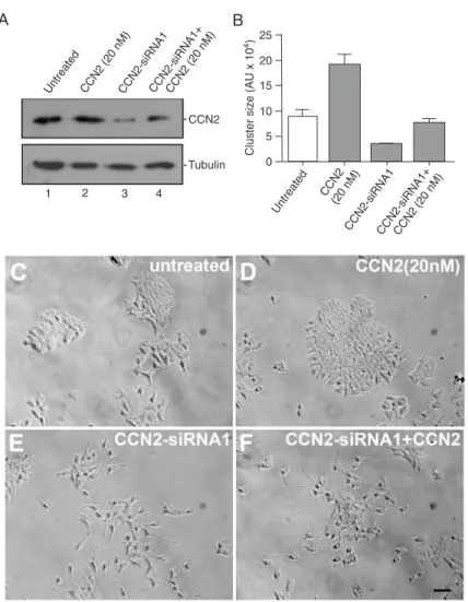
To examine the requirement of CCN2 for P19 cell aggregation, CCN2-siRNA silencing (CCN2-siRNA) was performed. We silenced CCN2 for 24 h by transfecting CCN2-siRNA1. CCN2 protein silencing in P19 cells was de-tected by Western blotting of the cell extracts using an anti-CCN2 antibody (Figure 2A). The endogenous CCN2 protein was silenced by CCN2-siRNA1 (Figure 2A, lane 3). P19 cells transfected with CCN2-siRNA2, whose oligonucleotide sequences were not able to block CCN2 protein translation (data not shown), display morphology and aggregation pattern similar to those of untreated P19 cells (Table 1, see Methods for details). The
addi-tion of 20 nM CCN2 recombinant protein allowed us to detect CCN2 (Figure 2A, lane 4). CCN2-siRNA1-transfected and -untransfected P19 cells were analyzed in terms of cluster formation (Figure 2B, C and E). CCN2-siRNA1-transfected P19 cells present clusters that were smaller compared to untransfected and CCN2-siRNA2-transfected cells (nega-tive control, data not shown). In addition cells appeared to be dissociated in CCN2-siRNA1-transfected cultures (Figure 2E). Interestingly, the addition of 20 nM recombinant CCN2 protein to CCN2-siRNA1-transfected P19 cells was able to achieve the normal cluster size observed in untreated cells, but did not reach the phenotype observed in 20 nM CCN2-treated cells (Figure 2C, D and F). The cluster size of CCN2-siRNA1 P19 cells treated with 20 nM CCN2 indicated that the cells had retained their natural state of aggregation since the cluster sizes were similar to those found in untreated P19 cells (Figure 2B).
We quantified the percentage of cells in the clusters dur -ing the aggregat-ing phenomenon promoted by CCN2. Four categories of clusters were established based on the number of cells per cluster (Table 1). In untreated cultures, almost 60% of the clusters are formed by 25 to 50 cells per cluster (Table 1). Conversely, in cultures treated with 0.1 and 5 nM CCN2, the percentage of cells per cluster increases and in cultures treated with 20 nM CCN2 approximately 70% of the clusters consist of >75 cells per cluster (Table 1). In CCN2-siRNA1-transfected cultures, only clusters containing less
CCN2/CTGF silencing blocks P19 cell aggregation 203
than 25 cells were observed. The addition of CCN2 protein partially reversed the inhibition of cluster formation by CCN2-siRNA1 and no effect was observed on cluster formation in CCN2-siRNA2 (scramble siRNA)-treated cells (Table 1). Since proliferation was not modified by CCN2, these results strongly suggest that CCN2 induces P19 cell ag-gregation.
Discussion
The present study addressed the ag-gregation induced by CCN2/CTGF of embryonic carcinoma P19 cells in culture. Our data show that CCN2/CTGF induces cluster formation when added exogenously to P19 culture media. Conversely, silencing of CCN2/CTGF protein by siRNA inhibits P19 cluster formation (Table 1). Therefore, our data indicate CCN2/CTGF as a pivotal component of the P19 cell aggregation ma-chinery.
Several reports have documented CCN2-mediated adhesion in adult mesen-chyme-derived cells such as fibroblasts, os -teoblasts, chondrocytes, myoblasts, stellate and mesangial cells (9,26-30). However, the role of CCN2 in the aggregation of embryonic carcinoma cells has not been investigated. P19 cells have the potential to differentiate into ectoderm or mesoderm cell lineages by adopting embryonic body-like structures or monolayer morphology, respectively, de-pending on the treatment (11,31-33). These features suggest that the aggregation of these cells is an important step during the establishment of their differentiation pro-gram. Thus, these cells provide an excellent tool to understand the events controlling cell aggregation, proliferation and differentiation. We have shown that monolayers of P19
Figure 2. CCN2 is required for P19 cell aggregation.A, Immunoblotting detec-tion of CCN2 protein in P19 extracts of untreated cells (lane 1) and of cells treated with 20 nM CCN2 (lane 2), CCN2-siRNA1 (lane 3), and CCN2-siRNA1 plus 20 nM CCN2 (lane 4). Tubulin detection was used as a loading control. B, Cluster size of P19 cells and of cells treated with 20 nM CCN2, CCN2-siRNA1, and CCN2-siRNA1 plus 20 nM CCN2 protein. Phase contrast microscopy of untreated P19 cells (C), in the presence of 20 nM recombinant CCN2 (D), trans-fected with CCN2-siRNA1 (E) and with CCN2-siRNA1 plus 20 nM CCN2 protein (F). Each experiment was carried out in triplicate. N = 4; scale bar 50 μm for all panels.
Table 1. Cell cluster analysis showing different percentages of cells per cluster in each experimental condition.
Number of cells per cluster
Untreated CCN2 CCN2-siRNA1 CCN2-siRNA1 + CCN2 (20 nM) CCN2-siRNA2
0.1 nM 5 nM 20 nM
<25 22.4% 9.5% 3.3% 4.3% 100% 65.2% 36.0%
25-50 58.6% 4.5% 10.0% 4.3% 0 34.8% 47.6%
51-75 15.5% 26.2% 26.6% 17.4% 0 0 14.3%
and Mv1Lu cells form spherical aggregates when CCN2 and TGFß-1 are added together to the culture medium (3). Under these conditions, the spherical aggregates were able to express endothelial markers and to adopt an ag-gregate morphology during their differentiation. The CCN2 protein is highly expressed during early development in morphogenetic areas where migratory behavior, adhesion properties and pluripotency state are cellular characteristics for tissue organization, as shown in Xenopus leavis and in mice (3,4,34). Furthermore, cell adhesion is also critical for tumor survival and progression. In addition, some tumor cells can adopt an embryonic behavior and express pluripotency markers, including octamer-binding transcription factor 4 and Nanog (35). Thus, acquisition of adhesion potential could explain why CCN2 has been implicated in metas-tasis (36,37). Our findings have brought more information regarding chemoadhesive and aggregation behavior upon CCN2 stimulation; therefore, P19 cells combine properties of cell pluripotency as well as tumor behavior.
Cell condensation results from different interactions between cell surface receptors and molecules of the microenvironment such as extracellular matrix-related proteins and glycoproteins. In fact, it has been reported that CCN2 interacts with fibronectin (25,38) and that, during migration, cells can perform attachment/detachment and undergo cytoskeletal actin rearrangement to move upon the extracellular matrix network (9,11). In our analysis of the cluster number and size of untreated versus CCN2-treated cells, we noticed an inverse correlation, i.e., the larger the cluster, the lower the number of clusters, suggesting that cells reorganize the culture topology probably by migrating and aggregating toward colony formation. One could argue that small colonies of untreated P19 cells could proliferate in response to CCN2. However, this does not seem to be the case since the [3H]-thymidine incorporation assay did
not reveal differences in proliferation between CCN2-treated
and -untreated P19 cells. Since CCN2 addition did not affect cell proliferation, the increase in size followed by a decrease in cluster number could be interpreted as cells migrating from clusters to form larger clusters by aggregation. Alternatively and less likely, CCN2 addition could also favor cluster ag-gregation, which indeed, in any way would decrease the number of clusters. Indeed, this result is consistent with previous findings showing that CCN2 induces cell migration and cytoskeleton actin rearrangement in human mesangial cells (9,11). Silencing of CCN2 by siRNA on P19 cells had a noticeable effect on the inhibition of cluster formation. Interestingly, the addition of CCN2 recombinant protein to CCN2-siRNA1 P19 cells did not induce a large cluster, but restored the basal capacity of P19 cell to form aggregates. A possible explanation for this would be that the exogenous source of CCN2 induced cell reaggregation to the normal phenotype in culture. Further experiments are needed to determine whether CCN2 can modulate the expression of other CCN members such as CCN1 that could mediate cell aggregation in P19 cells (39).
The current study addressed the chemoadhesive and aggregant properties of CCN2 also for embryonic carcinoma P19 cells, showing that CCN2 induces cell aggregation and that this phenomenon occurs in a CCN2 dose-dependent manner. Since P19 cells have been used extensively as a model/tool in stem cell differentiation research (40), our data may contribute to a better understanding of P19 adhesion properties, which are crucial in most of the approaches employed in such studies.
Acknowledgments
We thank Dr. Sophie Creuzet and Dr. Vivaldo Moura Neto for their comments about the manuscript. Research supported by CNPq, CAPES, PRONEX, and FAPERJ.
1. Bork P. The modular architecture of a new family of growth regulators related to connective tissue growth factor. FEBS Lett 1993; 327: 125-130.
2. Garcia Abreu J, Coffinier C, Larrain J, Oelgeschlager M, De Robertis EM. Chordin-like CR domains and the regulation of evolutionarily conserved extracellular signaling systems.
Gene 2002; 287: 39-47.
3. Abreu JG, Ketpura NI, Reversade B, De Robertis EM. Connective-tissue growth factor (CTGF) modulates cell signalling by BMP and TGF-beta. Nat Cell Biol 2002; 4: 599-604.
4. Ivkovic S, Yoon BS, Popoff SN, Safadi FF, Libuda DE, Stephenson RC, et al. Connective tissue growth factor co-ordinates chondrogenesis and angiogenesis during skeletal development. Development 2003; 130: 2779-2791. 5. Shimo T, Kanyama M, Wu C, Sugito H, Billings PC, Abrams
References
WR, et al. Expression and roles of connective tissue growth factor in Meckel’s cartilage development. Dev Dyn 2004; 231: 136-147.
6. Song JJ, Aswad R, Kanaan RA, Rico MC, Owen TA, Barbe MF, et al. Connective tissue growth factor (CTGF) acts as a downstream mediator of TGF-beta1 to induce mesenchymal cell condensation. J Cell Physiol 2007; 210: 398-410. 7. Sodek KL, Ringuette MJ, Brown TJ. Compact spheroid
for-mation by ovarian cancer cells is associated with contractile behavior and an invasive phenotype. Int J Cancer 2009; 124: 2060-2070.
8. Soprano DR, Teets BW, Soprano KJ. Role of retinoic acid in the differentiation of embryonal carcinoma and embryonic stem cells. Vitam Horm 2007; 75: 69-95.
aggrega-CCN2/CTGF silencing blocks P19 cell aggregation 205
tion during neural differentiation of P19 mouse embryonic carcinoma cells. J Proteome Res 2009; 8: 1765-1781. 10. Rossant J, Papaioannou VE. Outgrowth of embryonal
carci-noma cells from injected blastocysts in vitro correlates with abnormal chimera development in vivo. Exp Cell Res 1985; 156: 213-220.
11. McBurney MW, Jones-Villeneuve EM, Edwards MK, Ander-son PJ. Control of muscle and neuronal differentiation in a cultured embryonal carcinoma cell line. Nature 1982; 299: 165-167.
12. Leask A, Abraham DJ. The role of connective tissue growth factor, a multifunctional matricellular protein, in fibroblast biology. Biochem Cell Biol 2003; 81: 355-363.
13. Steinberg MS. Differential adhesion in morphogenesis: a modern view. Curr Opin Genet Dev 2007; 17: 281-286. 14. Nishida T, Maeda A, Kubota S, Takigawa M. Role of
mechan-ical-stress inducible protein Hcs24/CTGF/CCN2 in cartilage growth and regeneration: mechanical stress induces expres-sion of Hcs24/CTGF/CCN2 in a human chondrocytic cell line HCS-2/8, rabbit costal chondrocytes and meniscus tissue cells. Biorheology 2008; 45: 289-299.
15. Ono M, Kubota S, Fujisawa T, Sonoyama W, Kawaki H, Aki-yama K, et al. Promotion of hydroxyapatite-associated, stem cell-based bone regeneration by CCN2. Cell Transplant
2008; 17: 231-240.
16. Tan TW, Lai CH, Huang CY, Yang WH, Chen HT, Hsu HC, et al. CTGF enhances migration and MMP-13 up-regulation via alphavbeta3 integrin, FAK, ERK, and NF-kappaB-dependent pathway in human chondrosarcoma cells. J Cell Biochem
2009; 107: 345-356.
17. Aoyama E, Hattori T, Hoshijima M, Araki D, Nishida T, Kubota S, et al. N-terminal domains of CCN family 2/connective tis-sue growth factor bind to aggrecan. Biochem J 2009; 420: 413-420.
18. Wang MY, Chen PS, Prakash E, Hsu HC, Huang HY, Lin MT, et al. Connective tissue growth factor confers drug re-sistance in breast cancer through concomitant up-regulation of Bcl-xL and cIAP1. Cancer Res 2009; 69: 3482-3491. 19. Korkaya H, Paulson A, Charafe-Jauffret E, Ginestier C,
Brown M, Dutcher J, et al. Regulation of mammary stem/ progenitor cells by PTEN/Akt/beta-catenin signaling. PLoS Biol 2009; 7: e1000121.
20. Lowry OH, Rosebrough NJ, Farr AL, Randall RJ. Protein measurement with the Folin phenol reagent. J Biol Chem
1951; 193: 265-275.
21. Rober RA, Sauter H, Weber K, Osborn M. Cells of the cel-lular immune and hemopoietic system of the mouse lack lamins A/C: distinction versus other somatic cells. J Cell Sci
1990; 95 (Part 4): 587-598.
22. Amado NG, Cerqueira DM, Menezes FS, da Silva JF, Neto VM, Abreu JG. Isoquercitrin isolated from Hyptis fasciculata
reduces glioblastoma cell proliferation and changes beta-catenin cellular localization. Anticancer Drugs 2009; 20: 543-552.
23. Vistica DT, Skehan P, Scudiero D, Monks A, Pittman A, Boyd MR. Tetrazolium-based assays for cellular viability: a criti-cal examination of selected parameters affecting formazan production. Cancer Res 1991; 51: 2515-2520.
24. Siegel S. Non-parametric statistics for behavioral sciences. New York: McGraw Hill; 1956.
25. Chen XM, Qi W, Pollock CA. CTGF and chronic kidney fibrosis. Front Biosci 2009; 1: 132-141.
26. Babic AM, Chen CC, Lau LF. Fisp12/mouse connective tis-sue growth factor mediates endothelial cell adhesion and migration through integrin alphavbeta3, promotes endothe-lial cell survival, and induces angiogenesis in vivo. Mol Cell Biol 1999; 19: 2958-2966.
27. Brigstock DR. Strategies for blocking the fibrogenic actions of connective tissue growth factor (CCN2): From pharmaco-logical inhibition in vitro to targeted siRNA therapy in vivo. J Cell Commun Signal 2009; 3: 5-18.
28. Perbal B. Ten years later. J Cell Commun Signal 2009; 3: 1-3.
29. Schutze N, Noth U, Schneidereit J, Hendrich C, Jakob F. Differential expression of CCN-family members in primary human bone marrow-derived mesenchymal stem cells dur-ing osteogenic, chondrogenic and adipogenic differentiation.
Cell Commun Signal 2005; 3: 5.
30. Maeda A, Nishida T, Aoyama E, Kubota S, Lyons KM, Kuboki T, et al. CCN family 2/connective tissue growth fac-tor modulates BMP signalling as a signal conducfac-tor, which action regulates the proliferation and differentiation of chon-drocytes. J Biochem 2009; 145: 207-216.
31. Okamoto K, Okazawa H, Okuda A, Sakai M, Muramatsu M, Hamada H. A novel octamer binding transcription factor is differentially expressed in mouse embryonic cells. Cell 1990; 60: 461-472.
32. Suzuki T, Kim HS, Kurabayashi M, Hamada H, Fujii H, Aikawa M, et al. Preferential differentiation of P19 mouse embryonal carcinoma cells into smooth muscle cells. Use of retinoic acid and antisense against the central nervous system-specific POU transcription factor Brn-2. Circ Res
1996; 78: 395-404.
33. Santiago MF, Liour SS, Mendez-Otero R, Yu RK. Glial-guided neuronal migration in P19 embryonal carcinoma stem cell aggregates. J Neurosci Res 2005; 81: 9-20. 34. Surveyor GA, Brigstock DR. Immunohistochemical
localiza-tion of connective tissue growth factor (CTGF) in the mouse embryo between days 7.5 and 14.5 of gestation. Growth Factors 1999; 17: 115-124.
35. Ponticos M, Holmes AM, Shi-wen X, Leoni P, Khan K, Rajku-mar VS, et al. Pivotal role of connective tissue growth factor in lung fibrosis: MAPK-dependent transcriptional activation of type I collagen. Arthritis Rheum 2009; 60: 2142-2155. 36. Kang Y, Siegel PM, Shu W, Drobnjak M, Kakonen SM,
Cor-don-Cardo C, et al. A multigenic program mediating breast cancer metastasis to bone. Cancer Cell 2003; 3: 537-549. 37. Aikawa T, Gunn J, Spong SM, Klaus SJ, Korc M.
Connec-tive tissue growth factor-specific antibody attenuates tumor growth, metastasis, and angiogenesis in an orthotopic mouse model of pancreatic cancer. Mol Cancer Ther 2006; 5: 1108-1116.
38. Pi L, Ding X, Jorgensen M, Pan JJ, Oh SH, Pintilie D, et al. Connective tissue growth factor with a novel fibronectin binding site promotes cell adhesion and migration during rat oval cell activation. Hepatology 2008; 47: 996-1004. 39. Chaqour B, Goppelt-Struebe M. Mechanical regulation of the
Cyr61/CCN1 and CTGF/CCN2 proteins. FEBS J 2006; 273: 3639-3649.