Bassi DG etal.
520
Rev Assoc Med Bras 2010; 56(5): 520-1gist
-
induced
upper
gastrointestinal
bleeding
deomir germano bassi1, Fernanda perez adornoda silva2, Flavia mariade oliveira sousa3 Study conducted at Hospital Universitário de Taubaté, São Paulo, SP, Brazil
1- Professor Titular - Doutor da Disciplina de Clínica Cirúrgica da Universidade de Taubaté e membro do Colégio Brasileiro de Cirurgia, São Paulo, SP 2- Acadêmica de Medicina da Universidade de Taubaté, São Paulo, SP
3- Residente de Cirurgia Geral do Hospital Universitário de Taubaté, São Paulo, SP
Imaging in Medicine
Gastrointestinal stromal tumors (GIST) are rare neoplasms that mostly affect patients over the age of 50, with no gender predilection1,2. The diagnosis of GIST is made by means of
immu-nohistochemistry testing for CD117 (c-kit), and combination treatment with surgical intervention and adjuvant chemotherapy has produced good outcomes3. We report a case of gastric GIST
diagnosed after an episode of upper gastrointestinal bleeding and successfully treated by subtotal gastrectomy.
c
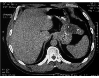
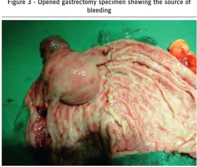
ase reportA 51-year-old male presented with a 5-day history of mode-rately intense, crampy abdominal pain in the periumbilical region associated with nausea, dizziness, and melena. One day prior to seeking care, the patient developed hematemesis and lighthea-dedness. Upper GI endoscopy showed an ulcerated submucosal gastric lesion with signs of recent bleeding and an overlying clot, mild duodenitis, and two well-healed chronic duodenal ulcers. The initial diagnostic hypothesis was leiomyoma. A CT scan of the abdomen showed a mass lesion on the anterior wall of the stomach (Figure 1). The patient underwent subtotal central gastrectomy on the fourth hospital day. Intraoperatively, a tumor roughly 8 cm long at the largest axis was found on the anterior wall of the stomach, approximately 6 cm from the cardia (Figures 2 and 3).
Anatomic pathology showed a mesenchymal spindle cell neoplasm with slight pleomorphism, a mitotic index of zero,
necrosis and hyaline degeneration, and conirmed tumor-free
margins.
Immunohistochemical testing was diffusely positive for
CD117, leading to a inal diagnosis of intermediate-risk GIST.
The patient had an uneventful clinical course and was discharged
on the ifth postoperative day.
d
iscussionGastrointestinal stromal tumor (GIST) is a nonspeciic desig -nation given to mesenchymal tumors arising from the interstitial cells of Cajal, pacemaker cells associated with Auerbach’s plexus that express CD117 (c-kit), a proto-oncogenic protein. Expression of c-kit distinguishes GIST from leiomyoma, leiomyoblastoma, and other mesenchymal tumors of the gastrointestinal tract1,3,4.
Most GISTs are symptomatic, with 60% to 70% occur-ring in the stomach, small intestine, or rectum; the most common clinical manifestations are gastrointestinal bleeding,
bowel obstruction, and abdominal pain2. Preoperative laboratory
diagnosis is still elusive, although research is underway to identify
speciic biomarkers2,4.
Despite substantial improvement in the understanding of GIST over the past few years, doubts remain as to possible
prognostic factors. Identiication of these factors is tied to the
importance of stratifying patients into risk groups and, conse-quently, pinpointing those with a higher likelihood of recurrence or shorter survival after surgical resection, thus enabling the use of adjuvant targeted therapy in cases with a worse prognosis3.
Treatment consists of surgical resection with margins of at least 2 to 4 cm1,2,3,4. Tumor site is an important factor in planning
surgery, as anterior lesions are best treated with wedge resection and posterior ones with gastrectomy2. In very low and low-grade
tumors, surgery is followed by outpatient follow-up, whereas high-risk patients receive adjuvant imatinib therapy. There is no consensus as to the optimal management of intermediate-risk tumors.
We conclude that the dificulty in obtaining an accurate
preoperative diagnosis has an impact on surgical treatment of GIST, and that management of patients with “intermediate-risk”
Gist-inDuceDupperGastrointestinalBleeDinG
521
Rev Assoc Med Bras 2010; 56(5): 520-1tumors is still challenging. We suggest that GIST be included in the differential diagnosis of all patients with upper gastrointes-tinal bleeding.
r
eFerences1. John SK, Basu S, Lawrance RJ, Davies N. An unusual presentation of a Gastrointestinal stromal tumour (GIST). World J Surg Oncol. 2007;5:78. 2. Consolo FS, Cardoso AAP, Branco OM, Lamego H, Arruda L. Hemorragia
digestiva por tumor estromal gástrico: relato de caso. Rev Col Bras Cir. 2007; 34(1).
3. Basilio PRO, Pannain VL, Portari P, Iglesias CA, Basilio ACO. GIST: avaliações morfológica e imuno-histoquímica do prognóstico. J Bras Patol. Med Lab. 2009; 45:49-54
Figure 2 - Subtotal (central) gastrectomy specimen showing neoplastic lesion
Figure 3 - Opened gastrectomy specimen showing the source of bleeding
4. Cruz RJ, Vincenzi R, Ketzer BM, Cecilio AL, Cepeda LA. Spontaneous intra-tumoral bleeding and rupture of giant gastric stromal tumor (> 30 cm) in a young patient. World J Surg Oncol. 2008;6:76.
*Correspondence:
Av. Tiradentes, 500 - Bom Conselho Taubaté - SP, Brazil
CEP: 12030-180