Gastrointestinal Stromal Tumor
Gastrointestinal Stromal Tumor of the Esophagus:
Report of a Case
Özofagusun Gastrointestinal Stromal Tümörü:
Bir Olgu Sunumu
DOI: 10.4328/JCAM.1276 Received: 28.08.2012 Accepted: 30.09.2012 Printed: 01.01.2016 J Clin Anal Med 2016;7(1): 111-3 Corresponding Author: M.Muharrem Erol, Uludağ Üniversitesi Tıp Fakültesi, Göğüs Cerrahisi AD. Nilüfer, Bursa, Türkiye.
T.: +90 2242952340 F.: +90 2244428698 E-Mail:[email protected]
Özet
Gastrointestinal stromal tümörler (GIST) gastrointestinal sistemin mezenkimal hücrelerinden gelişen nadir tümörlerdir. Özofagus kaynaklı GIST ise mide ve ince bağırsakla karşılaştırıldığında oldukça nadirdir. Bu olgu sunumunda geniş cerrahi rezeksiyonla çıkardığımız bir GIST olgusunu sunuyoruz.
Anahtar Kelimeler
Gastrointestinal Stromal Tümör; Özofagus; Cerrahi
Abstract
Gastrointestinal stromal tumors are rare neoplasms to be thought to arise from mesenchymal cells of the gastrointestinal tract. Gastrointestinal stromal tumors (GIST) of the esophagus are well documented but are very much rarer than gas-trointestinal stromal tumors of the stomach and small bowel. We describe a case of GIST of the esophagus that was resected with wide surgical resection.
Keywords
Gastrointestinal Stromal Tumor; Esophagus; Surgery
M.Muharrem Erol¹, Hüseyin Melek¹, A.Sami Bayram¹, Elif Ülker Akyıldız², Cengiz Gebitekin¹ ¹Göğüs Cerrahisi AD., ²Patoloji AD., Uludağ Üniversitesi Tıp Fakültesi, Bursa, Türkiye
| Journal of Clinical and Analytical Medicine Introduction
The term gastrointestinal stromal tumor (GIST) refers to all mesenchymal tumors in the gastrointestinal tract [1]. These tumors demonstrate a pathobiology and clinical behavior dif
-ferent from those of smooth muscle and Schwann cell tumors. GISTs account for 0.1% to 3% of all tumors in the gastrointes
-tinal tract. The majority of GISTs occur in the subdiaphragmatic gastrointestinal tract, but small number of cases has been in the esophagus [2, 3]. Here in we present a patient with a esoph
-ageal GIST treated with wide surgical excision.
Case Report
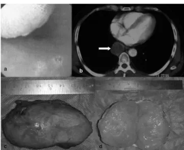
51 years old male patient had dysphagia and stomach ache for 6 months. We performed endoscopic examination and found external compression at 4 cm cranial to the gastroesophageal junction (Figure-1a). The computerized tomography showed a mass surrounding the esophagus at the same level (Figure-1b). We performed a right thoracotomy. We found a mass that be
-gan 4 cm below the azygous vein and lied distally. The mass was settled between muscular and mucosal layers of esopha
-gus. We opened the muscular layer and completely resected the mass that was 3x7 cm in diameter and had thin capsule and a smooth border (Figure-1c,d) . Result of frozen section was benign esophageal tumour so that ater enucleating the muscu
-lar layers were sutured one by one over the mucosal layer. We discharged the patient at postoperative 3rd day. No mitosis was detected during pathological examination at 50 times magni
-ication. Ki67 labeling index was 2/ 1000. CD117, Vimentin, CD34 (Figure-2), Desmin and SMA were positive and S100 was negative. The mass had the diagnosis of middle degree malign esophageal gastrointestinal stromal tumor. The follow up at 3rd month was uneventful.
Discussion
Gastrointestinal stromal tumors (GISTs) are uncommon
mes-enchymal tumors that arise in the wall of the gastrointestinal tract (GI). They account for approximately 0.1% to 3% of GI neoplasms [4]. GISTs were previously thought to be smooth muscle neoplasms, and most were classiied as leiomyoma or leiomyosarcoma [2].
In 1983, Mazur and Clark challenged the longstanding concept that most mesenchymal tumors of the stomach were of smooth muscle origin, and introduced the concept of stromal tumor [3- 5]. With the advent of immunohistochemistry and electron mi
-croscopy, it became apparent that GISTs might have myogenic features, neural attributes or characteristics of both muscle and nerve. Two thirds of these tumors arise from the stomach, 25% arise from the small intestine and 5% arise from the esophagus [2]. GISTs occur in an older patient population (50-60 years) [2, 3]. Approximately one half of patients with an esophageal GIST are asymptomatic; the remainder exhibit symptoms, which may include dysphagia, retrosternal pain, pyrosis, cough, odynopha
-gia and weight loss [1]. Our case was 51 year – old male and had dysphagia and stomach ache for 6 months.
Most esophageal GISTs are endocentric (intraluminal polypoid mass); on endoscopy the mucosa is usually intact and the mass appears as a rounded, smooth, raised lesion rarely showing cen
-tral umblication or ulceration [1]. Esophageal GISTs originate
from between the walls of the esophagus. They are kinds of proliferation of spindle cells or epitheloid cells [5].
The majority of GISTs are benign (60-80%). The most consis
-tent prognostic factors are site of presentation and tumor size [3]. Kimiyashi suggested a criterion for diferentiation between
benign and malignant GISTs; hemorrhage or necrosis, the
diam-eter of the tumor > 5cm, Ki-67 labeling index > 3%. If the tumor has only one of the items above it is malignant. If none of the items above can be found, then it is benign. The diameter of our mass 3x7cm, Ki-67 labeling index was 2/ 1000 and no mi
-tosis was detected during pathological examination at 50 times magniication, so that our mass had the diagnosis of middle
degree malign esophageal gastrointestinal stromal tumor.
GIST; there was focal positivity for pancytokeratin marker and for CD56 and difuse strong positivity for CD117 and CD34. CD117 is sensitive and speciic . Although CD34 is a sensi
-tive immunochemistry marker of GISTs, CD34 is expressed in 60-70% cases of GISTs [5]. Our mass had positivity for CD34,
CD117.
These tumors seem to be resistant to chemoradiation. Because of the lack of any efective alternative therapies, surgical resec
-tion should be considered for all patients with GISTs [2].
Figure 1. Endoscopic appearance of mass (A), Mass appearance in computerized tomography (B), Appearance of mass (C, D).
Figure 2. Pathological photographs of mass. With Hex200 (A), With +CD34 (B), With +CD117 (C), With +vimentine (D).
| Journal of Clinical and Analytical Medicine
112
| Journal of Clinical and Analytical Medicine
Competing interests
The authors declare that they have no competing interests.
References
1. Manu N, Richard P, Howard S. Bleeding esophageal GIST. Dis Esophagus 2005; 18: 281-82.
2. Basoglu A, Kaya E, Celik B. Giant gastrointestinal stromal tumor of the esopha-gus presenting with dispnea. J Thorac Cardiovasc Surg 2006; 131(5): 1198-9. 3. Lee JR, Anstadt MP, Khwoja S. gastrointestinal stromal tumor of the posterior mediastinum. Eur J Cardiothorac Surg 2002; 22(6): 1014-16.
4. Cabrero IA, Vazques G, Santiesteban FIS. Clinicopathologic study of 275 cases of gastrointestinal stromal tumors: the experience at 3 large medical centers in Mexico. Ann Diag Path 2007; 11(1): 39-45.
5. Zhu X, Zhang XO, Li BM. Esophageal mesenchymal tumors: endoscopy, pathol-ogy and immunohistochemistry. World J Gastroenterol 2007; 7(5): 768-73.
How to cite this article:
Erol MM, Melek H, Bayram AS, Akyıldız EÜ, Gebitekin C. Gastrointestinal Stromal Tumor of the Esophagus: Report of a Case. J Clin Anal Med 2016;7(1): 111-3.
Journal of Clinical and Analytical Medicine | 113