Hypomagnesemia and its relation with chronic low-grade
inflammation in obesity
ANA RAQUEL SOARESDE OLIVEIRA1, KYRIA JAYANNE CLÍMACO CRUZ1, JULIANA SOARES SEVERO2, JENNIFER BEATRIZ SILVA MORAIS2,
TAYNÁH EMANNUELLE COELHODE FREITAS3, ROGÉRIO SANTIAGO ARAÚJO4, DILINADO NASCIMENTO MARREIRO5*
1PhD Student in Food and Nutrition, Universidade Federal do Piauí (UFPI), Teresina, PI, Brazil 2MSc Student in Food and Nutrition, UFPI, Teresina, PI, Brazil
3Nutritionist, UFPI, Teresina, PI, Brazil
4MD, Endocrinologist, PhD Professor, Department of General Practice, UFPI, Teresina, PI, Brazil 5PhD Professor, Department of Nutrition, UFPI, Teresina, PI, Brazil
S
UMMARYStudy conducted at Department of Nutrition, Universidade Federal do Piauí
(UFPI), Teresina, PI, Brazil
Article received: 5/23/2016
Accepted for publication: 5/31/2016
*Correspondence:
Address: Rua Hugo Napoleão, 665, Ed. Palazzo Reale, apto. 2001
Teresina, PI – Brazil Postal code: 64048-320
http://dx.doi.org/10.1590/1806-9282.63.02.156
Introduction: The accumulation of visceral fat in obesity is associated with excessive production of proinlammatory adipokines, which contributes to low--grade chronic inlammation state. Moreover, the literature has shown that mineral deiciency, in particular of magnesium, has important role in the patho-genesis of this metabolic disorder with relevant clinical repercussions.
Objective: To bring updated information about the participation of hypomagnesemia in the manifestation of low-grade chronic inlammation in obese individuals.
Method: Articles published in PubMed, SciELO, LILACS and ScienceDirect, using the following keywords: “obesity,” “magnesium” and “low grade inlammation.”
Results: Scientiic evidence suggests that magnesium deiciency favors the manifestation of low-grade chronic inlammation in obese subjects.
Conclusion: From literature data, it is evident the participation of magnesium through biochemical and metabolic reactions in protecting against this metabolic disorder present in obesity.
Keywords: obesity, magnesium, low-grade inlammation.
I
NTRODUCTIONWhite adipose tissue is the main energy source in the body, mobilizing fatty acids according to metabolic need.1 In
excessive amounts, this tissue produces proinlamma-tory adipokines, a process inluenced by the anatomical location of fat deposits. Visceral fat, being metabolically more active, favors an increase in the production of these substances, contributing to chronic low-grade inlamma-tion in obesity.2,3
Low-grade chronic inlammation differs from other types of inlammation as it leads to latent tissue damage for extended periods of time, lasting for decades, silently.1,2
Studies have shown that in obese individuals the inlam-matory state favors an increase in the formation of reactive oxygen species that can lead to an overload of the antioxi-dant defense system, contributing to the manifestation of oxidative stress and, consequently, cell damage and death.4,5
Biochemical and nutritional disorders present in obese individuals are being extensively investigated in
order to elucidate the mechanisms involved in the patho-genesis of obesity. In this sense, minerals have been the subject of extensive research in order to identify their relation with metabolic disorders.
Magnesium in particular has attracted great inter-est from researchers as it plays a role in glucose me-tabolism, insulin homeostasis, synthesis of adenosine triphosphate, proteins and nucleic acids, as well as in membrane stability and regulation of hormonal and immunological function.6,7
Magnesium deiciency is characterized as a nutrition-al problem that leads to changes in the cellular function and biological activity of the molecules, and may contribute to the onset of metabolic disorders related to the inlam-matory process, especially in obese individuals, who present low serum and dietary concentrations of this mineral.8-10
the pathogenesis of chronic diseases such as obesity, the objective of this review was to bring updated information on the participation of hypomagnesemia in the manifesta-tion of low-grade chronic inlammamanifesta-tion in obese individuals.
M
ETHODThe literature search was carried out in PubMed, SciELO, LILACS and ScienceDirect databases with no restrictions as to year of publication, considering the following inclu-sion criterion: studies on the metabolic and physiological aspects of magnesium, which presented relevant aspects on the role of this mineral in the manifestation of chron-ic low-grade inlammation in obese individuals. The ar-ticles were selected based on originality and relevance, taking into account the accuracy and adequacy of the experimental design and the sample number. Established and recent works were preferably used.
The search for bibliographic references was performed using the following keywords: “obesity,” “magnesium” and “low grade inlammation.” The literature search in-cluded the following types of studies: randomized or quasi-randomized controlled clinical trials, case-control study, and review articles.
M
ETABOLIC AND PHYSIOLOGICAL ASPECTSOF MAGNESIUM
Magnesium is the second most abundant intracellular cation and is involved in about 300 biochemical reactions related to anabolic and catabolic actions in the body, such as glycolysis and protein and lipid metabolism.11 This
min-eral contributes to increase the production of intracellular adenosine triphosphate and the use of glucose, acting as a cofactor in all reactions that involve energy transfer.12
On average, the body of an adult contains 1 mole of magnesium. About half of the mineral content is present in the bone and the other half in soft tissues. More pre-cisely, 0.3% of the total is found in serum, 0.5% in eryth-rocytes, 19.3% in soft tissues, 27% in muscles, and 52.9% in bones. In serum about one-third of the magnesium is bound to proteins. Of this total, 25% is bound to albumin and 8% to globulins. Of the remaining magnesium, about 80% is in the form of free ion (55% of total magnesium) and about 20% is combined with phosphate, citrate and other compounds.13
Magnesium homeostasis in the body is dependent on the amount ingested, intestinal absorption, renal ex-cretion and need presented by various tissues.11 About 25
to 60% of ingested magnesium is absorbed into the gas-trointestinal tract by passive or active transport. The transport of this nutrient through the paracellular
path-way is responsible for 80 to 90% of its absorption, which occurs predominantly between microvilli of the small intestine through simple diffusion, and this process is stimulated when intraluminal concentrations of this mineral are high. This absorption pathway occurs main-ly in the ileum and distal parts of the jejunum, where the permeability to this ion is greater. This is because in these sites there is a low expression of claudin proteins 1, 3, 4, 5 and 8, which participate in the formation of paracel-lular barriers and pores, regulating the passage of sub-stances through the epithelium.14-16
However, in the case of low intraluminal concentrations, the magnesium is absorbed through the action of speciic transporters belonging to the family called transient recep-tor potential channel of melastatin type (TRPM6 and 7), and this process occurs by the active absorption of sodium ions, followed by water.17 This transport requires strict
regulation since magnesium ions cross two cell membranes. The active absorption of the mineral occurs mainly in the colon and, to a lesser extent, in the jejunum and ileum.14-16
It is important to emphasize that excessive calorie intake promotes an increase in the intestinal absorption of magnesium, since the mechanism involved in this process is energy dependent. However, the absorption of this mineral can be impaired in the presence of lipids, phosphorus, phytates and oxalate. Diets low in protein (< 30 g/day) also slow the absorption of magnesium.18,19
The kidneys are the main excreting organs involved in magnesium homeostasis, and 70% of the entire content of iltered mineral is reabsorbed in the thick ascending branch of the loop of Henle via the paracellular route. The driving force for magnesium reabsorption is positive transluminal epithelial tension generated by the recycling of potassium through the apical membrane, which is linked with sodium, water and calcium. In the distal convoluted tubule, mag-nesium transport mainly occurs by active process medi-ated by TRPM6, and is characterized by negative and highly resistant luminal tension, a speciic process that does not depend on calcium absorption.20,21
In a situation of reduced oral intake of magnesium, the kidneys are able to reduce their excretion. The other routes of magnesium excretion are feces and sweat, with the fecal concentration of the mineral being about 150 to 200 mg/day, while sweating contributes about 15 mg dai-ly loss.18,22 The balance of magnesium in the body is
The evaluation of nutritional status relative to mag-nesium can be obtained by assessing its contents in plasma, erythrocyte, urine and diet. Plasma magnesium has been widely used. However, this marker does not relect its total content since, even after reduction in mineral intake, plas-ma concentrations replas-main constant for a long period of time.6,11 The reference values for normal plasma magnesium
concentrations are between 0.75 and 1.05 mmol/L.18,19
Erythrocyte magnesium concentration is approxi-mately 2.5 mmol/L and since it has a half-life of 120 days, medium and long-term evaluations of the mineral’s stock in the body can be performed.24,25 As for urinary
magne-sium, approximately 3 to 4 mmol of the nutrient is lost daily through this excretion route. Urine is considered a good indicator for recent changes in nutritional status regarding magnesium, because in cases of stock depletion, excretion is reduced by renal reabsorption mechanisms to maintain its homeostasis in the body.24
The main food sources of magnesium are whole grains, dark green vegetables, legumes, walnuts, seeds, chestnuts and almonds.26 The dietary recommendation of this
min-eral is 400 to 420 and 310 to 320 mg daily for adult men and women, respectively.27
H
YPOMAGNESEMIA AND LOW-
GRADECHRONIC INFLAMMATION
The literature has shown that the diet of obese individu-als has reduced magnesium content, which is a nutri-tional problem of great relevance.28,29 Huang et al.30 and
Song et al.31 found that dietary intake of magnesium is
inversely proportional to body mass index, waist circum-ference, and body fat percentage.
The reduced intake of magnesium by obese individu-als can be explained mainly by the high consumption of processed foods containing low magnesium and by the reduced intake of food sources of magnesium, which seems to contribute to the reduction of its concentrations in the blood compartments.15
Studies have found reduced plasma concentrations of magnesium in obese individuals.32,33 Guerrero-Romero and
Rodríguez-Morán34 have shown that individuals with
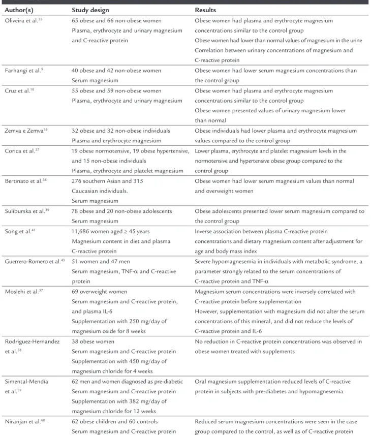
nor-mal body weight but metabolically obese exhibit reduced serum magnesium concentrations compared to the obese who are metabolically healthy. Table 1 shows data on the status of magnesium in obese individuals, as well as its participation in chronic low-grade inlammation.
Magnesium deiciency seems to affect the activation of proinlammatory pathways in obese individuals.40 In
this regard, several researchers have observed that the reduced intake of this mineral and its low serum
concen-tration are strongly related to the increase in the plasma concentration of inlammatory biomarkers, such as C-reactive protein, tumor necrosis factor alpha (TNF-α) and interleukin 6 (IL-6).26,33,41
Nielsen et al.42 found that magnesium intake in
amounts below estimated average requirement (EAR) shows a positive correlation with plasma C-reactive protein and body mass index in adults. Guerrero-Romero et al.43 found
severe hypomagnesemia in individuals with metabolic syndrome, being this parameter strongly related to serum concentrations of C-reactive protein and TNF-α.
A study conducted by Oliveira et al.35 revealed reduced
dietary magnesium content and urinary excretion in obese women. In addition, a positive correlation was observed between urinary magnesium and serum concentrations of C-reactive protein in these patients, suggesting the inlu-ence of hypomagnesuria on this inlammatory marker.
Reduced concentrations of magnesium in plasma compromise its intracellular homeostasis and contribute to the development of a proinlammatory state through overproduction and release of cytokines such as interleu-kin 1β (IL-1β) and TNF-α, and increased serum concen-trations of neuropeptides.11,44,45
It is important to mention that the mechanisms in-volved in the inlammatory response present in magnesium deicient obese individuals are not yet clearly elucidated. However, according to the literature, the opening of cal-cium channels and the activation of N-methyl-D-aspartate (NMDA) receptors, as well as the priming of phagocytic cells, induce the entry of calcium into the cell, release of neurotransmitters, such as substance P, membrane oxida-tion and activaoxida-tion of nuclear transcripoxida-tion factor kappa B (NF-kB), which favors the inlammatory process.22,45,46
The inlammatory response is mainly related to the change in the extracellular concentration of magnesium, since the deiciency of this mineral reduces its plasma concentrations but does not alter its intracellular con-centration. Thus, it is important to emphasize the action of magnesium as a natural calcium antagonist and that the reduction of magnesium in the extracellular compart-ment induces an increase in the concentration of intracel-lular calcium, favoring the activation of phagocytic cells and the production of cytokines.35,46
TABLE 1 Studies evaluating the status of magnesium in obese individuals or their relationship to chronic low-grade inlammation.
Author(s) Study design Results
Oliveira et al.35 65 obese and 66 non-obese women
Plasma, erythrocyte and urinary magnesium and C-reactive protein
Obese women had plasma and erythrocyte magnesium concentrations similar to the control group
Obese women had lower than normal values of magnesium in the urine Correlation between urinary concentrations of magnesium and C-reactive protein
Farhangi et al.9 40 obese and 42 non-obese women
Serum magnesium
Obese women had lower serum magnesium concentrations than the control group
Cruz et al.10 55 obese and 59 non-obese women
Plasma, erythrocyte and urinary magnesium
Obese women had plasma and erythrocyte magnesium concentrations similar to the control group
Obese women presented values of urinary magnesium lower than normal
Zemva e Zemva36 32 obese and 32 non-obese individuals
Plasma and erythrocyte magnesium
Obese individuals had lower plasma and erythrocyte magnesium values compared to the control group
Corica et al.37 19 obese normotensive, 19 obese hypertensive,
and 15 non-obese individuals
Plasma, erythrocyte and platelet magnesium
Lower plasma, erythrocyte and platelet magnesium levels in the normotensive and hypertensive obese group compared to the control group
Bertinato et al.38 276 southern Asian and 315
Caucasian individuals. Serum magnesium
Obese women had lower serum magnesium values than normal and overweight women
Suliburska et al.39 78 obese and 20 non-obese adolescents
Serum magnesium
Obese adolescents presented lower serum magnesium compared to the control group
Song et al.41 11,686 women aged ≥ 45 years
Magnesium content in diet and plasma C-reactive protein
Inverse association between plasma C-reactive protein
concentrations and dietary magnesium content after adjustment for age and body mass index
Guerrero-Romero et al.43 51 women and 47 men
Serum magnesium, TNF-α and C-reactive protein
Severe hypomagnesemia in individuals with metabolic syndrome, a parameter strongly related to the serum concentrations of C-reactive protein and TNF-α
Moslehi et al.57 69 overweight women
Serum magnesium and C-reactive protein, and plasma IL-6
Supplementation with 250 mg/day of magnesium oxide for 8 weeks
Magnesium serum concentrations were inversely correlated with C-reactive protein before supplementation
However, supplementation with magnesium did not alter the serum concentrations of this mineral, and did not reduce the levels of C-reactive protein and IL-6
Rodriguez-Hernandez et al.58
38 obese women
Serum magnesium and C-reactive protein Supplementation with 450 mg/day of magnesium chloride for 4 weeks
No reduction in C-reactive protein concentrations was observed in obese women treated with supplements
Simental-Mendía et al.59
62 men and women diagnosed as pre-diabetic Serum magnesium and C-reactive protein Supplementation with 382 mg/day of magnesium chloride for 12 weeks
Oral magnesium supplementation reduced levels of C-reactive protein in subjects with pre-diabetes and hypomagnesemia
Niranjan et al.60 62 obese children and 60 controls
Serum magnesium and C-reactive protein
by leptin, a hormone that also favors the receptor’s activa-tion.40 Thus, excessive calcium inlux into the neuronal
tissue promotes the release of neurotransmitters, such as substance P, which triggers an inlammatory response through the release of cytokines, histamine and free radicals47 (Figure 1).
In adipose tissue, the increase of the intracellular calcium content derives from the opening of the L-type calcium channels, which is regulated by magnesium bind-ing sites. In the presence of deiciency of this nutrient, the blockage of these channels is compromised, increasing the inlux of calcium to the adipose cells. Excess intracel-lular calcium, in turn, results in the activation of calcium--dependent processes, such as the release of proinlamma-tory cytokines. Note that one of the major events in the calcium-mediated inlammatory process is the activation of NF-kB.46,48,49
NF-kB is a potent proinlammatory gene transcrip-tion factor. When activated, it binds to speciic genes,
stimulating the production and release of the proinlam-matory cytokines, namely TNF-α and IL-6, and adhesion molecules. These cytokines, when released in excess, favor the secretion of C-reactive protein by the liver.35
It is important to say that TRPM7 channels appear to regulate magnesium concentrations in tissues, and are therefore important in the homeostasis of this mineral.50
Note, however, that TRPM7 is not selective for magnesium, and its expression may also mediate calcium inlux and consequently inlammation, which depends on the serum concentrations of both minerals. That is, in situations of magnesium deiciency, calcium competes with magnesium and enters the cells.51,52
Magnesium deiciency appears to increase the produc-tion of free radicals and the sensitivity of cells to the attack of reactive oxygen species.53,54 Hypomagnesemia favors
the iniltration of neutrophils and macrophages in the affected cells, which potentiates the activity of the enzyme nicotinamide adenine dinucleotide phosphate oxidase
FIGURE 1 Action of magnesium as an anti-inlammatory nutrient in the brain. The increase of calcium in the intracellular medium promotes the transcription of inlammatory mediators through the release of substance P. Magnesium can inhibit this inlammatory pathway by its action as a natural calcium antagonist, blocking the increase of intracellular concentrations of this mineral.
Ca2+: calcium; Mg2+: magnesium; NMDAR: N-methyl-D-aspartate receptor; GLU: glutamate. Ca2+
Ca2+
Mg2+
Leptin
NMDAR
Substance P
Transcription of inlammatory mediators
and free radicals
Plasma membrane
FIGURE 2 Action of magnesium as an anti-inlammatory nutrient in adipose tissue. The increase of calcium in the intracellular medium
promotes the oxidation of cell membranes and the transcription of inlammatory mediators through the activation of NF-κB and its transloca-tion into the nucleus, and increases the oxidative stress through the activatransloca-tion of the NO synthase and NADPH oxidase. Magnesium can inhibit this inlammatory pathway by its action as a natural calcium antagonist, blocking the increase of intracellular concentrations of this mineral.
Ca2+: calcium; Mg2+: magnesium; NF-κB: nuclear factor kappa B; NO synthase: nitric oxide synthase.
Transcription of inlammatory mediators Ca2+
Ca2+
NO O2
-NF-kB
Nucleus
Plasma membrane L-type calcium channels
NO synthase NADPH oxidase
NO + OH-→ NOOO
-Mg2+
(NADPH oxidase), increasing the production of the su-peroxide radical.46,55
Hypomagnesemia also contributes to reduce the ex-pression and activity of antioxidant enzymes, such as glutathione peroxidase (GPx), superoxide dismutase (SOD) and catalase (CAT), and cellular and tissue antioxidant concentrations, as well as increases the production of hydrogen peroxide by inlammatory cells.11,56
Combined with this, in the presence of hypomagne-semia, intracellular ionic calcium contributes to the exces-sive production of uric acid and hydroxyl radical, which reacts with nitric oxide, which is also high in hypomag-nesemia, forming peroxynitrite.22,57 Thus, excessive
pro-duction of reactive species in magnesium-deicient indi-viduals also contributes to the inlammatory state present in obese individuals (Figure 2).
Some studies have been conducted to evaluate the effect of magnesium supplementation in obese or over-weight individuals. However, no reduction in the
concen-tration of inlammatory biomarkers was observed. Mosle-hi et al.57 found that supplementation with 250 mg/day
of magnesium oxide for 8 weeks was not able to reduce levels of C-reactive protein in overweight women. Rodri-guez-Hernandez et al.58 did not observe reduced
concen-trations of this inlammatory protein in obese women supplemented with 450 mg of magnesium chloride for 4 weeks, either.
F
INAL CONSIDERATIONSbases to explain the action of this nutrient as a protection against chronic inlammation present in obesity.
C
ONFLICT OF INTERESTThe authors declare no conlict of interest.
R
ESUMOHipomagnesemia e sua relação com a inlamação crônica de baixo grau na obesidade
Introdução: O acúmulo de gordura visceral na obesida-de está associado à produção excessiva obesida-de adipocinas pró-inlamatórias, o que contribui para o estado de inla-mação crônica de baixo grau. A literatura também tem mostrado que a deiciência de minerais, em particular do magnésio, possui papel importante na patogênese desse distúrbio metabólico com repercussões clínicas relevantes.
Objetivo: Trazer informações atualizadas sobre a parti-cipação da hipomagnesemia na inlamação crônica de baixo grau em indivíduos obesos.
Método: Bases de dados Pubmed, SciELO, Lilacs e Scien-ceDirect, utilizando as palavras-chave: “obesity”, “mag-nesium” e “low grade inlammation”.
Resultados: As evidências cientíicas sugerem que a de-iciência de magnésio favorece a manifestação da inla-mação crônica de baixo grau em indivíduos obesos.
Conclusão: É evidente a participação do magnésio, por meio de reações bioquímicas e metabólicas, na proteção contra esse distúrbio metabólico presente na obesidade.
Palavras-chave: obesidade, magnésio, inlamação crôni-ca de baixo grau.
R
EFERENCES1. Lay SL, Simard G, Martinez MC, Andriantsitohaina R. Oxidative stress and metabolic pathologies: from an adipocentric point of view. Oxid Med Cell Longev. 2014; 2014:908539.
2. Ikeoka D, Mader JK, Pieber TR. Adipose tissue, inlammation and cardio-vascular disease. Rev Assoc Med Bras (1992). 2010; 56(1):116-21. 3. França AKTC, Santos AM, Salgado JV, Hortegal EV, Silva AAM, Salgado
Filho N. Estimated visceral adipose tissue, but not body mass index, is associated with reductions in glomerular iltration rate based on cystatin C in the early stages of chronic kidney disease. Int J Nephrol. 2014; 2014:574267.
4. Tunc O, Bakos HW, Tremellen K. Impact of body mass index on seminal oxidative stress. Andrologia. 2010; 43(2):121-8.
5. Zaki ME, El-Bassyouni H, Kamal S, El-Gammal M, Youness E. Association of serum paraoxonase enzyme activity and oxidative stress markers with dyslipidemia in obese adolescents. Indian J Endocrinol Metab. 2014; 18(3):340-4.
6. Elin RJ. Assessment of magnesium status for diagnosis and therapy. Magnes Res. 2010; 23(4):194-8.
7. Volpe SL. Magnesium in disease prevention and overall health. Adv Nutr. 2013; 4(3):378S-83S.
8. Sales CH, Santos AR, Cintra DE, Colli C. Magnesium-deicient high-fat diet: effects on adiposity, lipid proile and insulin sensitivity in growing rats. Clin Nutr. 2014; 33(5):879-88.
9. Farhangi MA, Ostadrahimi A, Mahboob S. Serum calcium, magnesium, phosphorous and lipid proile in healthy Iranian premenopausal women. Biochem Med. 2011; 21(3):312-20.
10. Cruz KJC, Oliveira ARS, Pinto DP, Morais JBS, Lima FS, Colli C, et al. Inluence of magnesium on insulin resistance in obese women. Biol Trace Elem Res. 2014; 160(3):305-10.
11. Barbagallo M, Dominguez LJ. Magnesium and aging. Curr Pharm Des. 2010; 16(7):832-9.
12. Khan AM, Sullivan L, McCabe E, Levy D, Vasan RS, Wang TJ. Lack of association between serum magnesium and the risks of hypertension and cardiovascular disease. Am Heart J. 2010; 160(4):715-20.
13. Elin RJ. Assessment of magnesium status. Clin Chem. 1987; 33(11):1965-70. 14. Baaij JHF, Hoenderop JGJ, Bindels RJM. Regulation of magnesium balance: lessons learned from human genetic disease. Clin Kidney J. 2012; 5(Suppl 1):i15-i24.
15. Jahnen-Dechent W, Ketteler M. Magnesium basics. Clin Kidney J. 2012; 5(Suppl 1):i3-i14.
16. Houillier P. Mechanisms and regulation of renal magnesium transport. Annu Rev Physiol. 2014; 76:411-30.
17. Pham PC, Pham PM, Pham SV, Miller JM, Pham PT. Hypomagnesemia in patients with type 2 diabetes. Clin J Am Soc Nephrol. 2007; 2(2):366-73. 18. Bohl CH, Volpe SL. Magnesium and exercise. Crit Rev Food Sci Nutr. 2002;
42(6):533-63.
19. Martin KJ, González EA, Slatopolsky E. Clinical consequences and management of hypomagnesemia. J Am Soc Nephrol. 2009; 20(11):2291-5. 20. Silva RF, Beserra BTS, Oliveira ARS, Barbosa AM, Coelho JS, Poltronieri F,
et al. Relação entre exercício físico, estresse oxidativo e magnésio. Nutrição em Pauta. 2013; 21:15-9.
21. Vetter T, Lohse MJ. Magnesium and the parathyroid. Curr Opin Nephrol Hypertens. 2002; 11(4):403-10.
22. Weglicki WB. Hypomagnesemia and inlammation: clinical and basic aspects. Annu Rev Nutr. 2012; 32:55-71.
23. Sales CH, Pedrosa LFC. Magnesium and diabetes mellitus: their relation. Clin Nutr. 2006; 25(4):554-62.
24. Gibson RS. Principles of nutrition assessment. 2. ed. New York: Oxford University Press; 2004.
25. Rocha VS. Avaliação bioquímica e do consumo alimentar de magnésio em mulheres saudáveis no terceiro trimestre gestacional [dissertation]. São Paulo: Faculdade de Ciências Farmacêuticas, Universidade de São Paulo; 2009. 26. Evangelopoulos AA, Vallianou NG, Panagiotakos DB, Eorgiou A, Zacharias
GA, Alevra A, et al. An inverse relationship between cumulating components of the metabolic syndrome and serum magnesium levels. Nutr Res. 2008; 28(10):659-63.
27. Institute of Medicine. Dietary reference intakes for calcium, phosphorus, magnesium, vitamin D, and luoride. Washington: National Academy Press; 1997.
28. Jarvandi S, Gougeon R, Bader A, Dasgupta K. Differences in food intake among obese and nonobese women and men with type 2 diabetes. J Am Coll Nutr. 2011; 30(4):225-32.
29. López-Alarcón M, Perichart-Perera O, Flores-Huerta S, Inda-Icaza P, Rodríguez-Cruz M, Armenta-Álvarez A, et al. Excessive reined carbohydrates and scarce micronutrients intakes increase inlammatory mediators and insulin resistance in prepubertal and pubertal obese children independently of obesity. Mediators Inlamm. 2014; 2014:849031.
30. Huang JH, Lu YF, Cheng FC, Lee JN, Tsai LC. Correlation of magnesium intake with metabolic parameters, depression and physical activity in elderly type 2 diabetes patients: a cross-sectional study. Nutr J. 2012; 11:41. 31. Song CH, Choi WS, Oh HJ, Kim K. Associations of serum minerals with
body mass index in adult women. Eur J Clin Nutr. 2007; 61(5):682-5. 32. Lecube A, Baena-Fustegueras JÁ, Fort JM, Pelegrí D, Hernández C, Simó R.
Diabetes is the main factor accounting for hypomagnesemia in obese subjects. PLoS One. 2012; 7(1):e30599.
33. Song Y, Li TY, van Dam RM, Manson JE, Hu FB. Magnesium intake and plasma concentrations of markers of systemic inlammation and endothelial dysfunction in women. Am J Clin Nutr. 2007; 85(4):1068-74.
35. Oliveira AR, Crua KJ, Morais JB, Severo JS, Freitas TE, Veras AL, et al. Magnesium status and its relationship with c-reactive protein in obese women. Biol Trace Elem Res. 2015; 168(2):296-302.
36. Zemva A, Zemva Z. Ventricular ectopic activity, left ventricular mass, hyperinsulinemia, and intracellular magnesium in normotensive patients with obesity. Angiology. 2000; 51(2):101-6.
37. Corica F, Allegra A, Ientile R, Buemi M. Magnesium concentrations in plasma, erythrocytes, and platelets in hypertensive and normotensive obese patients. Am J Hypertens. 1997; 10(11):1311-3.
38. Bertinato J, Wu Xiao C, Ratnayake WM, Fernandez L, Lavergne C, Wood C, et al. Lower serum magnesium concentration is associated with diabetes, insulin resistance, and obesity in South Asian and white Canadian women but not men. Food Nutr Res. 2015; 59:25974.
39. Suliburska J, Cofta S, Gajewska E, Kalmus G, Sobieska M, Samborski W, et al. The evaluation of selected serum mineral concentrations and their association with insulin resistance in obese adolescents. Eur Rev Med Pharmacol Sci. 2013; 17(17):2396-400.
40. Nielsen FH. Magnesium, inlammation, and obesity in chronic disease. Nutr Rev. 2010; 68(6):333-40.
41. Song Y, Ridker PM, Manson JE, Cook NR, Buring JE, Liu S. Magnesium intake, C-reactive protein, and the prevalence of metabolic syndrome in middle-aged and older U.S. women. Diabetes Care. 2005; 28(6):1438-44. 42. Nielsen FH, Johnson LK, Zeng H. Magnesium supplementation improves
indicators of low magnesium status and inlammatory stress in adults older than 51 years with poor quality sleep. Magnes Res. 2010; 23(4):158-68. 43. Guerrero-Romero F, Bermudez-Peña C, Rodríguez-Morán M. Severe
hypomagnesemia and low-grade inlammation in metabolic syndrome. Magnes Res. 2011; 24(2):45-53.
44. Chacko SA., Song Y, Nathan L, Tinker L, Boer IH, Tylavsky F, et al. Relations of dietary magnesium intake to biomarkers of inlammation and endothelial dysfunction in an ethnically diverse cohort of postmenopausal women. Diabetes Care. 2010; 33(2):304-10.
45. Dibaba DT, Xun P, He K. Dietary magnesium intake is inversely associated with serum C-reactive protein levels: meta-analysis and systematic review. Eur J Clin Nutr. 2014; 68(4):510-6.
46. Mazur A, Maier JAM, Rock E, Gueux E, Nowacki W, Rayssiguier Y. Magnesium and the inlammatory response: potential physiopathological implications. Arch Biochem Biophys. 2007; 458(1):48-56.
47. Weglicki WB, Phillips TM. Pathobiology of magnesium deiciency: a cytokine/neurogenic inlammation hypothesis. Am J Physiol. 1992; 263(3 Pt 2):R734-7.
48. Nielsen FH, Milne DB, Gallagher S, Johnson L, Hoverson B. Moderate magne-sium deprivation results in calcium retention and altered potasmagne-sium and phos-phorus excretion by postmenopausal women. Magnes Res. 2007; 20(1):19-31. 49. Rayssiguier Y, Libako P, Nowacki B, Rock E. Magnesium deiciency and
metabolic syndrome: stress and inlammation may relect calcium activation. Magnes Res. 2010; 23(2):73-80.
50. Severo JS, Morais JBS, Freitas TEC, Cruz, KJC, Oliveira, ARS, Poltronieri F, et al. Aspectos metabólicos e nutricionais do magnésio. Nutr Clín Diet Hosp. 2015; 35(2):67-74.
51. Sarmiento D, Montorfano I, Cáceres M, Echeverría C, Fernández R, Cabel-lo-Verrugio C, et al. Endotoxin-induced vascular endothelial cell migration is dependent on TLR4/NF-B pathway, NAD(P)H oxidase activation, and transient receptor potential melastatin 7 calcium channel activity. Int J Bio-chem Cell Biol. 2014; 55:11-23.
52. Huang L, Ng MN, Chen M, Lin X, Tang T, Cheng H, et al. Inhibition of TRPM7 channels reduces degranulation and release of cytokines in rat bone marrow-derived mast cells. Int J Mol Sci. 2014; 15(7):11817-31.
53. Bae YJ, Choi MK. The estimated daily manganese intake of Korean children aged 11-12. Nutr Res Pract. 2011; 5(6):548-52.
54. Patrick L. Nonalcoholic fatty liver disease: relationship to insulin sensitivity and oxidative stress. Treatment approaches using vitamin E, magnesium, and betaine. Altern Med Rev. 2002; 7(4):276-91.
55. Amorim, AG, Tirapegui J. Aspectos atuais da relação entre exercício físico, estresse oxidativo e magnésio. Rev Nutr. 2008; 21(5):563-75.
56. Belin RJ, He K. Magnesium physiology and pathogenic mechanisms that contribute to the development of the metabolic syndrome. Magnes Res. 2007; 20(2):107-29.
57. Moslehi N, Vafa M, Rahimi-Foroushani A, Golestan B. Effects of oral magnesium supplementation on inlammatory markers in middle-aged overweight women. J Res Med Sci. 2012; 17(7):607-14.
58. Rodriguez-Hernandez H, Cervantes-Huerta M, Rodriguez-Moran M, Guerrero-Romero F. Oral magnesium supplementation decreases alanine aminotransferase levels in obese women. Magnes Res. 2010; 23(2):90-6. 59. Simental-Mendía LE, Rodríguez-Morán M, Guerrero-Romero F. Oral
magnesium supplementation decreases C-reactive protein levels in subjects with prediabetes and hypomagnesemia: a clinical randomized double-blind placebo-controlled trial. Arch Med Res. 2014; 45(4):325-30.