Weakness acquired in the intensive care unit.
Incidence, risk factors and their association with
inspiratory weakness. Observational cohort study
INTRODUCTION
Intensive care unit (ICU)-acquired weakness represents an important clinical problem, and it is increasingly common among patients admitted to the ICU.(1) his condition is characterized by a decrease in muscular strength; is
Ladislao Pablo Diaz Ballve1,2, Nahuel Dargains1, José García Urrutia Inchaustegui1,3, Antonella Bratos1, Maria de los Milagros Percaz1, Cesar Bueno Ardariz1, Sabrina Cagide1, Carolina Balestrieri1, Claudio Gamarra1, Dario Paz1, Eliana Rotela1, Sebastian Muller1, Fernando Bustos1, Ricard Aranda Castro1, Esteban Settembrino1
1. Hospital Nacional Profesor Alejandro Posadas - Buenos Aires, Argentina.
2. Universidad Nacional de la Matanza - Buenos Aires, Argentina.
3. Clínica Olivos - SMG - Olivos, Buenos Aires, Argentina.
Objective: his paper sought to
determine the accumulated incidence and analyze the risk factors associated with the development of weakness acquired in the intensive care unit and its relationship to inspiratory weakness.
Methods: We conducted a
prospective cohort study at a single center, multipurpose medical-surgical intensive care unit. We included adult patients who required mechanical ventilation ≥ 24 hours between July 2014 and January 2016. No interventions were performed. Demographic data, clinical diagnoses, the factors related to the development of intensive care unit -acquired weakness, and maximal inspiratory pressure were recorded.
Results: Of the 111 patients
included, 66 developed intensive care unit -acquired weakness, with a cumulative incidence of 40.5% over 18 months. he group with intensive care unit-acquired weakness were older (55.9 ± 17.6 versus 45.8 ± 16.7), required more mechanical ventilation (7 [4 - 10] days
versus 4 [2 - 7.3] days), and spent more time in the intensive care unit (15.5 [9.2 - 22.8] days versus 9 [6 - 14] days). More patients presented with delirium (68%
Conflicts of interest: None.
Submitted on April 11, 2017 Accepted on June 19, 2017
Corresponding author:
Ladislao Pablo Diaz Ballve
Hospital Nacional Profesor Alejandro Posadas Av. Pres. Arturo U. Illia, s/n - El Palomar Buenos Aires 1684
Argentina
E-mail: [email protected]
Responsible editor: Gilberto Friedman
Debilidad adquirida en la unidad de cuidados intensivos.
Incidencia, factores de riesgo y su asociación con la debilidad
inspiratoria. Estudio de cohorte observacional
ABSTRACT
Keywords: Muscle weakness; Respiration, artiicial; Delirium; Maximal respiratory pressures; Hyperglycemia
versus 39%), hyperglycemia > 3 days (84% versus 59%), and positive balance > 3 days (73.3% versus 37%). All comparisons were signiicant at p < 0.05. A multiple logistic regression identiied age, hyperglycemia ≥ 3 days, delirium, and mechanical ventilation > 5 days as independent predictors of intensive care unit-acquired weakness. Low maximal inspiratory pressure was associated with intensive care unit-acquired weakness (p < 0.001), and the maximum inspiratory pressure cut-of value of < 36cmH2O had sensitivity and speciicity values of 31.8% and 95.5%, respectively, when classifying patients with intensive care unit-acquired weakness.
Conclusion: he intensive care unit
acquired weakness is a condition with a high incidence in our environment. he development of intensive care unit-acquired weakness was associated with age, delirium, hyperglycemia, and mechanical ventilation > 5 days. he maximum inspiratory pressure value of ≥ 36cmH2O was associated with a high diagnostic value to exclude the presence of intensive care unit -acquired weakness.
generally associated with atrophy; has an acute onset; and is difuse, symmetrical, and generalized. It develops after the onset of a critical illness, with no other identiiable cause. Intensive care unit-acquired weakness usually manifests bilaterally in the limbs with hyporelexia or arrelexia and the preservation of the cranial nerves.(2-4)
Other common indings include a reduced cross-sectional area of muscle, decreased muscle protein synthesis with increased proinlammatory cytokine production, proteolysis, and muscle catabolism. In addition, the deterioration of the microvascular function, which is associated with resistance to insulin, is usually described.(5)
Intensive care unit-acquired weakness and its associated neuromuscular dysfunctions are detected in 25-50% of patients who require more than 5 days of invasive mechanical ventilation (MV),(6) which is associated with diiculty in weaning, a prolonged stay in the ICU, and increases in morbidity and mortality.(7-9) In turn, it can persist for years after discharge and afect patient quality of life.(10,11)
he etiology of ICU-acquired weakness is multifactorial and related to various risk factors such as prolonged MV, ICU stay, prolonged immobility, the use of neuromuscular blockers or corticoid therapy, hyperglycemia, shock, sepsis, and renal failure.(2,10,12,13)
Intensive care unit-acquired weakness is not limited to the muscles of the extremities. Powers et al. observed that atrophy of the diaphragmatic musculature occurs 18 hours after the initiation of controlled MV and has been described as a cause of delayed ventilatory weaning; conversely, the same level of atrophy occurs in the skeletal muscles of the extremities after 96 hours of controlled MV.(8)
Currently, no consensus exists regarding the gold standard for the diagnosis of ICU-acquired weakness.(14) Diferent methods are used to identify this clinical picture, including muscular biopsy, electromyogram, and the skeletal muscle strength assessment of the Medical Research Council (mss-MRC). Both muscular biopsies and electromyograms are invasive tests with limitations for application in the ICU and should be used to deine or clarify a diagnostic suspicion; however, their usefulness as a research method is limited.(12,14,15) he simplest and most widely accepted tool for diagnosing ICU-acquired weakness is the mss-MRC.(12,16-18) he force of inspiratory muscles is measured via maximum inspiratory pressure (Pimax).(19,20)
he current study sought to calculate the cumulative 18-month incidence of ICU-acquired weakness among patients admitted to a medical/surgical ICU. In addition, we analyzed whether the variables identiied as risk factors were associated, both jointly and independently, with the development of ICU-acquired weakness. Secondarily, we assessed the relationship between ICU-acquired weakness and inspiratory muscle weakness via Pimax.
METHODS
A prospective cohort study was conducted at a single institution. he study protocol was presented to and approved by the Teaching and Research Committee and the “Dr. Vicente Federico del Giudice” Bioethics Committee of the Hospital Nacional Profesor Alejandro
Posadas.
he study was performed at a multipurpose ICU with 26 beds. his unit receives patients with both medical and postoperative pathologies from a general acute care hospital. Patients > 18 years of age hospitalized in the ICU who required invasive MV for > 24 hours were included between July 2014 and January 2016. he patient or relative in charge provided informed consent to participate in this study. Patients with central or peripheral nervous system injury, motor sequelae as a reason for admission, histories of neuromuscular disease, antecedents of cognitive disorders that prevented the understanding of simple orders, orthopedic or traumatic limitations upon admission, or a Barthel score < 35 points the week prior to admission to the ICU (referred by the patient or family member) were excluded.
value ≥ 150mg/dL per glucose test that required correction with intravenous insulin), prolonged corticosteroid therapy (≥ 3 days using any corticoid), positive balance (≥ 3 consecutive days with total excretion less than ingestion), the positive presence of delirium ([Confusion Assessment Method of the Intensive Care Unit - CAM-ICU] at least once a day),(11) Pimax (in cmH
2O) and the lower limit of normality (minimum theoretical value of Pimax for each patient in cmH2O, calculated using the Evans formula).(12)
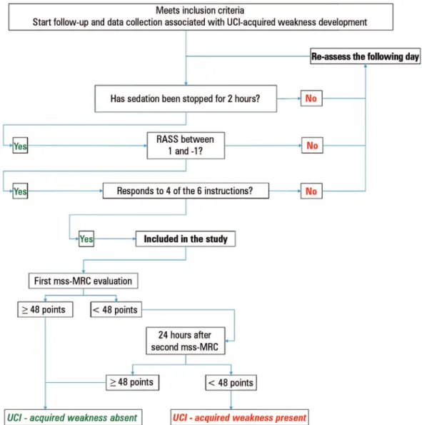
Prior to using the mss-MRC, the state of alertness was assessed using the Richmond Agitation-Sedation Scale (RASS), the values of which should range between 1 and -1. he infusion of sedatives was discontinued at least 30 minutes prior to applying the mss-MRC. he compression capacity was assessed by asking the patient to perform between 4 and 6 simple commands: “Open your eyes” or “Close your eyes” (as appropriate), “Lift your eyebrows”, “Move your head to one side (or the other)”, “Squeeze my hand”, “Open your mouth”, and “Stick out your tongue”. After four of these commands were performed, muscle force was evaluated using the mss-MRC (Appendix 1).
Figure 1 shows the method for arriving at a diagnosis. he patient was classiied as “without ICU-acquired weakness” when he or she reached ≥ 48 points or was considered as “re-assessable” when the cut-of point was not reached (i.e., mss-MRC < 48). During the morning of the following day, those who were “re-assessable” were given a second mss-MRC, which was performed by a diferent operator (who did not know the result of the irst measurement). If the patient exceeded the cut-of point, then they were considered “without ICU-acquired weakness”; if, however, the blind evaluator obtained a value of < 48 points a second time, then the patient was considered to have ICU-acquired weakness.
hirty minutes after the irst mss-MRC measurement, the Pimax was determined. Patients sat at 45º, and a unidirectional valve aneroid manovacuometer was used to measure pressure. A nozzle interface was used for those without an artiicial airway in place, and a 15mm adapter was used for patients with an orotracheal or tracheostomy tube. We quantiied the Pimax achieved in 20 seconds,(21) and the highest value of three replicates was reported. he inter-observer reliability of diferent consecutive operators was measured using a subsample of the irst 10 patients whose mss-MRC and Pimax assessments were repeated.
Statistical analyses
he results of the categorical variables are presented as counts and proportions within their categories. he numerical variables, whether continuous or discrete, are presented according to their distribution as the means and standard deviations or medians and interquartile ranges.
he chi-square test or Fisher’s exact test was used as appropriate to compare the association between categorical variables, and Student’s t-test or Mann-Whitney U-test was used for numerical variables according to the distribution. he inter-observer reliability for the performance of the mss-MRC in the diagnosis of ICU-acquired weakness (mss-MRC ≥ 48) was assessed using the agreement index for nominal variables (Cohen’s Kappa), and the intra-class correlation coeicient (ICC) index was used for the Pimax.
To estimate the simultaneous efect of the variables identiied as possible risk factors on the incidence of weakness, a conditional binary logistic regression model was used. Inclusion of the variables in the model was decided based on a p-value of < 0.1 in the univariate comparison. In addition, numerical variables that were signiicant in the univariate analysis and were previously individualized as clinically relevant subgroups were included dichotomously in the multivariate analysis (days of invasive MV > 5 days) for a better interpretation. A backward stepwise selection was used with Wald’s method. he result of the multivariate binary logistic regression was expressed as an odds ratio (OR) with its corresponding 95% conidence intervals (95%CI).
he inal calibration of the model was evaluated using the Hosmer-Lemeshow test, and the discriminating power was established based on an area under the curve (AUC) analysis.
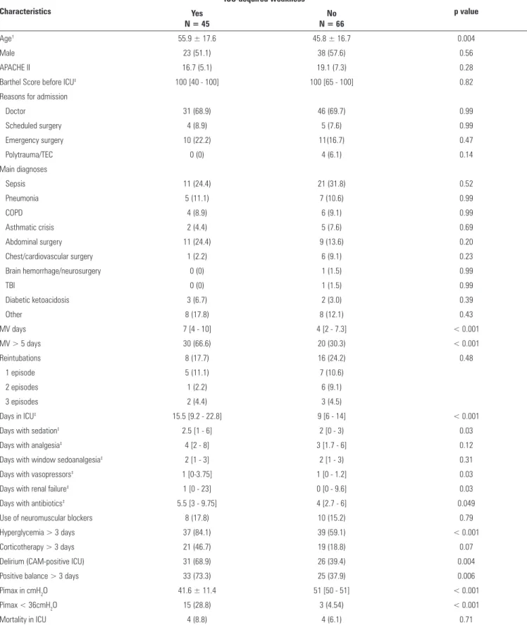
A survival analysis using a Kaplan-Meier curve was used for the variables time to event (ICU-acquired weakness), and the subgroups with or without delirium as well as those with or without hyperglycemia were compared (i.e., the signiicant variables in the binary logistic regression analysis) relative to the development of ICU-acquired weakness over time. he log-rank test was used for comparisons among the subgroups.
Figure 1 - Flowchart of the procedures performed in this study. ICU - intensive care unit; mss-MRC - muscular strength scale of the Medical Research Council.
performance of this cut-of point as well as the sensitivity, speciicity, and positive and negative likelihood ratio (LR+ and LR-, respectively) of this parameter as a method to classify patients with ICU-acquired weakness was analyzed. he LR+ and LR- are reported because of their stability with respect to the possible variability in the prevalence of ICU-acquired weakness. Finally, the lower limit of normality was calculated to individualize the number of patients who did not reach the theoretical values for their age.
A value of p = 0.05 was considered signiicant. R version 3.1.3 was used to analyze the data.(22)
RESULTS
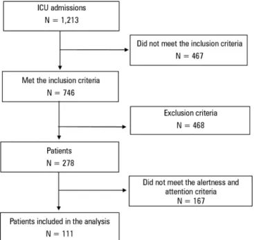
A total of 111 consecutive patients were included (Figure 2), 66 of which were classiied with “ICU-acquired weakness”. A cumulative incidence of ICU-acquired weakness of 40.5% was observed after an 18-month follow-up period (95%CI = 31.8% - 49.8%). he incidence rate or density of ICU-acquired weakness was 0.0038 per patient per day of follow up. he maximum follow-up period for a patient was 156 days.
weakness were age (OR = 1.03, 95%CI = 1.002 - 1.03, p = 0.035), hyperglycemia > 3 days (OR = 3.85, 95%CI = 1.28 - 11.54, p = 0.016), the presence of delirium (OR = 3.34, 95%CI = 1.31 - 8.50, p = 0.011), and invasive MV use > 5 days (OR = 2.83, 95%CI = 1.00 - 7.97, p = 0.049).
he regression model showed a correct classiication power of 73.6% regarding the events in the response variable.
he inal logistic regression model obtained a correct calibration measured by the Hosmer-Lemeshow test (p = 0.854). Discrimination was classiied as “good” assessed by the area under the ROC curve (AUC = 0.815, 95%CI = 0.73 - 0.89, p < 0.001).
he Kaplan-Meier curve (Figure 3) showed the probability of having ICU-acquired weakness depending on whether the patient had delirium during follow up. he groups that presented with delirium (dotted line) versus those that did not (dashed line) are shown. he comparison using the log-rank test was signiicant (p = 0.03). he probability of presenting with ICU-acquired weakness according to whether the patient had sustained hyperglycemia (> 3 days), a survival analysis, and a between-group comparison were not signiicant (log-rank test, p = 0.159).
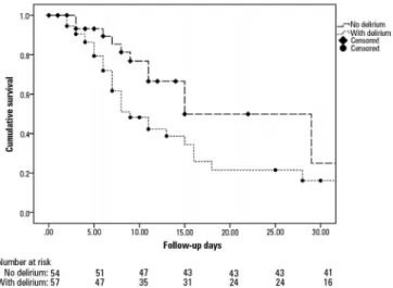
Regarding inspiratory muscle strength, the absolute Pimax values were compared between the group that developed ICU weakness, 41.6 (± 11.4) cmH2O, and the group that did not, 48.8 (± 4.67) cmH2O (p < 0.0001; Figure 4). he cut-of value described above (Pimax < 36cmH2O versus Pimax ≥ 36 cmH2O) showed that 30 (66%) of the 45 patients who developed ICU-acquired weakness fell below the cut-of point; on the other hand, only 15 (22%) patients in the group that did not have a clinical diagnosis of ICU-acquired weakness obtained a Pimax value of < 36cmH2O (p < 0.001). he OR of presenting with ICU-acquired weakness and not reaching 36cmH2O was 9.48 (95%CI = 2.53 - 35.4; p < 0.001). According to the cut-of value chosen (< 36cmH2O), a sensitivity value of 31.8% (95%CI = 18.1 - 45.6) was obtained, a speciicity value of 96.6% (95%CI = 91 - 100), an LR+ of 7.11 (95%CI = 2.17 - 23.3), and an LR- of 0.71 (95%CI = 0.57 - 0.90) were needed to correctly classify the patients with ICU-acquired weakness as diagnosed using the mss-MRC.
he mean lower limit of normal was 60.3 (± 9.8)
cmH2O, and the maximum and minimum predicted
values were 84.7 and 46.5cmHO, respectively. No
Figure 2 - Flowchart of patients under study. ICU - intensive care unit.
as follows: age 55.9 (± 17.6) years versus 45.8 (± 16.7) years, respectively; median time with invasive MV 7 [4 - 10] days versus 4 [2 - 7.3] days, respectively; median time in the ICU 15.5 [9.2 - 22.8] days versus 9 [6 - 14] days, respectively; median time with sedation 2.5 [1 - 6] days versus 2 [0 - 3] days, respectively; median time with vasopressors 1 [0 - 3.75] day versus 1 [0 - 1.2] day, respectively; median time to renal failure 1 [0 - 23] days
versus 0 [0 - 9.6] days, respectively; and median time
receiving antibiotics 5.5 [3-9.75] days versus 4 [2.7 - 6] days, respectively. In addition, more patients had delirium (31 [68.9) versus 26 [39.4]], hyperglycemia > 3 days (37 [84.1) versus 39 [59.1]), corticosteroid therapy > 3 days (21 [46.7) versus 19 [18.8]], and positive balance > 3 days (33 [73.3) versus 25 [37.9]] in the ICU-acquired weakness group.
he reliability between the ive evaluators of the mss-MRC was measured using the data of the irst 15 patients evaluated, and a Kappa value of 0.74 (95%CI = 0.51 - 0.97; p < 0.001) was obtained, showing “substantial”(23) agreement to conirm or exclude ICU-acquired weakness. Likewise, the degree of agreement among the ive evaluators for Pimax (in cmH2O) was measured, and an “excellent”(24) agreement was obtained (ICC = 0.97; 95%CI = 0.93 - 0.99; p < 0.001).
Table 1 - Characteristics of the sample
Characteristics
ICU-acquired weakness
p value Yes
N = 45
No N = 66
Age† 55.9 ± 17.6 45.8 ± 16.7 0.004
Male 23 (51.1) 38 (57.6) 0.56
APACHE II 16.7 (5.1) 19.1 (7.3) 0.28
Barthel Score before ICU‡ 100 [40 - 100] 100 [65 - 100] 0.82
Reasons for admission
Doctor 31 (68.9) 46 (69.7) 0.99
Scheduled surgery 4 (8.9) 5 (7.6) 0.99
Emergency surgery 10 (22.2) 11(16.7) 0.47
Polytrauma/TEC 0 (0) 4 (6.1) 0.14
Main diagnoses
Sepsis 11 (24.4) 21 (31.8) 0.52
Pneumonia 5 (11.1) 7 (10.6) 0.99
COPD 4 (8.9) 6 (9.1) 0.99
Asthmatic crisis 2 (4.4) 5 (7.6) 0.69
Abdominal surgery 11 (24.4) 9 (13.6) 0.20
Chest/cardiovascular surgery 1 (2.2) 6 (9.1) 0.23
Brain hemorrhage/neurosurgery 0 (0) 1 (1.5) 0.99
TBI 0 (0) 1 (1.5) 0.99
Diabetic ketoacidosis 3 (6.7) 2 (3.0) 0.39
Other 8 (17.8) 8 (12.1) 0.43
MV days 7 [4 - 10] 4 [2 - 7.3] < 0.001
MV > 5 days 30 (66.6) 20 (30.3) < 0.001
Reintubations 8 (17.7) 16 (24.2) 0.48
1 episode 5 (11.1) 7 (10.6)
2 episodes 1 (2.2) 6 (9.1)
3 episodes 2 (4.4) 3 (4.5)
Days in ICU‡ 15.5 [9.2 - 22.8] 9 [6 - 14] < 0.001
Days with sedation‡ 2.5 [1 - 6] 2 [0 - 3) 0.03
Days with analgesia‡ 4 [2 - 8] 3 [1.7 - 6] 0.12
Days with window sedoanalgesia‡ 2 [1 - 3] 2 [1 - 3) 0.31
Days with vasopressors‡ 1 [0-3.75] 1 [0 - 1.2] 0.03
Days with renal failure‡ 1 [0 - 23] 0 [0 - 9.6] 0.03
Days with antibiotics‡ 5.5 [3 - 9.75] 4 [2.7 - 6] 0.049
Use of neuromuscular blockers 8 (17.8) 10 (15.2) 0.79
Hyperglycemia > 3 days 37 (84.1) 39 (59.1) < 0.001
Corticotherapy > 3 days 21 (46.7) 19 (18.8) 0.07
Delirium (CAM-positive ICU) 31 (68.9) 26 (39.4) 0.004
Positive balance > 3 days 33 (73.3) 25 (37.9) 0.006
Pimax in cmH2O 41.6 ± 11.4 51 [50 - 51] < 0.001
Pimax < 36cmH2O 15 (28.8) 3 (4.54) < 0.001
Mortality in ICU 4 (8.8) 4 (6.1) 0.71
Table 2 - Multivariate binomial logistic regression
Variables OR 95%CI p value
Age (years) 1.03 1.002 - 1.03 0.035
MV > 5 days 2.83 1.005 - 7.97 0.049
Delirium (CAM-positive ICU) 3.34 1.31 - 8.50 0.011
Hyperglycemia > 3 days 3.85 1.28 - 11.54 0.016
OR - odds ratio; 95%CI - 95% confidence intervals; MV - mechanical ventilation; CAM-ICU - Confusion Assessment Method for the intensive care unit.
Figure 3 - Kaplan-Meier curve. The likelihood of developing weakness in the presence of delirium (dotted line) versus no delirium (dashed line) after a 30-day
follow-up period. (log-rank, p = 0.03).
Figure 4 - Error bars with means and 95% CIs of maximal inspiratory pressure regarding patients with Medical Research Council muscle strength scale values of
≥ 48 and < 48. 95%CI - 95% confidence intervals; Pimax - maximum inspiratory pressure; mss-MRC - muscular strength scale of the Medical Research Council.
predicted Pimax value was below the cut-of point deined in the literature (Pimax < 36cmH2O) for any patient.(19)
DISCUSSION
he most relevant inding of the current study was the independent association between delirium and the development of ICU-acquired weakness. hus far, no evidence has directly linked delirium with weakness.(25) Despite the lack of direct data, increasing evidence has described common factors and outcomes among both conditions. hus, patients who are delusional or develop ICU-acquired weakness are more likely to have a greater use of sedation, more days of invasive MV, longer stays in the ICU and hospital, and higher mortality rates in the ICU and hospital 1 year after discharge.(26-30) his inding acquires a greater importance considering that the muscle strength assessment was performed only in patients who were alert (i.e., those with RASS from 1 to -1) and aware (i.e., those who fulilled 4 of 6 commands). As such, we believe that unidentiied delirium precluded the possibility of obtaining a low mss-MRC value. Another meeting point exists between both conditions: early ICU mobility as a treatment strategy to avoid the development of ICU-acquired weakness and the onset of delirium to reduce its impact.(31) his meeting point supports the proposed theory in which we suggest that both conditions can be causally associated and should be studied in greater detail together.
As expected, the mean age of patients who had ICU-acquired weakness was signiicantly higher and was an independent factor that favored the development of this clinical picture. Elderly people can develop sarcopenia, which is further aggravated in those admitted to the ICU(32) and can act as the cause or aggravating factor with regard to the weakness found.(33)
the ICU.(35,36) he observed relationship between insulin therapy and the lower development of ICU-acquired weakness might justify the association between hyperglycemia and the increased risk for developing ICU-acquired weakness observed among our patients.
his study found a lower mortality rate among patients with or without ICU-acquired weakness than that published by other studies.(37,38) Similarly, the Acute Physiology and Chronic Health Examination (APACHE) score was also lower than those of other similar studies.(29,39) his inding might explain the low mortality rate associated with patients with ICU-acquired weakness. We also believe, as suggested by several authors, that the diagnosis of weakness based on the mss-MRC is applicable to patients who achieve a certain degree of alertness and comprehension, whereas its application is limited in comatose patients or those with sedoanalgesia.(25,40)
On the other hand, similar to what other authors have reported, we observed a signiicant association between patients with inspiratory muscle weakness and ICU-acquired weakness.(19,37) Because assessment via the mss-MRC requires co-workers and conscious patients, an alternative might be the assessment of respiratory muscles because this method can be dispensed at will (see the maneuver described by Marini to evaluate Pimax with a unidirectional valve).(21)
he association between limb weakness and respiratory muscle weakness was explored in two previous studies. De Jonghe et al.(17) used the median of their sample and established a value of 30cmH2O, which was associated with ICU-acquired weakness. Tzanis et al.(19) deined Pimax as 36cmH2O and diagnosed inspiratory weakness in patients with ICU-acquired weakness, with a sensitivity of 88% and a speciicity of 76%.
In our patients, the sensitivity was considerably lower, but the speciicity values were higher. According to our indings, this diference suggests that a Pimax of ≥ 36cmH2O is more useful to exclude respiratory weakness and less useful as a monitoring method for the early diagnosis of ICU-acquired weakness.
In conclusion, the incidence found is similar to that reported so far and varies according to the adopted
deinition of ICU-acquired weakness, the diagnostic modality, and the characteristics of the included population.(3,6,41,42) he relatively high-incidence density suggests a phenomenon that must be monitored daily. For this purpose, we suggest using simple, non-invasive diagnostic methods and reserving the most invasive methods only for those who cannot have their peripheral muscles assessed using the mss-MRC.
he results found should be validated in the general population to discern possible local biases and the reproducibility of the phenomena found.
he study has limitations. he irst is the design; being a single center study, the indings might be due to local biases. For example, poor adherence to protocols might prevent the development of ICU-acquired weakness. he indings must be replicated before generalizing them to the general population. Another clear limitation arises from the tool chosen to diagnosis ICU-acquired weakness (i.e., the mss-MRC), which cannot be using among patients with altered consciousness or those who cannot execute simple instructions. As a result, we believe that the incidence of ICU-acquired weakness might have been underestimated because of this diiculty.
Another limitation was the lack of diagnostic conirmation via diagnostic scaling (muscle biopsy or electromyogram) as suggested by Latronico et al.(9) to discern the type of condition and diferentiate muscular involvement from neural involvement or both; these methods can be used to identify the origin of the weakness in more detail.
CONCLUSION
Objetivo: Conocer la incidencia acumulada y analizar los factores riesgo asociados al desarrollo de debilidad adquirida en la unidad de cuidados intensivos y su asociación con la debilidad inspiratoria.
Métodos: Estudio de cohorte prospectivo en un solo centro,
unidad de cuidados intensivos médico-quirúrgica polivalente. Se incluyeron pacientes adultos, que hayan requerido ventilación mecánica ≥ 24 horas entre julio de 2014 y enero de 2016. No hubo intervenciones. Se registraron datos demográicos, diagnóstico clínico y factores relacionados con el desarrollo de debilidad adquirida en la unidad de cuidados intensivos y Presión inspiratoria máxima.
Resultados: Ciento once pacientes incluidos, 66
desarrollaron debilidad adquirida en la unidad de cuidados intensivos, con una incidencia acumulada del 40,5% en 18 meses. El grupo con debilidad adquirida en la unidad de cuidados intensivos presentó mayor edad (55,9 ± 17,6 versus
45.8 ± 16.7), además de más días con ventilación mecánica (7 [4 - 10] versus 4 [2 - 7,3]), más días en unidad de cuidados intensivos (15,5 [9,2 - 22,8] versus 9 [6 - 14]). Hubo más pacientes con delirio (68% versus 39%), con hiperglucemia > 3
días (84% versus 59%); y con balance positivo > 3 días (73,3%
versus 37%). Todas las comparaciones fueron signiicativas con p < 0,05. La regresión logística múltiple identiicó a la edad, la hiperglucemia ≥ 3 días, el delirio y la ventilación mecánica > 5 días como predictores independientes para debilidad adquirida en la unidad de cuidados intensivos. La presión inspiratoria máxima baja se asoció a debilidad adquirida en la unidad de cuidados intensivos (p < 0,001) y el punto de corte presión inspiratoria máxima < 36cmH2O obtuvo una sensibilidad y especiicidad del 31,8% y 95,5% para clasiicar al grupo con debilidad adquirida en la unidad de cuidados intensivos.
Conclusión: La debilidad adquirida en la unidad de cuidados
intensivos es una condición con un alta incidencia en nuestro medio. El desarrollo de debilidad adquirida en la unidad de cuidados intensivos se asoció a la edad, delirio, hiperglucemia y la ventilación mecánica > 5 días. La presión inspiratoria máxima ≥ 36cmH2O demostró un alto valor diagnóstico para descartar la presencia de debilidad adquirida en la unidad de cuidados intensivos.
RESUMEN
Descriptores: Debilidad muscular; Respiración artiicial;
Delirio; Presiones inspiratorias máximas; Hiperglucemia
REFERENCES
1. de Jonghe B, Lacherade JC, Sharshar T, Outin H. Intensive care unit-acquired weakness: risk factors and prevention. Crit Care Med. 2009;37(10 Suppl):S309-15.
2 Deem S. Intensive-care-unit-acquired muscle weakness. Respir Care. 2006;51(9):1042-52; discussion 1052-3.
3. Stevens RD, Marshall SA, Cornblath DR, Hoke A, Needham DM, de Jonghe B, et al. A framework for diagnosing and classifying intensive care unit-acquired weakness. Crit Care Med. 2009;37(10 Suppl):S299-308. 4. Schweickert WD, Hall J. ICU-acquired weakness. Chest.
2007;131(5):1541-9.
5. Fan E. Critical illness neuromyopathy and the role of physical therapy and rehabilitation in critically ill patients. Respir Care. 2012;57(6):933-44; discussion 944-6.
6. Lipshutz AK, Gropper MA. Acquired neuromuscular weakness and early mobilization in the intensive care unit. Anesthesiology. 2013;118(1):202-15. 7. Mendez-Tellez PA, Needham DM. Early physical rehabilitation in the ICU
and ventilator liberation. Respir Care. 2012;57(10):1663-9.
8. Powers SK, Kavazis AN, Levine S. Prolonged mechanical ventilation alters diaphragmatic structure and function. Crit Care Med. 2009;37(10 Suppl):S347-53.
9. Latronico N, Bolton CF. Critical illness polyneuropathy and myopathy : a major cause of muscle weakness and paralysis. Lancet Neurol. 2011;10(10):931-41.
10. Truong AD, Fan E, Brower RG, Needham DM. Bench-to-bedside review: mobilizing patients in the intensive care unit--from pathophysiology to clinical trials. Crit Care. 2009;13(4):216.
11. Busico M, Intile D, Sívori M, Irastorza N, Alvarez AL, Quintana J, et al. Risk factors for worsened quality of life in patients on mechanical ventilation. A prospective multicenter study. Med Intensiva. 2016;40(7):422-30. 12. Ibarra-Estrada MA, Briseño-Ramírez J, Chiquete E, Ruiz-Sandoval JL. Debilidad
adquirida en la Unidad de Cuidados Intensivos: Polineuropatía y miopatía del
13. Rodriguez PO, Setten M, Maskin LP, Bonelli I, Vidomlansky SR, Attie S, et al. Muscle weakness in septic patients requiring mechanical ventilation: protective effect of transcutaneous neuromuscular electrical stimulation. J Crit Care. 2012;27(3):319.e1-8.
14. Latronico N, Gosselink R. A guided approach to diagnose severe muscle weakness in the intensive care unit. Rev Bras Ter Intensiva. 2015;27(3):199-201.
15. Jolley SE, Bunnell A, Hough CL. ICU-acquired weakness. Chest. 2016;150(5):1129-40.
16. Nordon-Craft A, Moss M, Quan D, Schenkman M. Intensive care unit-acquired weakness: implications for physical therapist management. Phys Ther. 2012;92(12):1494-506.
17. De Jonghe B, Bastuji-Garin S, Durand MC, Malissin I, Rodrigues P, Cerf C, Outin H, Sharshar T; Groupe de Réflexion et d’Etude des Neuromyopathies en Réanimation. Respiratory weakness is associated with limb weakness and delayed weaning in critical illness. Crit Care Med. 2007;35(9):2007-15.
18. Bates B. A guide to physical examination and history taking. 5th ed. Philadelphia: Lippencott Silliams and Wilkins; 1991. p. 500-60.
19. Tzanis G, Vasileiadis I, Zervakis D, Karatzanos E, Dimopoulos S, Pitsolis T, et al. Maximum inspiratory pressure, a surrogate parameter for the assessment of ICU-acquired weakness. BMC Anesthesiol. 2011;11:14. 20. Evans JA, Whitelaw WA. The assessment of maximal respiratory mouth
pressures in adults. Respir Care. 2009;54(10):1348-59.
21. Marini JJ, Smith TC, Lamb V. Estimation of inspiratory muscle strength in mechanically ventilated patients: the measurement of maximal inspiratory pressure. J Crit Care. 1986;1(1):32-8.
22. R Development Core Team. R: A language and environment for statistical computing [Internet]. Vienna, Austria; 2015. Available from: https:// www.r-project.org
23. Landis JR, Koch GG. The measurement of observer agreement for categorical data. Biometrics. 1977;33(1):159-74.
Appendix 1 - The muscle strength scale of the Medical Research Council(18)
0: No muscle contraction detected
1: Fasciculation barely noticeable or traces of contraction
2: Active motion with gravity removed
3: Active movement against gravity
4: Active movement against gravity and some resistance
5: Active movement against gravity and full resistance
25. Latronico N, Herridge M, Hopkins RO, Angus D, Hart N, Hermans G, et al. The ICM research agenda on intensive care unit - acquired weakness. Intensive Care Med. 2017 Mar 13. [Epub ahead of print].
26. Garnacho-Montero J, Madrazo-Osuna J, García-Garmendia JL, Ortiz-Leyba C, Jiménez-Jiménez FJ, Barrero-Almodóvar A, et al. Critical illness polyneuropathy: risk factors and clinical consequences. A cohort study in septic patients. Intensive Care Med. 2001;27(8):1288-96.
27. De Jonghe B, Bastuji-Garin S, Sharshar T, Outin H, Brochard L. Does ICU-acquired paresis lengthen weaning from mechanical ventilation? Intensive Care Med. 2004;30(6):1117-21.
28. Shinotsuka CR, Salluh JI. Perceptions and practices regarding delirium, sedation and analgesia in critically ill patients: a narrative review. Rev Bras Ter intensiva. 2013;25(2):155-61.
29. Mehta S, Cook D, Devlin JW, Skrobik Y, Meade M, Fergusson D, Herridge M, Steinberg M, Granton J, Ferguson N, Tanios M, Dodek P, Fowler R, Burns K, Jacka M, Olafson K, Mallick R, Reynolds S, Keenan S, Burry L; SLEAP Investigators; Canadian Critical Care Trials Group. Prevalence, risk factors, and outcomes of delirium in mechanically ventilated adults. Crit Care Med. 2015;43(3):557-66.
30. Hermans G, Van Mechelen H, Bruyninckx F, Vanhullebusch T, Clerckx B, Meersseman P, et al. Predictive value for weakness and 1-year mortality of screening electrophysiology tests in the ICU. Intensive Care Med. 2015;41(12):2138-48.
31. Schweickert WD, Pohlman MC, Pohlman AS, Nigos C, Pawlik AJ, Esbrook CL, et al. Early physical and occupational therapy in mechanically ventilated, critically ill patients: a randomised controlled trial. Lancet. 2009;373(9678):1874-82.
32. Kizilarslanoglu MC, Kuyumcu ME, Yesil Y, Halil M. Sarcopenia in critically ill patients. J Anesth. 2016;30(5):884-90.
33. Janssen I, Heymsfield SB, Ross R. Low relative skeletal muscle mass (sarcopenia) in older persons is associated with functional impairment and physical disability. J Am Geriatr Soc. 2002;50(5):889-96.
34. Bercker S, Weber-Carstens S, Deja M, Grimm C, Wolf S, Behse F, et al. Critical illness polyneuropathy and myopathy in patients with acute respiratory distress syndrome. Crit Care Med. 2005;33(4):711-5. 35. Hermans G, Wilmer A, Meersseman W, Milants I, Wouters PJ, Bobbaers H,
et al. Impact of intensive insulin therapy on neuromuscular complications and ventilator dependency in the medical intensive care unit. Am J Respir Crit Care Med. 2007;175(5):480-9.
36. van den Berghe G, Wouters P, Weekers F, Verwaest C, Bruyninckx F, Schetz M, et al. Intensive insulin therapy in critically ill patients. N Engl J Med. 2001;345(19):1359-67.
37. Peñuelas O, Muriel A, Frutos-Vivar F, Fan E, Raymondos K, Rios F, et al. Prediction and outcome of intensive care unit-acquired paresis. J Intensive Care Med. 2016 Apr 13. [Epub ahead of print].
38. Ali NA, O’Brien JM Jr, Hoffmann SP, Phillips G, Garland A, Finley JC, Almoosa K, Hejal R, Wolf KM, Lemeshow S, Connors AF Jr, Marsh CB; Midwest Critical Care Consortium. Acquired weakness, handgrip strength, and mortality in critically ill patients. Am J Respir Crit Care Med. 2008;178(3):261-8.
39. Morris PE, Goad A, Thompson C, Taylor K, Harry B, Passmore L, et al. Early intensive care unit mobility therapy in the treatment of acute respiratory failure. Crit Care Med. 2008;36(8):2238-43.
40. Hough CL, Lieu BK, Caldwell ES. Manual muscle strength testing of critically ill patients: feasibility and interobserver agreement. Crit Care. 2011;15(1):R43.
41. De Jonghe B, Sharshar T, Lefaucheur JP, Authier FJ, Durand-Zaleski I, Boussarsar M, Cerf C, Renaud E, Mesrati F, Carlet J, Raphaël JC, Outin H, Bastuji-Garin S; Groupe de Réflexion et d’Etude des Neuromyopathies en Réanimation. Paresis acquired in the intensive care unit: a prospective multicenter study. JAMA. 2002;288(22):2859-67.
42. de Letter MA, Schmitz PI, Visser LH, Verheul FA, Schellens RL, Op de Coul DA, et al. Risk factors for the development of polyneuropathy and myopathy in critically ill patients. Crit Care Med. 2001;29(12):2281-6.