Heart conduction system defects and sustained
ventricular tachycardia complications in a patient
with granulomatosis with polyangiitis. A case
report and literature review
INTRODUCTION
Granulomatosis with polyangiitis (also known as Wegener’s granulomatosis) is a rare systemic inlammatory disorder of unknown etiology(1,2) that is characterized by vasculitis of the small arteries, the arterioles and the capillaries as well as necrotizing granulomatous lesions and has a wide clinical presentation. he upper and lower respiratory tracts and the kidneys are the most commonly afected sites.(1,3-5)
Cardiac involvement has long been regarded as rare, but it can vary from a subclinical disease to a wide spectrum of abnormalities, including myocarditis, valvar lesions, conduction system defects, coronary arteritis and pericarditis.(1,3)
CLINICAL CASE
his report presents a case of a 44-year-old female patient diagnosed with granulomatosis with polyangiitis ive months ago. he condition was characterized by involvement of the upper and lower respiratory tracts, a positive antineutrophil cytoplasmic antibodies (c-ANCA), a nasal mucosa Laryssa Passos Sarmento Santos1, Victor
Guerreiro Bomfim1, Camila Fagundes Bezerra1,
Natália Vieira Costa2, Rafael Barreto Paes
de Carvalho1, Ricardo Sobral de Carvalho1,
Rogério da Hora Passos1, Olivia Carla Bomfim
Boaventura1,3, André Luiz Nunes Gobatto1,2,3
1. Hospital São Rafael - Salvador (BA), Brazil. 2. Universidade Federal da Bahia - Salvador (BA), Brazil.
3. Universidade de Salvador - Salvador (BA),
Brazil. a rare systemic inlammatory disorder Granulomatosis with polyangiitis is
characterized by vasculitis of the small arteries, the arterioles and the capillaries together with necrotizing granulomatous lesions. his case reports on a young female patient, previously diagnosed with granulomatosis with polyangiitis, who was admitted to the intensive care unit with seizures and hemodynamic instability due to a complete atrioventricular heart block. he event was associated with multiple episodes of sustained ventricular tachycardia without any structural heart changes or electrolyte disturbances. In the intensive
Conflicts of interest: None.
Submitted on October 16, 2016 Accepted on March 14, 2017
Corresponding author: André Luiz Nunes Gobatto Unidade de Terapia Intensiva Geral I Hospital São Rafael
Avenida São Rafael, 2.152, 5º andar Zip code: 41253-190 - Salvador (BA), Brazil E-mail: andregobatto@gmail.com
Responsible editor: Luciano César Pontes de Azevedo
Distúrbios de condução cardíaca e taquicardia ventricular como
complicação da granulomatose com poliangiíte em uma paciente.
Relato de caso e revisão da literatura
ABSTRACT
Keywords: Granulomatosis with
polyangiitis; Atrioventricular block; Heart conduction system; Pacemaker, artiicial; Bradycardia; Case reports care unit, the patient was itted with a provisory pacemaker, followed by immunosuppression with corticosteroids and immunobiological therapy, resulting in a total hemodynamic improvement. Severe conduction disorders in patients presenting granulomatosis with polyangiitis are rare but can contribute to increased morbidity. Early detection and speciic intervention can prevent unfavorable outcomes, speciically in the intensive care unit.
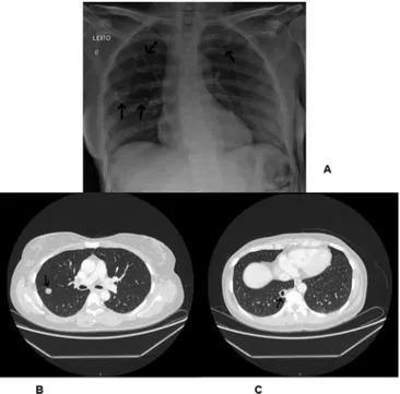
biopsy, chronic unspeciic inlammation, and no renal involvement. Bilateral nodules identiied in the lung and a neck with imagem exams showed an irregular mucosal thickening of the paranasal sinuses, heavy padding of the bilateral ethmoid cells, partial absence of a turbinate, and preservation of the nasal septum along with a bilateral illing of the mastoid and the middle ear cells. Computed tomography (CT) of the skull and sinuses revealed an increased thickening of the maxillary and the sphenoid, a septum erosion, bilateral mastoiditis and pansinusitis (Figures 1 and 2). hus, immunosuppressive therapy (cyclophosphamide and methylprednisolone) was administered, resulting in an unsatisfactory response to the treatment.
Figure 1 - (A) Chest radiography in PA view showing bilateral nodules (patient previously diagnosed with Poliangiitis granulomatosis). (B and C) Same nodules showing thorax taken by computed tomography.
Over time, the symptoms did not improve, and the patient exhibited a worsening exophthalmia, drowsiness and apathy. he patient was admitted for hospitalization and started on an immunobiological therapy with rituximab. Overall, the patient’s condition was poor. was dehydrated but did not exhibit signs of any respiratory or cardiac pathological abnormalities nor signs of an infection or metabolic or electrolyte disorders.
On the irst day of hospitalization, the patient presented with drowsiness followed by a tonic-clonic seizure episode that lasted roughly one minute. When the seizure subsided, she was bradycardic (32bpm) and hypotensive (84 x 48mmHg). he patient was immediately transferred to the cardiovascular intensive care unit, where a complete atrioventricular heart block was identiied by bedside electrocardiogram, which was further associated with multiple episodes of sustained ventricular tachycardia (SVT) upon monitoring (Figure 3). he intensive care unit medical team performed a bedside transthoracic echocardiogram; however, this test did not show any important cardiac morphological abnormalities with the exception of a mild mitral regurgitation with 62% of an ejection fraction. No thrombi or myocardial wall dyskinesias were identiied. he patient was given a provisory transvenous pacemaker (ultrasonically guided), resulting in an immediate improvement of the cardiac symptoms. he levels of myocardial necrosis markers such as troponi and pro-BNP were normal. No signs of an infection, electrolyte disturbances or organic dysfunctions were observed. he C-reactive protein and the erythrocyte sedimentation rates were elevated (Table 1). A cardiac magnetic resonance imaging (MRI) or a myocardial biopsy was not performed due to the recent pacemaker implantation and the risk of hemodynamic instability. An endotracheal intubation was determined to be unnecessary because she was able to protect her airways, even with drowsiness (Glasgow coma score of 13). She recovered a level of consciousness soon after the pacemaker’s introduction.
As the patient recovered hemodynamic stability, she was started on therapy with rituximab at one dose every week for four weeks. After six days, no further episodes of SVT were noted; however, the patient was still dependent on the provisory pacemaker, and a permanent pacemaker was then implanted (Figure 4). he patient was discharged after treatment with broad-spectrum antibiotics due to pneumonia.
Table 1 - Laboratory values
Day 1 Day 3 Day 5 Day 19
CPK (U/L) 20 20
CKMB (ng/mL) 0.32 0.77
Troponin I (ng/mL) 0.022 0.012
Hemoglobin (g/dL) 9.3 9 9.5 9.7
Hematocrit (%) 29.7 28.5 30 32.7
Leukogram (mil/mm) 10,900 8,300 12,000 11,000
Platelets (mil/mm) 682,000 575,000 494,000 648,000
Sodium (mmol/L) 135 138 136 136
Potassium (mmol/L) 4.2 3.2 4.2 4.6
Chloride (mmol/L) 95 100 95 102
Calcium (mmol/L) 2.6 2.3 2.2 2.5
Phosphor (mmol/L) 1.2 1.2 0.9
Magnesium (mmol/L) 0.7 0.7 1 0.8
C reactive protein (mg/L) 425 263 70.7 34.7
ESR (mm/hour) 120 120 50 30
Creatinine (mg/dL) 0.5 0.5 0.7 0.5
Urea (mg/dL) 9 17 22 40
CPK - creatinofosfoquinase; CKMB - creatine kinase MB isoenzyme; ESR - eritrocyte sendimentation rate.
Figure 3 - (A) Electrocardiogram showing a total atrioventricular block and a Torsades de points. (B) Multiple ventricular tachycardias in intensive care unit monitoring. (C) Same patient post-transvenous temporary pacing. We can see a complete atrioventricular dissociation.
B lymphocytes using the marker CD19 resulted in near zero levels upon the follow-up visit. After 6 months of immunobiological therapy, normalization of all disease activity markers was observed, and the pacemaker was turned of, as an asymptomatic sinus rhythm was maintained. he patient continues to be followed-up with by the Rheumatology and Cardiology departments, waiting for removal of the permanent pacemaker (Figure 5).
DISCUSSION
Granulomatosis with polyangiitis is a systemic inlammatory disorder of unknown etiology(1,3) that is characterized by a vasculitis of the small arteries, the arterioles and the capillaries, together with necrotizing
Figure 4 - (A) Chest radiography in AP view showing bilateral nodules of the entry on the cardiovascular intensive care unit. (B) Chest radiography in PA view showing location and positioning of the pacemaker.
granulomatous lesions. Clinical presentation of granulomatosis with polyangiitis depends upon the afected organ and the degree of progression from local to systemic arteritis.(3) he upper and lower respiratory tracts, the kidneys and the eyes are typical sites of occurrence and observed in 50 to 60% of clinical cases.(1,5) In approximately 8% to 16% of the cases, the eyes can be the only site of aliction at the initial presentation, while persistent symptoms are recorded in 87% of all of patients.(4) A positive c-ANCA or tissue biopsy are important for the initial diagnosis(1) in order to exclude other diseases with a similar presentation.
Several types of ANCA can be recognized, but the two subtypes relevant to the onset of systemic vasculitis are those that are directed towards proteinase-3 (PR3) and myeloperoxidase. c-ANCA is related to the PR3 ANCA and has a high speciicity (> 90%), which helps in the diagnosis of granulomatosis with polyangiitis and other closely related diseases.(3)
Early treatment is crucial in order to prevent severe complications and often to preserve life. For those patients with a severe disease, there are now two co-treatment options for inducing a remission: cyclophosphamide plus corticosteroids or rituximab plus corticosteroids. Remission can be induced in greater than 90% of the patients who are treated with either of these two therapies.(5)
Cardiac involvement in granulomatosis with polyangiitis occurs in 6% to 44% of the cases and is secondary to necrotizing vasculitis with granulomatous iniltrates.(6) Pericarditis is the most common cardiac manifestation (35%), followed by cardiomyopathy (30%), coronary artery disease (CAD) (12%), valvar disease (6%), concomitant CAD and valvar disease (6%), concomitant pericarditis and cardiomyopathy (1.6%), and severe conduction disorders (1.6%).(3)
Atrial tachycardia, atrial ibrillation and lutter are the most common arrhythmias that are found in patients diagnosed with granulomatosis with poliangiitis. Ventricular arrhythmias are usually noted in association with dilated cardiomyopathy, cardiac ischemia or secondary to cardiac masses and are uncommon in hearts with no structural damage.(6) All conduction defects varying in severity can be recognized, including intraventricular conduction defects, irst and second degree heart blocks and a complete heart block. Treatment for these types of heart conduction system dysfunctions may require
a transient or a permanent pacemaker depending on whether the arrhythmia is induced by reversible causes such as hydro-electrolytic disturbances or drugs.(1)
A bedside echocardiogram may be a valuable tool to assist intensive care physicians with diferential diagnoses of shock and to assess structural heart diseases or ventricular dysfunctions in cases of unexplained arrhythmias. In this case, bedside echocardiography testing was useful in determination of shock diagnosis and subsequently the decision of a quickly introduced pacemaker.(7)
Only ten reported cases of granulomatosis with poliangiitis patients with a concurrent atrioventricular blockage have been reported within the last ten years.(8-19) What made our patient diferent was that she presented with SVT, a life threatening condition that can be associated with an atrioventricular block, leading the patient to state of hemodynamic instability. hese complications are common in patients with dilated cardiomyopathy, ischemia, and electrolyte disturbances and are secondary to cardiac masses; however, our patient did not have any of the above stated diagnoses or macroscopical cardiac structural disease as determined via echocardiography.
Cardiac MRI and myocardial biopsy would be beneicial in order to diagnose any cardiac involvement of granulomatosis with polyangiitis disease. An MRI was not performed in this case due to the presence of the pacemaker, and a biopsy was not performed due the risks inherent to the procedure. he elevated levels of inlammatory markers, along with clear signs of the disease activity on the day of the heat block and SVT, were associated with no other indings of a structural heart disease, ventricular dysfunction, electrolyte disturbances, or drugs; thus, the diagnosis of granulomatosis with polyangiitis with a cardiac involvement was determined to be the most likely cause. Furthermore, the immunosuppression therapy resulted in complete improvement of the patient, and the pacemaker was turned of and withdrawn from use.
CONCLUSION
REFERENCES
1. Goodfield NE, Bhandari S, Plant WD, Morley-Davies A, Sutherland GR. Cardiac involvement in Wegener’s granulomatosis. Br Heart J. 1995;73(2):110-5.
2. Jennette JC. Nomenclature and classification of vasculitis: lessons learned from granulomatosis with polyangiitis (Wegener’s granulomatosis). Clin Exp Immunol. 2011;164 Suppl 1:7-10.
3. McGeoch L, Carette S, Cuthbertson D, Hoffman GS, Khalidi N, Koening CL, Langford CA, McAlear CA, Moreland L, Monach PA, Seo P, Specks U, Ytterberg SR, Merkel PA, Pagnoux C; Vasculitis Clinical Research Consortium. Cardiac involvement in granulomatosis with polyangiitis. J Rheumatol. 2015;42(7):1209-12.
4. Franco CM, Oliveira GM, Fidelix TS, Vieira LA, Trevisani VF. Nodular scleritis and granulomatous polyangiitis (Wegener) mimicking tuberculosis. Rev Bras Oftalmol. 2015;74(2):106-9.
5. Comarmond C, Cacoub P. Granulomatosis with polyangiitis (Wegener): clinical aspects and treatment. Autoimmun Rev. 2014;13(11):1121-5. 6. Kallenberg CG. Pathophysiology of ANCA-associated small vessel
vasculitis. Curr Rheumatol Rep. 2010;12(6):399-405.
7. Flato UA, Campos AL, Trindade MR, Guimarães HP, Vieira ML, Brunori F. Intensive care bedside echocardiography: true or a distant dream? Rev Bras Ter Intensiva. 2009;21(4):437-45.
8. Handa R, Wali JP, Aggarwal P, Wig N, Biswas A, Kumar AK. Wegener’s granulomatosis with complete heart block. Clin Exp Rheumatol. 1997;15(1):97-9.
9. Ohkawa S, Miyao M, Chida K, Mizuuchi T, Kida K, Takubo K, et al. Extensive involvement of the myocardium and the cardiac conduction system in a case of Wegener’s granulomatosis. Jpn Heart J. 1999;40(4):509-15.
10. Suleymenlar G, Sarikaya M, Sari R, Tuncer M, Sevinc A. Complete heart block in a patient with Wegener’s granulomatosis in remission--a case report. Angiology. 2002;53(3):337-40.
11. Kouba DJ, Kirsch DG, Mimouni D, Nousari CH. Wegener’s granulomatosis with cardiac involvement masquerading as Lyme disease. Clin Exp Rheumatol. 2003;21(5):647-9.
12. Wilcke JT, Nielsen PK, Jacobsen TN. Reversible complete heart block due to Wegener’s granulomatosis. Int J Cardiol. 2003;89(2-3):297-8. 13. Ghaussy NO, Du Clos TW, Ashley PA. Limited Wegener’s granulomatosis
presenting with complete heart block. Scand J Rheumatol. 2004;33(2):115-8.
14. Elikowski W, Baszko A, Puszczewicz M, Stachura E. [Complete heart block due to Wegener’s granulomatosis: a case report and literature review]. Kardiol Pol. 2006;64(6):622-7. Polish.
15. Lim HE, Lee YH, Ahn JC. Wegener’s granulomatosis with progressive conduction disturbances and atrial fibrillation. Heart. 2007;93(7):777. 16. Sarlon G, Durant C, Grandgeorge Y, Bernit E, Veit V, Hamidou M, et al.
[Cardiac involvement in Wegener’s granulomatosis: report of four cases and review of the literature]. Rev Med Interne. 2010;31(2):135-9. French. 17. Ruisi M, Ruisi P, Finkielstein D. Cardiac manifestations of Wegener’s
granulomatosis: Case report and review of the literature. J Cardiol Cases. 2010;2(2):e99-e102.
18. Cassidy CJ, Sowden E, Brockbank J, Teh LS, Ho E. A patient with Wegener’s granulomatosis in apparent remission presenting with complete atrioventricular block. J Cardiol Cases. 2011;3(2):e71-e74. 19. Lin CH, Tsai SH, Chen HC, Chen SJ. Heart blockage in a patient with
cavitary lung lesions. Am J Emerg Med. 2012;30(8):1663.e1-3.
A granulomatose com poliangiíte é um raro distúrbio inla-matório sistêmico que se caracteriza por vasculite de pequenas artérias, arteríolas e capilares, associada a lesões granulomatosas necrotizantes. Este artigo relata o caso de uma paciente com diagnóstico prévio de granulomatose com poliangiíte, admitida à unidade de terapia intensiva com quadro de crises convulsivas e instabilidade hemodinâmica em razão de bloqueio atrioventri-cular completo. Estas manifestações se associaram a múltiplos episódios de taquicardia ventricular sustentada; não havia alte-rações estruturais cardíacas, nem se detectaram distúrbios hi-droeletrolíticos. Na unidade de terapia intensiva, a paciente foi
submetida à implantação de marca-passo provisório, imunossu-pressão com uso de corticosteroides e terapia imunobiológica, resultando em melhora hemodinâmica completa. Distúrbios graves da condução cardíaca em pacientes com granulomatose com poliangiíte são raros, mas associam-se à grande morbidade. O reconhecimento precoce e o uso de intervenções especíicas são capazes de prevenir a ocorrência de desfechos desfavoráveis, especialmente na unidade de terapia intensiva.
RESUMO
Descritores: Granulomatose com poliangiíte; Bloqueio

