Brazilian recommendations of mechanical
ventilation 2013. Part I
INTRODUCTION
Invasive or non-invasive mechanical ventilation (MV) must be performed in an adequate and safe manner to avoid the occurrence of ventilation-induced lung injury. Based on physiological principles, evidence collected in laboratory experiments, and randomized clinical or observational studies involving actual patients that were available in the literature, current MV recommendations indicate that ventilatory support should be performed at a tidal volume (Vt) of 6mL/Kg predicted body weight, with a delta between plateau pressure and positive end-expiratory pressure (PEEP) not greater than 15cmH2O, and end-expiratory pressure levels suicient to avoid airway and alveolar collapse and ensure adequate gas exchange. Other recommendations include positioning the patient to guarantee adequate and harmless ventilation (such as prone
Carmen Sílvia Valente Barbas, Alexandre Marini Ísola, Augusto Manoel de Carvalho Farias, Alexandre Biasi Cavalcanti, Ana Maria Casati Gama, Antonio Carlos Magalhães Duarte, Arthur Vianna, Ary Serpa Neto, Bruno de Arruda Bravim, Bruno do Valle Pinheiro, Bruno Franco Mazza, Carlos Roberto Ribeiro de Carvalho, Carlos Toufen Júnior, Cid Marcos Nascimento David, Corine Taniguchi, Débora Dutra da Silveira Mazza, Desanka Dragosavac, Diogo Oliveira Toledo, Eduardo Leite Costa, Eliana Bernardete Caser, Eliezer Silva, Fabio Ferreira Amorim, Felipe Saddy, Filomena Regina Barbosa Gomes Galas, Gisele Sampaio Silva, Gustavo Faissol Janot de Matos, João Claudio Emmerich, Jorge Luis dos Santos Valiatti, José Mario Meira Teles, Josué Almeida Victorino, Juliana Carvalho Ferreira, Luciana Passuello do Vale Prodomo, Ludhmila Abrahão Hajjar, Luiz Cláudio Martins, Luiz Marcelo Sá Malbouisson, Mara Ambrosina de Oliveira Vargas, Marco Antonio Soares Reis, Marcelo Brito Passos Amato, Marcelo Alcântara Holanda, Marcelo Park, Marcia Jacomelli, Marcos Tavares, Marta Cristina Paulette Damasceno, Murillo Santucci César Assunção, Moyzes Pinto Coelho Duarte Damasceno, Nazah Cherif Mohamad Youssef, Paulo José Zimmermann Teixeira, Pedro Caruso, Péricles Almeida Delfino Duarte, Octavio Messeder, Raquel Caserta Eid, Ricardo Goulart Rodrigues, Rodrigo Francisco de Jesus, Ronaldo Adib Kairalla, Sandra Justino, Sérgio Nogueira Nemer, Simone Barbosa Romero, Verônica Moreira Amado
Perspectives on invasive and noninvasive ventilatory support for critically ill patients are evolving, as much evidence indicates that ventilation may have positive efects on patient survival and the quality of the care provided in intensive care units in Brazil. For those reasons, the Brazilian Association of Intensive Care Medicine (Associação de Medicina Intensiva Brasileira - AMIB) and the Brazilian horacic Society (Sociedade Brasileira de Pneumologia e Tisiologia - SBPT), represented by the Mechanical Ventilation Committee and the Commission of Intensive herapy, respectively, decided to review the literature and draft recommendations for mechanical ventilation with the goal of creating a document for bedside guidance as to the best practices on mechanical ventilation available to their members. The present recommendations are a joint initiative of
the Mechanical Ventilation Committee of the Brazilian Intensive Care Medicine Association (Associação de Medicina Intensiva Brasileira - AMIB) and the Commission of Intensive Therapy of the Brazilian Thoracic Society (Sociedade Brasileira de Pneumologia e Tisiologia - SBPT).
Completion of the drafting of the document:
October 20, 2013
Conflicts of interest: With the help of the Brazilian Thoracic Society, the AMIB Division of Scientific Issues procured financial support from industrial companies and laboratories, distributed as sponsorship quotas, to cover part of the event costs (participants’ air tickets, food and lodging). None of those companies participated in the drafting of the present document, nor had access to its content until it was disclosed (after its final format was approved) as brochures distributed at the Brazilian Congress of Intensive Care Medicine in Rio de Janeiro in 2013. The companies that collaborated with the present project are: Air Liquide, Covidien, GE, Intermed, Magnamed, Mindray and Philips.
Corresponding author:
Carmen Silvia Valente Barbas
Disicplina de Pneumologia, Hospital das Clínicas da Faculdade de Medicina da Universidade de São Paulo Avenida Dr. Eneas de Carvalho Aguiar, 44 Zip code - 05403-900 - São Paulo (SP), Brazil E-mail: [email protected]
Recomendações brasileiras de ventilação mecânica 2013. Parte I
ABSTRACT he document was based on the available
evidence regarding 29 subtopics selected as the most relevant for the subject of interest. he project was developed in several stages, during which the selected topics were distributed among experts recommended by both societies with recent publications on the subject of interest and/or signiicant teaching and research activity in the ield of mechanical ventilation in Brazil. he experts were divided into pairs that were charged with performing a thorough review of the international literature on each topic. All the experts met at the Forum on Mechanical Ventilation, which was held at the headquarters of AMIB in São Paulo on August 3 and 4, 2013, to collaboratively draft the inal text corresponding to each sub-topic, which was presented to, appraised, discussed and approved in a plenary session that included all 58 participants and aimed to create the inal document.
positioning in cases of severe acute respiratory distress syndrome - ARDS) and the use of advanced support techniques (such as extracorporeal carbon dioxide (CO2) removal) in cases of refractory ARDS. he development of increasingly more sophisticated ventilators allow for ine adjustment of sensitivity and include several trigger mechanisms, diferent inspiratory low speeds, acceleration, mechanisms for ending inspiratory time, and monitoring options, which enable adjustment of the patient-ventilator synchrony and MV as a function of the patient’s disease. In this regard, the possibility of providing diferential ventilatory support for restrictive and obstructive conditions stands out.
For that reason, joint analysis of the available evidence on ventilatory support by Brazilian experts who deal with mechanical ventilation like anesthesiologists, intensivists, pneumonologists, physical therapists, nurses, nutritionists and speech therapists was necessary. Such evidence, taken together with experience gathered by the various specialties, may provide guidance to health care professionals in Brazilian intensive care units (ICU) on how to provide safe and efective respiratory support for patients with respiratory failure, based on the best evidence available, in order to avoid the occurrence of ventilator-associated lung injury.
herefore, the aim of the present study was to review the available literature on 29 subtopics related to ventilatory support for individuals with respiratory failure, and following presentation, discussion, and approval at a plenary session including all 58 participating specialists, to present the results in the form of recommendations and suggestions.
METHODS
Literature available from MedLine (2003-2013) and the Cochrane Central Register of Controlled Trials (CENTRAL) was reviewed by specialists with a higher education (intensivists, anesthetists, pulmonary specialists, physical therapists, and nurses) who were distributed in pairs for review of each of the 29 selected subtopics related to non-invasive and invasive ventilatory support for patients with respiratory failure.
After reviewing the articles available in the literature, each pair answered the questions formulated by the organizing commission (composed by Carmen Silvia Valente Barbas, President of the Committee of Respiratory Failure and Mechanical Ventilation of AMIB, Alexandre Marini Isola, National Coordinator of the Course of MV in ICU
- VENUTI, and Augusto Manoel de Carvalho Farias, Coordinator of the Department of Intensive Care of the SBPT) according to criteria previously suggested by other authors.(1-4) hus, the term recommendation was used when
the level of evidence was high, i.e., derived from randomized studies conducted with more than 100 participants, meta-analyses, all-or-nothing efect, or patient safety. he term suggestion was used when the available evidence was weak, i.e., based on observational or case-control studies, case series, or on the experience of specialists to provide guidance for eicient and safe ventilatory support in Brazil. We therefore hoped that these evidence-based recommendations would help to avoid potential deleterious efects associated with inadequate ventilatory support in our patients.
he 58 participating specialists were requested to answer the proposed questions during an eight-hour session conducted at the Brazilian Intensive Care Medicine Association (Associação de Medicina Intensiva Brasileira - AMIB) on August 3, 2013. he answers were formulated based on the evidence available in the literature and on the experience of the specialists and were then presented at a plenary session that included all 58 participating specialists, which was held on August 4, 2013 at AMIB headquarters. During that session, the answers were discussed, modiied when needed, voted on, and approved in accordance with the suggestions and observations of the specialists who attended the meeting.
he reports made by all the pairs of specialists were gathered by the project organizing commission, which revised, formatted and drafted the inal document, following the authors’ revisions. he document was then printed in the form of a bedside manual of recommendations to be distributed to ICUs all across Brazil, and it was also sent for publication in the Brazilian Journal of Intensive Care (Revista Brasileira de Terapia Intensiva - RBTI) and the Brazilian Journal of Pneumology (Jornal Brasileiro de Pneumologia).
INDICATIONS FOR NONINVASIVE AND INVASIVE VENTILATORY SUPPORT
an endotracheal or a tracheostomy tube. Noninvasive ventilation (NIV) consists of the application of inspiratory pressure to ventilate the patient through a nasal/facial interface (inspiratory positive airway pressure (IPAP) and/or pressure support ventilation (PSV)) or of positive expiratory pressure to keep the airway and alveoli open and thus improve oxygenation (expiratory positive airway pressure (EPAP or PEEP)). he continuous positive airway pressure (CPAP) mode consists of the exclusive application of continuous end-expiratory pressure to the airway through a nasal/facial interface, while the patient’s ventilation is fully spontaneous.
Noninvasive positive pressure mechanical ventilation: when to start
Recommendation - In the absence of contraindications (Table 1), patients unable to maintain spontaneous ventilation (minute ventilation >4Lpm, PaCO2<50mmHg, and pH>7.25) should start bi-level NIV, with a suicient inspiratory pressure to maintain adequate ventilation; the goal is to avoid progression to muscle fatigue and/or respiratory arrest.(5)
Noninvasive positive pressure mechanical ventilation: when to discontinue
Recommendation - Use of NIV should be monitored at bedside by a health care professional within thirty minutes to two hours. For NIV to be considered successful, the following criteria should be met: reduction of the respiratory rate (f), increase in the tidal volume (Vt), improvement of the level of consciousness, reduction or cessation of the use of accessory muscles, increase in the partial pressure of oxygen (PaO2) and/or the peripheral oxygen saturation (SpO2), and reduction of PaCO2 without signiicant abdominal distension. When NIV is unsuccessful, orotracheal intubation (OTI) with initiation of invasive ventilation should immediately be performed. Successful NIV is expected in 75% of hypercapnia cases and approximately 50% of hypoxia cases.(5)
Noninvasive mechanical ventilation in asthma exacerbations
Suggestion - NIV may be used together with pharmacological treatment to improve airlow obstruction and reduce respiratory efort in individuals with moderate and severe asthma attacks.(5,7)
Noninvasive mechanical ventilation in acute exacerbations of chronic obstructive pulmonary disease
Recommendation - NIV should be used in COPD exacerbations to reduce the need for intubation (relative risk - RR: 0.41 [95% conidence interval - 95%CI: 0.33-0.53]), reduce hospital length of stay and reduce mortality rates (RR: 0.52 [95%CI: 0.35-0.76).(5,6)
Acute cardiogenic pulmonary edema
Recommendation - NIV (bilevel positive airway pressure (BIPAP) with EPAP at 5 to 10 and IPAP at up to 15cmH2O) or CPAP at 5 to 10cmH2O must be used in individuals with acute cardiogenic pulmonary edema to reduce the need for endotracheal intubation (RR: 0.53 [95%CI: 0.34-0.83]), as well as the in-hospital mortality rate (RR: 0.6 [95%CI: 0.45-0.84]).(5,8,9)
Noninvasive mechanical ventilation in acute respiratory distress syndrome
Suggestion - NIV may be used in ARDS, especially in cases of mild ARDS; the desired therapeutic goals should be achieved within thirty minutes to two hours. Avoid delaying intubation in unsuccessful cases.(5,10)
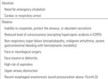
Table 1 - Contraindications to noninvasive ventilation Absolute
Need for emergency intubation Cardiac or respiratory arrest Relative
Inability to cooperate, protect the airways, or abundant secretions Reduced level of consciousness (excepting hypercapnic acidosis in COPD) Non-respiratory organ failure (encephalopathy, malignant arrhythmia, severe gastrointestinal bleeding with hemodynamic instability)
Face or neurological surgery Face trauma or deformity High risk of aspiration Upper airway obstruction
Recent esophageal anastomosis (avoid pressurization above 15cmH2O)
COPD - chronic obstructive pulmonary disease.
Suggestion - NIV may be used in patients with reduced consciousness levels due to hypercapnia in chronic obstructive pulmonary disease (COPD). he level of consciousness should clearly improve one or two hours after beginning NIV.(5,6)
Recommendation - NIV should be avoided in severe ARDS due to the high rate of respiratory failure and need for OTI, especially when PaO2/FIO2<140 and the Simpliied Acute Physiology Score (SAPS) II >35.(5,10)
Noninvasive mechanical ventilation in severe community-acquired pneumonia
Suggestion - NIV may be used in severe cases of community-acquired pneumonia (CAP) with hypoxemic respiratory failure, particularly in individuals with concomitant COPD; the desired therapeutic efect should be achieved within thirty minutes to two hours. Avoid delaying intubation in unsuccessful cases.(5,11)
Post-extubation
Recommendation - NIV should be used to shorten the duration of invasive ventilation (NIV weaning-facilitating action), reduce mortality, reduce the rate of ventilator-associated pneumonia (VAP), and shorten the ICU and hospital stay of individuals with COPD and hypercapnia.(5,12,13)
Recommendation - NIV should be started immediately in high-risk patients (Table 2) to avoid ARF and reintubation (prophylactic action).(5,12-15)
in atelectasis, decreased work of breathing, and reduction in the need for OTI; furthermore, NIV may possibly reduce the mortality rate. In such cases, NIV must be used cautiously, with a full understanding of the limitations of and contraindications for its use.(5,16-19)
Suggestion - In esophageal surgery, NIV may be used to avoid ARF by maintaining lower inspiratory pressures (EPAP <8 and IPAP <15). his same suggestion applies to thoracic, abdominal, cardiac, or bariatric surgery.(5,17-19)
Bronchoscopy
Suggestion - NIV may be used during and after bronchoscopy to reduce the risk of complications in individuals with severe refractory hypoxemia, postoperative respiratory failure, or severe COPD.(5)
Special care must be provided to individuals subjected to transbronchial biopsy, which includes maintenance of the airway pressures at <20cmH2O and performance of chest radiographs in cases of clinical decompensation and approximately six hours after the procedure (in order to rule out pneumothorax).
MASKS AND VENTILATORS FOR PROVIDING NO-NINVASIVE VENTILATION
Ventilators available in Brazil: characteristics, ad-vantages and disadad-vantages
Suggestion - NIV may be performed using portable ventilators speciically designed for this purpose and that have leak compensation. he device should be coupled to a nasal/facial interface with a single-limb circuit and a built-in exhalation port. NIV may also be performed using microprocessor-controlled ventilators with software for this speciic purpose, which should be coupled to the nasal/facial interface by means of an elbow connector and the ventilator’s dual-limb circuit (Table 1 - electronic supplementary material). he CPAP mode may be generated using of low generators (20,21) (Table 3).
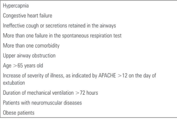
Table 2 - Patients considered to be at risk of extubation failure and who could benefit from noninvasive ventilation immediately after extubation (prophylactic use)
Hypercapnia Congestive heart failure
Ineffective cough or secretions retained in the airways More than one failure in the spontaneous respiration test More than one comorbidity
Upper airway obstruction Age >65 years old
Increase of severity of illness, as indicated by APACHE >12 on the day of extubation
Duration of mechanical ventilation >72 hours Patients with neuromuscular diseases Obese patients
Table 3 - Types of modes of ventilation for noninvasive support
Modes Description Indication*
CPAP
Constant airway pressure Spontaneous ventilation
Recommendation: in cardiogenic APE, PO of abdominal surgery, and mild/moderate sleep apnea
BIPAP (BILEVEL)
Two pressure levels (IPAP and EPAP)
Flow cycled
Recommendation: in acute hypercapnia, for respiratory muscle rest; in cardiogenic APE; and in immunosuppressed individuals with infection
CPAP - continuous positive airway pressure; BIPAP - bilevel positive airway pressure; APE - acute pulmonary edema; PO - postoperative period; IPAP - inspiratory positive airway pressure; EPAP - expiratory positive airway pressure. * except when contraindicated.
Recommendation - Avoid the use of NIV following the onset of a new respiratory failure event after extubation (curative action).(5,12-16)
Carbon dioxide rebreathing
Suggestion - Avoid or minimize CO2 rebreathing when single-limb circuit ventilators are used. he risk of CO2 rebreathing is lower with systems where the exhalation ports are built into the mask compared to ones where the exhalation ports are in the ventilator circuit. Other factors that might contribute to CO2 rebreathing are use of low PEEP and reduced pressure support; special attention is needed in such cases.(22)
Oxygen supplementation
Suggestion - In the case of ventilators with a gas blender, the device allow adjustments in the oxygen (O2) supplementation. When portable NIV devices without a gas blender are used, oxygen should be given straight to the mask beyond the exhalation port using an external O2 source. he supplemental FiO2 depends on the O2 low, position of the O2 connector in the circuit, degree of leak in the ventilator circuit, the type of interface used, and the level of IPAP and EPAP supplied.(23-26)
Monitoring during noninvasive ventilation
Recommendation - Monitor Vt, f and SpO2 during the use of NIV. Use a graphical monitoring system when available. Asynchrony, air leaks, auto-PEEP, eicacy of efort, and the leak compensation mechanism should be continuously monitored.(26,27)
Indications for the choice of interface in common clinical situations
Recommendation - Choose an appropriate interface, i.e., the one that adjusts best to the patient’s face to achieve the greatest clinical eiciency.
Recommendation - Use interfaces without nasal compression when the estimated duration of NIV is >24 to 48 hours.
Recommendation - Use interfaces with a PEEP valve when CPAP with low generator is used.
Recommendation - When NIV is performed with an ICU (conventional microprocessor-controlled) ventilator, use a mask connected to a dual-limb circuit. When NIV-speciic ventilators are used, use a mask for single-limb circuits(20,23-25) (Table 4).
Adaptation to and tolerance of interfaces
Nasal masks
Suggestion - Nasal masks may be used in cases of mild ARF for patients with claustrophobia or maladaptation to the facial mask.
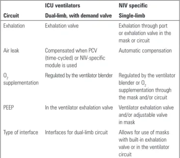
Table 4 - Differences between noninvasive ventilation using portable ventilators specific for noninvasive ventilation and intensive care unit microprocessor-controlled ventilators with a non-invasive ventilation module
ICU ventilators NIV specific
Circuit Dual-limb, with demand valve Single-limb Exhalation Exhalation valve Exhalation through port
or exhalation valve in the mask or circuit Air leak Compensated when PCV
(time-cycled) or NIV-specific module is used
Automatic compensation
O2
supplementation
Regulated by the ventilator blender Regulated by the ventilator blender or O2
supplementation through the mask and/or circuit PEEP In the ventilator exhalation valve Ventilator exhalation valve
and/or adjustable valve in mask
Type of interface Interfaces for dual-limb circuit Allows for use of masks with built-in exhalation valve or in the ventilator circuit
ICU - intensive care unit; PCV - pressure-controlled ventilation; NIV - noninvasive ventilation; O2 - oxygen; PEEP - positive end-expiratory pressure.
Suggestion - Several interfaces can be combined when patients need continuous ventilatory support to avoid the occurrence of ischemia due to reduction of blood low that is caused by the pressure of the mask on the patient’s face (Table 5).(25)
Oral-nasal (facial) masks
Recommendation - Use face masks in cases of mild to moderate ARF to achieve fast improvement of physiological parameters (gas exchange and work of breathing). Monitor the patient’s tolerance and the occurrence of side efects, such as ulcers at support points and gastric distension.
Full-face mask and Helmet
Recommendation - Use these interfaces in the most severe cases of hypoxemic respiratory failure because they allow for greater airway pressurization. As those devices cover the patient’s entire face, the pressure they exert on the skin is more widely distributed, and thus pressure points on the nose are minimized, consequently reducing the risk of skin injury (Table 5).
Table 5 - Advantages and disadvantages of the various types of interfaces
Interface Advantages Disadvantages Suggested ventilators and adjustments Nasal Less risk of aspiration
Facilitates expectoration Less claustrophobia Allows talking Allows eating Easy handling Less dead space
Mouth air leak
Depressurization through the mouth Nose irritation
Limited use in patients with nasal obstruction Mouth dryness
Continuous-flow single-limb circuit devices
Facial Less mouth air leak
More appropriate for acute conditions because it allows for greater flow rates and pressure levels
Higher risk of pressure ulcer on the nose or support points Greater claustrophobia
Greater risk of aspiration Hinders eating Hinders communication
Risk of asphyxia in case of ventilator malfunction Risk of bronchial aspiration
Continuous-flow or demand-flow devices Single- or dual-limb circuit
When dual-limb circuit devices are used, leak automatic compensation in the circuit is necessary
Total-face More comfortable for prolonged use Easy to adjust
Less risk of face skin injury Minimum air leak
Greater dead space
Should not be used in association with aerosol therapy Monitor for vomiting (attention to aspiration)
Continuous-flow devices Single-limb circuit
Use preferentially with NIV-specific ventilators or conventional ventilators with NIV module
Helmet More comfortable for prolonged use No risk of face or skin injury
Greater risk of CO2 rebreathing Favors patient-ventilator asynchrony
Risk of asphyxia in case of ventilator malfunction Should not be used in association with aerosol therapy High internal noise and greater feeling of pressure in the ears Need of higher pressures to compensate for the dead space Skin injury can occur in the axillae
Continuous-flow or demand-flow devices Dual- or single-limb circuit with PEEP valve in the helmet
NIV - noninvasive ventilation; CO2 - carbon dioxide; PEEP - positive end-expiratory pressure.
itself, and the points of contact are on the neck, shoulders and axillary region. However, as the dead space is large, the use of helmet-like masks in individuals with ventilatory disorders is limited; such patients may need requires correction by means of higher levels of pressure support. Internal noise is another cause of discomfort that should be taken into consideration. his type of interface may induce trigger asynchrony due to delayed release of the inspiratory low, with a consequent increase in the work of breathing(28-30) (Table 5).
INTUBATION AND TRACHEOSTOMY
Techniques for elective, semi-elective and emer-gency intubation
Recommendation - Use direct laryngoscopy with visualization of the larynx as the fastest and most reliable method for insertion of the orotracheal tube in elective or emergency cases. hree unsuccessful attempts at intubation by an experienced physician are considered to characterize a diicult airway, in which case the corresponding speciic guidelines should be followed.(31,32)
Elective intubation
Suggestion - Elective tracheal intubation is an intubation that is performed when there are no signs of imminent failure of airway protection, ventilation, and/or oxygenation. Under such conditions, the method of tracheal intubation that is most suited to each individual patient should be selected. Use direct laryngoscopy with OTI as the irst-choice method.(31,32)
Suggestion - Adequately prepare the patient for tracheal intubation, including pre-oxygenation, monitoring, and appropriate positioning during the procedure in order to achieve optimal laryngoscopy.(32,33)
Suggestion - A curved-blade laryngoscope of the appropriate size is preferred. A straight-blade laryngoscope may be used to achieve appropriate larynx exposure in cases where intubation is diicult.(31,32,34)
Emergency intubation
Suggestion - Use hypnotics (propofol, etomidate, ketamine or thiopental), opioids (fentanyl, alfentanil or remifentanil) and neuromuscular blocking drugs (rocuronium or succinylcholine). he Sellick maneuver (cricoid pressure) can be performed during the procedure to minimize the risk of gastric aspiration.(32,35-37)
Techniques and indications for tracheostomy: advantages and disadvantages
Timing of tracheostomy: recommendations based on the cause of respiratory failure
Spinal cord injury
Suggestion - Perform tracheostomy early (within seven days). High cervical spinal cord injury (C5 or above) is an independent predictor of the need for prolonged MV. Patients with injuries at lower levels should be assessed on an individual basis.(32,38)
Traumatic brain injury
Suggestion - Perform tracheostomy early (within seven days) in the most severe cases (Glasgow Coma Scale <8), as patients with traumatic brain injury usually require prolonged ventilatory support. he evidence regarding reductions in the VAP rate is contradictory, and there is no evidence that early tracheostomy reduces mortality, airway injury, or the length of hospital stay.(32,38,39)
Patients with trauma not affecting the central nervous system
Suggestion - Early tracheostomy is indicated when prolonged ventilatory support is anticipated.(32,38-40)
Patients admitted to the intensive care unit for clinical causes
Recommendation - Wait 14 days to perform a tracheostomy, as early use of this procedure does not reduce the 30-day mortality rate, length of stay in the ICU, or the need for sedation.(32,41-44)
Tracheostomy techniques
Recommendation - Perform percutaneous or conventional tracheostomy, depending on the available resources and the staf’s experience. Percutaneous tracheostomy can be performed at the bedside by ICU staf. Although it is more expensive and demands that
a bronchoscopy be performed to increase its safety, the associated rates of surgical wound infection are lower. Conventional tracheostomy must be performed in an operating room by specialized staf, except for the case of ICUs that are equipped with a room for surgical procedures. Both techniques have similar rates of major complications, such as bleeding, subcutaneous emphysema, pneumothorax and death.(32,45-47)
INITIAL ADJUSTMENT OF INVASIVE VENTILATION AND CONVENTIONAL VENTILATION MODES
Ventilation adjustment
Recommendation - Use the FIO2 needed to maintain SpO2 at 93 - 97%.(48,49)
Recommendation - Use a Vt of 6mL/kg/ predicted body weight. Reassess as a function of changes in the patient’s clinical condition.(48-52)
Recommendation - Use the assist-control mode (AC) as either volume-cycled (VCV) or time-cycled pressure-limited, known as pressure controlled ventilation mode (PCV), and reassess within the irst few hours based on the patient’s clinical condition.(48-51)
Recommendation - Adjust the initial f = 12 to 16 breaths per minute, with an inspiratory low rate or inspiratory time required to maintain the inspiration to expiration ratio (I:E) initially at 1:2 or 1:3. In patients with obstructive disease, the initial f can be lower (<12 breaths per minute), and in patients with restrictive disease it may be higher (e.g., >20 breaths per minute, if required by the patient’s clinical condition). Reassess as soon as the irst arterial blood gas results are available.(48,51-54)
Recommendation - Establish the type of ventilator triggering. he more widely available types of ventilator triggering are the time-triggered (ventilator-controlled mode) and the patient-triggered (low or pressure triggered, also known as pneumatically triggered) modes. he ventilator’s sensitivity should be adjusted to the most sensitive level to avoid auto-triggering. he ventilator can also be triggered by neural stimuli (neurally adjusted ventilatory assist-NAVA).(48,51-54)
Recommendation - Initially use a PEEP of 3 to 5cmH2O, except in cases of diseases such as ARDS, where the PEEP value should be assessed according to the speciic guidelines described in the each topic of the present recommendations.(48,49,55-57)
with thick secretions, and optimal humidiication should be maintained to avoid obstruction of the orotracheal tube.(58)
Recommendation - Set the alarms on an individual basis, using speciicity and sensitivity parameters appropriate for the patient’s clinical condition. Also, an apnea backup and the speciic parameters for apnea should be adjusted if they are available in the device.
Recommendation - After the initial parameters are deined, check the Vt, pressure and low curves to establish whether their values correspond to the expected parameters or if immediate readjustment is needed. Check pulse oximetry, which should be continuously monitored. Initially, set the maximum airway pressure at 40 cmH2O to avoid barotrauma, and adjust as soon as possible based on the patient’s clinical condition.(48,51-54)
Recommendation - Arterial blood gases must be assessed after 30 minutes of steady ventilation to check whether the ventilation and gas exchange goals were met. If they were not, perform necessary adjustments of the mode and cycling parameters.(48-51)
Recommendation - Assess the eventual hemodynamic repercussions of MV. Investigate the presence of hypovolemia, auto-PEEP and/or pneumothorax in patients with hypotension that is associated with positive pressure ventilation.
Recommendation - Maintain the most appropriate level of muscle work. In patients with high inspiratory low demands, use opioids to reduce the ventilatory drive and provide appropriate comfort for the patient. Induce muscle rest for 24 to 48 hours in patients with respiratory muscle fatigue or hemodynamic instability.
Recommendation - In patients who do not need muscle rest, start an assist mode of ventilation as soon as possible, with appropriate adjustment of the ventilator’s sensitivity. Avoid ventilator-induced diaphragmatic dysfunction, which usually occurs after 18 hours of controlled ventilation.
Suggestion - In older adults, patients who require prolonged use of controlled modes of ventilation, malnourished patients, patients using corticosteroids or neuromuscular blocking agents, and individuals with hypothyroidism, pay special attention to the assessment of respiratory muscle function.
Conventional modes of ventilation(59)
Suggestion - Use the volume assist-control mode (VCV) when the aim is to maintain a more stable minute volume (Vt x f). his mode of ventilation can be timed (controlled), and pressure- and low-triggered (assisted)
and is cycled of when the preset inspired Vt is achieved. he airway pressure is variable and depends on the patient’s ventilatory mechanics (special attention should be paid to monitoring the peak and plateau pressures when this mode is used, and it should be ensured that the maximum airway pressure alarm is properly set). his mode is also used to measure the peak and plateau pressures for calculating the compliance and resistance of the respiratory system under a constant square-wave inspiratory low pattern (see this speciic topic in the present recommendations).
Suggestion - Use the PCV assist-control mode when respiratory mechanics are impaired (low compliance and/ or high resistance), as it allows for better control of the airway and alveolar pressures. his mode characteristically limits pressure throughout all the inspiratory phase and is time-cycled. he inspiratory time is set in seconds by the caregiver. he low is free and decelerating waveform. In this mode, the Vt is variable and depends on the administered delta pressure and the patient’s ventilatory mechanics (special attention should be paid to monitoring the expired Vt and adjusting the maximum and minimum minute volume alarms). he inspiratory low speed (ramp, rise time or slope) can be increased or reduced. he rise time can be faster in patients with obstructive disease to obtain a better Vt. Special attention should be paid to the possible occurrence of peak low overshoot. In patients with restrictive disease, a slower rise timeshould be used.
Suggestion - PSV is considered the preferential mode during assisted/spontaneous ventilation. It should be started as soon as possible, based on the patient’s clinical condition. his is an exclusively patient-triggered mode, and can be low- or pressure-triggered. Characteristically, pressure is limited throughout all the inspiratory phase and is cycled of when the inspiratory low falls, typically to 25% of the peak inspiratory low. his cycling criterion (% of the peak inspiratory low) can be set between 5% and 80% in some of the most modern ventilators, which allows a reduction of the inspiratory time in patients with obstructive disease (% of the cycling of >25%) and an increase in the inspiratory time in patients with restrictive disease (% of the cycling of <25%). he rise time can be faster in patients with obstructive disease, thus decreasing inspiratory time and obtaining a better Vt. Special attention should be paid to the occurrence of peak low overshoot. In patients with restrictive disease, use a slower rise time, which may be accompanied by a Vt gain.
a ixed low rate until the airway pressure reaches the value predetermined by the caregiver (cycling). As a result, the Vt is unknown, and consequently the use of an external ventilometer (Wright’s ventilometer) is recommended; alternatively, arterial blood gases can be assessed after 20 minutes of steady ventilation to check whether the PaCO2 is compatible with the patient’s clinical condition (35 to 45 mmHg in most cases). his device usually does not have a built-in O2 blender or alarms. he multi-disciplinary staf must pay special attention to monitoring both ventilation and oxygenation.
Recommendation - Avoid the use of Synchronized Intermittent Mandatory Ventilation (SIMV) because it has been shown to be associated with a delay in MV weaning. Currently, the use of SIMV is restricted to patients in whom minimal minute volume is necessary at the beginning of MV weaning process (e.g., individuals with neuropathy, or upon immediate awakening from general anesthesia). As soon as the ventilatory drive stabilizes, SIMV should be shifted to PSV. A brief description of SIMV mode follows. Controlled cycles can be volume-cycled (V-SIMV) or pressure-limited (P-SIMV). Spontaneous cycles should be associated with PSV. SIMV is characterized by the fact that it allows for controlled, assisted and spontaneous cycles to occur within the same time window (TW), which is determined by the f of the controlled mode. Controlled cycles only occur when a patient assisted trigger did not occur in the immediately preceding TW. Otherwise, the ventilator waits for the next patient-trigger, i.e., an assisted cycle. Spontaneous cycles supported by PSV can occur in the remainder of the TW.
ASYNCHRONY AND NEW MODES OF MECHANICAL VENTILATION
Patient-ventilator asynchrony
Comment - Patient-ventilator asynchrony is a lack of coordination between the patient’s inspiratory efort and ventilatory needs and the support provided by the ventilator.(60) Asynchrony is a frequent event, occurring in
10% to 80% of all ventilator cycles, and is associated with prolonged of MV and ICU stays.(61)
Recommendation - he presence of asynchrony should be actively assessed during the assessment of patients subjected to MV, and it should be corrected.
Trigger asynchrony
Ineffective triggering
Comment - Inefective triggering occurs when the patient’s inspiratory efort is not enough to trigger the ventilator.(62) he reason might be a maladjustment
in ventilator sensitivity or patient-related factors such as respiratory muscle weakness, central respiratory depression, dynamic hyperinlation (auto-PEEP), or longer mechanical inspiratory time relative to the neurally stimulated inspiratory time.(62,63)
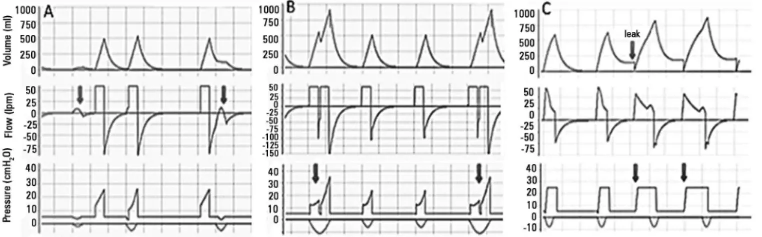
Identiication - Clinical examination of the patient’s chest and abdomen can reveal that the inspiratory efort is not accompanied by a ventilator cycle.(64,65) Figure 1A shows
how to identify this asynchrony in ventilator curves.(64,65)
Recommendation - To correct trigger asynchrony, the ventilator’s sensitivity should be adjusted to the most sensitive level possible, while avoiding auto-triggering; in addition, pressure triggering can be shifted to low triggering (which is usually more sensitive).
Suggestion - In the presence of auto-PEEP, extrinsic PEEP may be titrated up to 70 to 85% of the auto-PEEP; the efects of this adjustment on asynchrony must be checked.(62) During PSV, one might attempt to
reduce the pressure that is administered or to increase the percentage of the cycling criterion.(63) When
pressure-controlled ventilation (PCV) is used, one might attempt to reduce the inspiratory time, or in cases where VCV is used, to increase the inspiratory low rate or reduce the pause time.(62,63)
Double triggering
Comment - Two consecutive cycles are triggered by a single patient inspiratory efort. he ventilator’s mechanical inspiratory time is shorter than the patient’s neural inspiratory time.(3)
Identiication - Clinically two consecutive cycles without an interval between them can be observed; this pattern that may be repeated quite often. Figure 1B shows how to identify this asynchrony in the ventilator curves.(64-66)
double triggering occurs under PCV, the inspiratory time and/or delta of pression value could be increased. In PSV, one might try to increase the pressure level or reduce the percentage of the cycling criterion.(62,63)
Auto-triggering
Comment - he ventilator is triggered in the absence of a patient’s inspiratory efort. his can be caused by overly high ventilatory sensitivity, leaks in the system, low alterations due to presence of condensates in the circuit, detection of the heartbeat, or wide variations in chest pressure that are due to stroke volume (Figure 1C).(60,62)
Identiication - he observed respiratory frequency is higher than the adjusted one, and the cycles are not preceded by indicators of patient inspiratory efort.(64-67)
Recommendation - Once the presence of leak or condensate in the circuit is corrected or ruled out, gradually reduce the ventilator’s sensitivity to a level suicient for auto-triggering to stop.(62,64-66)
Flow asynchrony
Insufficient inspiratory flow
Comment - In insuicient inspiratory low, the low ofered is lower than patient ventilatory demands. his typically occurs when the low is set by the operator and cannot be increased by the patient’s inspiratory efort, as in VCV. Nevertheless, this phenomenon might also occur in
PCV and PSV, when the adjusted pressure is insuicient to ensure an appropriate balance between the patient’s ventilatory demands and mechanics.(67,68)
Identiication - he patient exhibits discomfort and uses the accessory respiratory muscles. Figure 2 shows how to identify this asynchrony in the ventilator curves.(67,68)
Recommendation - Correct the causes of the increased ventilatory demands, such as fever, pain, anxiety, or acidosis. In VCV, increase the inspiratory low rate and check for signs of patient comfort, as well as the shape of the pressure - time curve; shift to PCV or PSV, in which the low is not ixed;(68) adjust the
speed necessary to achieve the maximum airway pressure (rise time - speed of low rise, or increasing the controlled pressure value).(69)
Excessive inspiratory flow
Comment - Excessive inspiratory low can occur in VCV when the low is set above the level desired by the patient, or in PCV or PSV when high pressures or a faster rise time are set.
Identiication - In VCV, the pressure - time curve peak is achieved too early.(68,69) In PCV or PSV, the
airway pressure becomes higher than the adjusted level, a phenomenon known asovershoot.(69)
Recommendation - In VCV, reduce the low rate; in PCV and PSV, the rise time should be reduced until the overshoot disappears.(68)
Figure 2 - Flow asynchrony. In volume-controlled mode, the flow rate was adjusted below the patient’s demand; the patient thus maintained muscle effort throughout inspiration, and the curve consequently became concave and upward. The asynchrony exhibits increasing intensity from the first to the third cycle, as represented in the figure. The negative deflections in the pressure-time curve represent the patient’s inspiratory effort (muscle pressure) and are only visible when esophageal pressure is monitored. Figures obtained at Xlung.net, a virtual mechanical ventilation simulator. Available at: http//:www.xlung.net.
Cycling asynchrony
Premature cycling
Comment - In premature cycling, the ventilator interrupts the inspiratory low before the patient desired; in other words, the ventilator’s mechanical inspiratory time is shorter than the patient’s neurally controlled inspiratory time.(70) In VCV and PCV, the inspiratory time is adjusted
by the operator. In PSV, premature cycling occurs when a low pressure level and/or a high percentage of the cycling criterion are adjusted.(70) Figure 3 shows how to identify
this asynchrony in the ventilator curves. In some cases, the patient’s inspiratory efort may suice to trigger a new cycle (double cycling).(64,66,70)
Recommendation - In VCV, the inspiratory low rate may be reduced and/or Vt may be increased in compliance with the safety thresholds. Alternatively, one might shift to PCV or PSV, where the inspiratory low rate varies as a function of the patient’s inspiratory efort. When premature cycling occurs in PCV, the inspiratory time and/or the delta of inspiratory pressure value may be increased. In PSV, one could try to increase the pressure level or reduce the percentage of the cycling criterion.(62,63,70)
Delayed cycling
Comment - In delayed cycling, the ventilator’s mechanical inspiratory time is longer than the time desired by the patient; in other words, the ventilator cycling time is longer than the patient’s neurally controlled inspiratory
Figure 3 - Cycling asynchronies during pressure support ventilation. In the first cycle, the cutoff point of 25% of the peak inspiratory flow (percentage of the cycling criterion) was reached rapidly; the ventilator’s inspiratory time was therefore shorter than the time desired by the patient. This is shown in the expiratory segment of the flow curve, which tends to return to the baseline as a result of the patient’s inspiratory effort, which is still present. The last cycle represents the opposite situation, i.e., delayed cycling. The flow reduction occurs very slowly, which is typical of airway obstruction; the cycling threshold is therefore reached with some delay. Sometimes, the cycle is interrupted by a contraction of the respiratory muscles, which causes an increase above the support pressure adjusted at the end of inspiration (not shown in this figure). Figures obtained at Xlung.net, a virtual mechanical ventilation simulator. Available at: http//:www.xlung.net
time. In VCV, this can occur when the inspiratory time is extended by setting a high Vt or a low inspiratory low rate or if inadequate use is made of the inspiratory pause. In PCV, delayed cycling occurs when the inspiratory time is set beyond the time desired by the patient. In PSV, particularly in the case of obstructive diseases such as COPD, the increase in the resistance and compliance of the respiratory system gradually slows down the inspiratory low rate, thus increasing the inspiratory time.(70) Figure 3 shows how to
identify this asynchrony in the ventilator curves.(64,66)
Recommendation - In modes of ventilation in which the operator adjusts the inspiratory time, the latter should be reduced. In PSV, the percentage of the cycling criterion might be increased (e.g., from 25% to 40% or even higher).(70)
Suggestion - Patient-ventilator asynchrony should be treated by adjusting the ventilation parameters or shifting to other modes of ventilation (experts’ opinion).
Advanced modes of mechanical ventilation
Comment - he choice of the mode of ventilation should be based on the severity of the patient’s condition.(71) In
Suggestion - Use advanced modes of ventilation in speciic clinical situations, provided that the operator is thoroughly acquainted with the parameters of each mode and that the patient’s clinical condition can beneit from the resources speciic to each mode.
Pressure-regulated volume-control mode
Comment - his is a time-cycled pressure-limited ventilation mode. he ventilator readjusts the pressure limit at each cycle based on the Vt obtained in the previous one, until reaching a target Vt that has been preset by the operator.(72)
Suggestion - Indicate when limited-pressure Vt control is desired, aiming to automatically adjust the inspiratory pressure if the respiratory mechanics change.
Recommendation - Caution is required in adjusting the target Vt, as undesirable increases of the inspiratory pressure may result.
Airway pressure release ventilation and bilevel positive airway pressure ventilation
Comment - Airway pressure release ventilation (APRV) is pressure-limited and time-cycled, and is considered to be a spontaneous mode of ventilation. he operator adjusts the pressure high (PEEPhigh) and low (PEEPlow), the PEEPhigh to PEEPlow ratio, and the frequency of alternation between both PEEP levels; the time of PEEPhigh must be longer than the time of PEEPlow. he BIPAP mode also uses two PEEP levels, but the time of PEEPlow is longer than that of PEEPhigh. he patient can breathe spontaneously at both pressure levels.(73,74) Support pressure may also be applied, as its
value is added to the PEEPlow value, and the inal airway pressure (Paw) is the result of the sum of PSV + PEEPlow. When the PEEPhigh value is lower than PSV + PEEPlow value, during the PEEPhigh period the ventilator only complements the PSV value to reach the same level of Paw as in PEEPlow + PSV.
Suggestion - Use APRV when maintenance of spontaneous ventilation and alveolar recruitment is necessary; APRV may improve gas exchange and reduce dead space and asynchrony.
Recommendation - Caution is required when regulating the alternation between the two pressure levels because in this mode, the minute volume results from the sum of the obtained Vt, when the pressures are alternated, plus the Vt generated from PSV cicles.
Proportional assist ventilation
Comment - Proportional assist ventilation (PAV) is a spontaneous ventilation mode that follows the equation of motion to generate inspiration pressure (Pvent) in proportion to the patient’s inspiratory efort (Pmus). herefore, when the Pmus decreases, Pvent also decreases, and vice-versa.(71,75-79)
Some studies found better patient-ventilator synchrony when PAV, or its latest version, PAV plus (PAV+), is used compared to PSV. he PAV+ software estimates the work of breathing (WOB) of both patient and mechanical ventilator using the equation of motion, and calculates compliance and resistance through the application of 300-ms inspiratory micro-pauses every 4 to 10 ventilation cycles.
Indication - PAV is indicated for patients with respiratory drive and signiicant asynchrony under spontaneous modes of ventilation, PSV in particular. It is also indicated when one wants to determine the patient’s WOB and mechanical measurements during assisted ventilation, e.g., for obtaining real-time intrinsic PEEP estimates.(75-79)
Recommendation - Before starting the PAV+ mode, the operator should set the type and diameter of the tracheal prosthesis, the type of humidiier, maximum Vt and maximum allowed airway pressure (limits) in the ventilator.
Recommendation - Set the percentage of initial support at 50% to achieve a patient WOB of 0.3 - 0.7 J/L with adequate Vt and f. Pvent increases proportionally with the patient’s Pmus. he support percentage should not exceed 90%. If a greater percentage is needed, conventional assisted-controlled ventilation modes are recommended. Gradually reduce the support percentage in parallel with improvement of the patient’s clinical condition, to as low as 30%. When the (abovementioned) parameters are maintained, consider to extubate the patient.
Suggestion - PAV is an alternative to PSV in patients with signiicant asynchrony; it has the potential to improve the patient-ventilator interaction.
Recommendation - PAV should be avoided in patients without respiratory drive, as well as in MV with leaks that impair the measurements of resistance and compliance.
Automatic tube compensation
Suggestion - Use ATC plus or minus PSV to automatically compensate for the increase in the resistive work associated with the presence of a tracheal prosthesis (in PSV, the compensation should be calculated by the caregiver as a function of the prosthesis diameter; the smaller the diameter, the higher the PSV value should be, e.g., PSV=5cmH2O for 9-mm tubes, and PSV=9cmH2O for 6-mm tubes).
Recommendation - ATC is contraindicated for patients without respiratory drive, and care should be taken in patients who have excess secretions that interfere with inspiratory low; the airway pressure alarms should be properly set.
Neurally adjusted ventilatory assist
Comment - Neurally adjusted ventilatory assist (NAVA) is a mode of ventilation that captures the electrical activity of the diaphragm and uses it as a criterion for triggering and cycling-of cycling-of the ventilator, thus providing inspiratory support in proportion to the electrical activity of the diaphragm. Use of NAVA requires placement of an esophageal-gastric catheter, with sensors positioned on the distal third of the esophagus to detect the electrical activity of the diaphragm.(5,6) In clinical
studies, use of NAVA was associated with improved patient-ventilator synchrony when compared to PSV.
Indications - NAVA is indicated for patients with respiratory drive and signiicant asynchrony on spontaneous ventilation, and particularly in the case of loss of efort with PSV, as in patients with auto-PEEP (intrinsic PEEP).(77-79,83)
Recommendation - Special care is required in patients with oronasal or esophageal disorders that might hinder the passage or proper positioning of the NAVA catheter. he NAVA catheter should be properly placed and ixed, and its position should be checked on a regular basis. Once the probe is ixed, measure the electrical activity of the diaphragm (Edi), and adjust the NAVA gain as a function of the Vt, f and airway pressure (Edi versus NAVA gain). he ventilator is triggered by 0.5-µV variations in the Edi. From that point onwards, the ventilator delivers free low as a function of the Edi reading. he maximum airway pressure results from adding [maximum Edi - minimum Edi] multiplied by the NAVA gainto the extrinsic PEEP value. Cycling-of occurs when Edi falls to 70% of the maximum Edi peak detected.(77-79,83)
Recommendation - NAVA gain is adjusted as a function of the patient’s clinical condition, and should be assessed on an individual basis.
Suggestion - NAVA may be an alternative to PSV for patients with signiicant asynchrony; it may improve the
patient-ventilator interaction, especially in cases where there is loss of respiratory efort.
Adaptive support ventilation
Comment - Adaptive support ventilation (ASV) employs an algorithm to select the Vt and f combination necessary to reach the minute volume set by the caregiver by means of spontaneous and controlled cycles, with the lowest possible airway pressure. he version known as Intellivent-ASV employs an end-tidal CO2 (ETCO2) and a SpO2 sensor to adjust the PEEP and FIO2 automatically by means of a table.(83)
Indications - ASV is indicated for patients with severe respiratory failure when reductions of the work of breathing and stimulation of spontaneous respiration are desired.
Suggestion - Use ASV to ensure minute volume with appropriate lung protection in patients with unstable ventilatory drive, asynchrony or discomfort. Monitor for possible occurrence of leaks or excess secretions, which may impair the appropriate functioning of the ventilator.
VENTILATORS FOR INVASIVE VENTILATION Choice of mechanical ventilator
he following questions should be answered when choosing mechanical ventilators: in which patient population they will be used (adults, children, or newborn infants)? How often are patients with severe ventilation problems admitted (e.g., ARDS, severe obstructive disease, pulmonary istula, etc.)? What information do ventilators provide to contribute to decision-making about ventilatory support in that particular ICU? How will patients be weaned from MV? What mode of ventilation will be used? Which clinical and mechanical measurements contribute to decision-making? How often and in which situations will NIV be used?
Suggestion - Assess the particular characteristics of various ventilators as a function of the resources available to and the needs of your service:
Ventilators with basic resources. hese include one or more basic modes of ventilation without curves. As a rule, they are used for transportation of patients under MV.
Ventilators with basic resources and curves. hese include the basic modes of ventilation (VCV, PCV, SIMV and PSV) and the basic ventilation curves (volume, low and pressure).
and curves, these also include advanced ventilation modes, such as dual-control modes (e.g., PRVC), diferential modes for spontaneous ventilation (such as PAV+ and NAVA), and advanced monitoring methods (e.g., measuring the work of breathing, airway occlusion pressure [P 0.1], maximum inspiratory pressure [PImax], volumetric capnometry, and indirect calorimetry).
Recommendation - In the hospital setting, any ventilator should include at least the following features: (1) control of the expired tidal volume (eVt); (2) basic monitoring tools (at least inspiratory pressure); and (3) a gas blender coupled to the ventilator to avoid the use of O2 supplementation through the artiicial airway.
Recommendation - In addition to the requirements mentioned above, ventilators that are to be used in the ICU should also include the following: (1) curve monitoring (at least the pressure-time curve), (2) alarms (at least for the maximum and minimum airway pressure, for detection of apnea and disconnection from the ventilator).
Comment - he electronic supplementary material includes a list of the mechanical ventilators for adults available in Brazil (in August 2013) with a description of some of their features (Tables 2, 3, 4 and 5 in the supplementary material). his list does not include ventilators that are exclusively used in the following situations: (1) for NIV, (2) in children and newborn infants, (3) at home or for sleep apnea, and (4) in anesthesia.
MONITORING THE PATIENT UNDER VENTILATORY SUPPORT
Monitoring of gas exchange
How to perform bedside monitoring of the ventilatory mechanics
Recommendation - he ventilatory mechanics should be routinely monitored in all patients who are subjected to invasive mechanical ventilatory support, including the following parameters: eVt, peak pressure (maximum inspiratory pressure), plateau or inspiratory pause pressure (under controlled ventilation), extrinsic PEEP, auto-PEEP or intrinsic PEEP.(84-88)
Suggestion - Calculate the resistance of airways (Raw) and static compliance (Cst), and monitor the low-time, pressure-time, and volume-time curves in selected cases.(84-88)
Comment - In clinical practice, the alveolar pressure can be estimated by means of an inspiratory pause lasting at least two seconds. he pressure at the end of the pause
is known as plateau or pause pressure. For measurements to calculate the Raw, the inspiratory low rate must have a “square” wave pattern and be converted to liters/second.
Recommendation - he following are mandatory requirements for accurate measurement of the pause pressure: absence of respiratory muscle efort, pause duration of two to three seconds, and absence of leaks.(84-87)
Recommendation - Avoid alveolar pressure values >28 to 30cmH2O, which are indicative of low static lung compliance. In such case, the possible cause should be investigated (alteration of the lung parenchyma and/or the thoracic cage). In the former case, reduce the Vt and/or the driving pressure (also called distending pressure); in the latter, also other causes might be present, to wit, reduction of the chest wall compliance and/or intra-abdominal hypertension. In the latter case, the intra-abdominal pressure should be monitored and decompression should be started when needed.(84-88) Figure 4 shows how to
calculate Raw and Cst.
Figure 4 - Inspiratory pause maneuver and estimation of the airway resistance and pause (or plateau) pressure. VCV - volume-controlled ventilation. Paw - airway pressure; PEEP - positive end-expiratory pressure; Vt - tidal volume; Pel - elastic pressure; Palv - alveolar pressure.
Comment - Auto-PEEP, also called intrinsic PEEP (PEEPi), occurs when the end-expiratory pressure is higher than the airway pressure due to incomplete lung emptying.
Recommendation - Auto-PEEP is identiied on the low-time curve when the expiratory low does not return to zero at the end of expiration.(84-87)
Recommendation - In cases of ARDS, the distending pressure should be monitored; also known as driving pressure, this value is calculated by subtracting PEEP from the plateau pressure (Pplat). he distending pressure should always be ≤15cmH2O in cases of moderate or severe ARDS, when higher PEEP is necessary, resulting in an increase of Pplat to 30 - 40cmH2O (see topic: MV in ARDS in the present recommendations).(89-91)
Monitoring of gas exchange in mechanical ventilation
Arterial blood gas measurement
Recommendation - In order to ground clinical reasoning and therapeutic practice, arterial blood gas samples should be collected as soon as possible, preferably from the radial or the femoral artery, in all cases of ARF. Arterial blood gas assessments permit diagnostic assessment of the acid-base status and lung gas exchange through direct measurement of the pH, PaCO2, and PaO2, and calculation of the oxygen saturation (SaO2), bicarbonate (HCO3-) and base excess (BE). When intoxication causing methemoglobinemia and carboxyhemoglobinemia is suspected, SaO2 should be directly measured using co-oximetry.(92,93)
Recommendation - Collect samples for arterial blood gas measurement in all patients subjected to ventilatory support 20 minutes after the initial adjustment of the ventilator parameters, and then every day for the duration of the acute phase of the clinical problem. Samples should also be collected whenever the patient’s clinical condition changes.(92,93)
Recommendation - Avoid collecting samples for arterial blood gas measurement from areas irrigated by the artery to be punctured that are at risk of ischemia, and from infected sites. In patients with coagulopathy or thrombocytopenia, samples should only be collected when the test is fully necessary.(92,93)
Care in the performance of the blood gas measurement
Suggestion - Use standard kits or 5-mL syringes with a minimum amount of lithium or sodium heparin, and a ine needle (23 to 25G), preferentially with a safety mechanism.(92,93)
Recommendation - his procedure is invasive, and thus it must be performed under aseptic conditions. Whenever possible, the procedure should be explained to the patient and performed only with his or her consent.(92,93)
Recommendation - he puncture site should be compressed for at least ive minutes, or longer in cases of coagulopathy or use of anticoagulants.(92,93)
Recommendation - he sample should be analyzed as soon as possible. When analysis is performed outside the unit, the sample should be transported in a refrigerated container.(92,93)
Care in the interpretation of arterial blood gas measurements
Recommendation - Record the following parameters at the time of sample collection: FIO2, Vt, f, PEEP, SpO2, and ETCO2 (when capnography is performed).
Recommendation - he PaO2/FIO2 ratio should be calculated in all cases to assess the eiciency of oxygenation and the patient’s clinical progression.(92,93)
Suggestion - Record whether the patient is in the prone position, the mode of ventilation at the time of sample collection, and if alveolar recruitment maneuvers and PEEP titration were performed before sample collection.
Comment - he arterial blood gas measurement merely relects a speciic moment of the patient’s condition. Pulse oximetry and capnography are more adequate methods for continuous monitoring.
Pulse oximetry
Recommendation - Continuous monitoring by means of pulse oximetry should be performed in all patients who are receiving O2 supplementation, NIV, or invasive ventilatory support, as well as in patients with ARF.
Capnography
Recommendation - Perform capnography in patients with neurologic diseases who are receiving ventilatory support, to conirm the position of the ventilatory prosthesis, and whenever the CO2 level is above 50mmHg.
Suggestion - Capnography can be used for monitoring in patients with a ventilation-perfusion imbalance to detect acute alterations in status, as well as for monitoring of speciic therapies (e.g., thrombolytic therapy in pulmonary thromboembolism).
Regional monitoring
Monitoring by means of electrical impedance tomography
less resistant to the passage of the current. EIT is used for monitoring ventilation, and more recently, for bedside continuous monitoring of lung perfusion.(94-98)
Suggestion - Use EIT for detection of lung ventilation disorders, such as pneumothorax, as well as for evaluating changes in ventilation when placing the patient in speciic decubitus position, to check the position of the endotracheal tube, to assess pulmonary recruitment and collapse, and to assess the regional distribution of ventilation. In the future, EIT may be used for monitoring of lung perfusion.(94-98)
Computed tomography
Recommendation - Use computed tomography (CT) as a diagnostic method in cases of respiratory failure of unknown etiology; CT angiography should be used when pulmonary embolism is suspected.
Suggestion - In centers where CT is available, this method may be used to monitor alveolar recruitment and decremental PEEP titration in cases of moderate or severe ARDS, paying special attention to the care required in patient’s transportation, and taking the total radiation dose into consideration.(99,100)
Chest ultrasound
Recommendation - In centers where it is available, staf should be trained to use chest ultrasound for early detection of pneumothorax and pleural efusion, and as an aid in performing therapeutic procedures.
Suggestion - Chest ultrasound can be used to estimate alveolar re-aeration in patients treated for VAP, to assess pulmonary edema, to detect post-extubation atelectasis, and to estimate PEEP-induced pulmonary recruitment.(101-103)
SEDATION AND ANALGESIA DURING MECHANICAL VENTILATION
When are sedatives and analgesics indicated and how should they be administered?
Suggestion - Use sedation and analgesia in patients treated with MV in order to control anxiety, agitation and pain. Appropriate sedation helps the patient better tolerate the ventilator, diagnostic and therapeutic procedures.(104,105)
Recommendation - he sedation level should be mild to moderate to allow for early mobilization.(106)
Recommendation - Titrate propofol and midazolam for low, moderate and deep sedation. Dexmedetomidine should not be used to induce deep sedation. he recommended opioids are fentanyl, morphine, and remifentanil.(107)
Suggestion - Avoid using ketamine as the main sedative in patients undergoing MV. Ketamine may be useful in situations in which its opioid-sparing efect is required.(107,108)
Suggestion - Have a thorough knowledge of the main drugs used for analgesia and sedation in patients under ventilatory support:
Propofol - Its main action is as a gamma-aminobutyric acid (GABA) agonist. It has sedative, hypnotic, anxiolytic and anticonvulsant effects and promotes amnesia. It does not have an analgesic effect. It causes dose-dependent respiratory depression and hypotension secondary to systemic vasodilation, especially when administered by bolus. Prolonged infusion might make awakening unpredictable and cause propofol infusion syndrome (PRIS), which has an incidence of <1%. The mortality of PRIS is high, and the syndrome is characterized by worsening of metabolic acidosis, hypertriglyceridemia, arrhythmia, and hypotension with an increased need for vasopressors. The recommended initial dose is 5mcg/ kg/minute over five minutes, followed by continuous infusion at 5 to 50mcg/kg/minute.(107)
Midazolam - This is a GABA agonist that promotes anxiolysis, amnesia and hypnosis. It has anticonvulsant effects. It does not have an analgesic effect. The use of this benzodiazepine for hypnosis seems to be associated with a higher incidence of delirium. Compared to propofol, midazolam may increase the length under MV. Abstinence syndrome can occur after prolonged infusions, i.e., longer than seven days. The recommended initial dose is 0.01 to 0.05mg/kg, and the maintenance dose is 0.02 to 0.1mg/kg/h in continuous infusion.(109)
Dexmedetomidine - This is an alpha-2-adrenergic agonist with central action. It has sedative effects and helps to reduce the need for analgesics/opioids. It does not have an anticonvulsant effect. It is not associated with significant respiratory depression. It is not appropriate for inducing deep sedation. The prevalence of delirium is lower in patients treated with dexmedetomidine compared to benzodiazepines. In patients admitted to the ICU, the recommended loading dose is not used, and the drug is started as a continuous infusion. After the start of infusion, its action begins in 15 minutes, and the maximum effect is reached within one hour. The recommended dose is up to 1.4mcg/kg/hour.(107)