Red blood cell distribution width is associated with
myocardial injury in non-ST-elevation acute coronary
syndrome
Erhan Tenekecioglu,I*Mustafa Yilmaz,IOsman Can Yontar,IAdem Bekler,IITezcan Peker,IKemal Karaagac,I Ozlem Arican Ozluk,IFahriye Vatansever Agca,IMustafa Kuzeytemiz,IMuhammed Senturk,IBurhan Aslan,I Dursun TopalI
IBursa Yuksek Ihtisas Education and Research Hospital, Cardiology Clinic, Bursa, Turkey.IICanakkale Onsekiz Mart University, Medicine School, Cardiology
Clinic, Canakkale, Turkey.
OBJECTIVES:The red blood cell distribution width has been associated with an increased risk of cardiovascular events. In the present study, we assessed the relationship between red cell distribution width values and cardiac troponin I levels in patients admitted with non-ST-elevation acute coronary syndrome.
METHODS: We analyzed blood parameters in 251 adult patients who were consecutively admitted to the intensive coronary care unit with non-ST-elevation acute coronary syndrome over a 1-year period. For all patients, a baseline blood sample was collected for routine hematological testing. Cardiac troponin I was measured at baseline and after 6 h. The patients were diagnosed with non-ST-elevation myocardial infarction or unstable angina based on the elevation of cardiac troponin I levels.
RESULTS:The red cell distribution width was higher in the group with non-ST-elevation myocardial infarction compared with the patient group with unstable angina (14.6¡1.0 vs 13.06¡1.7, respectively; p= 0.006).
Coronary thrombus was detected more frequently in the group of patients with non-ST-elevation myocardial infarction than in the patients with unstable angina (72% vs 51%, respectively; p= 0.007). Using receiver
operating characteristic curve analysis for the prediction of non-ST-elevation myocardial infarction based on the red cell distribution width, the area under the curve was 0.649 (95% confidence interval: 0.546-0.753;
p= 0.006), suggesting a modest model for the prediction of non-ST-elevation myocardial infarction using the
red cell distribution width. At a cut-off value of 14%, the sensitivity and specificity of the red cell distribution width were 73% and 59%, respectively. Additionally, the red cell distribution width was positively correlated with cardiac troponin I (r = 0.19;p= 0.006).
CONCLUSION:A greater baseline red cell distribution width value was associated with myocardial injury and elevated cardiac troponin I levels in non-ST-elevation acute coronary syndrome. Therefore, the red cell distribution width could be considered for risk stratification of acute coronary syndrome patients admitted to emergency departments.
KEYWORDS: Atherosclerosis; Red Blood Cell; Myocardial Injury; Acute Coronary Syndrome; Cardiac Biomarker.
Tenekecioglu E, Yilmaz M, Yontar OC, Bekler A, Peker T, Karaagac K, et al. Red blood cell distribution width is associated with myocardial injury in non-ST-elevation acute coronary syndrome. Clinics. 2015;70(1):18-23.
Received for publication onAugust 4, 2014;First review completed onSeptember 1, 2014;Accepted for publication onNovember 7, 2014 E-mail: [email protected]
*corresponding author
& INTRODUCTION
Cardiac troponin I (cTnI) is the preferred biomarker for predicting not only short-term (30 days) but also long-term
(1 year and beyond) outcomes with respect to myocardial infarction (MI) and death (1,2). Elevated troponin levels are associated with increased risk and are additive to other risk factors, such as electrocardiography (ECG) changes and markers of inflammatory activity (3). The red cell distribu-tion width (RDW) has also been proposed as an indepen-dent predictor. It has been reported that the diagnostic accuracy of troponins within 2-4 h of symptom onset is limited due to the specific kinetics of this protein in the injured myocardium (4). The identification of patients with elevated troponin levels is especially useful for selecting the appropriate treatment in patients with non-ST-elevation acute coronary syndrome (NSTE-ACS).
Copyrightß2015CLINICS– This is an Open Access article distributed under the terms of the Creative Commons Attribution Non-Commercial License (http:// creativecommons.org/licenses/by-nc/3.0/) which permits unrestricted non-commercial use, distribution, and reproduction in any medium, provided the original work is properly cited.
No potential conflict of interest was reported.
The RDW is a measure of the heterogeneity of red blood cell size obtained from red blood cell size distribution curves. This parameter has been shown to be predictive of morbidity and mortality in various cardiovascular diseases, such as heart failure, stable coronary artery disease and acute myocardial infarction (AMI) (5,6). The RDW has also been proposed as an independent predictor of mortality in non-ST-elevation MI (NSTEMI) (7). In the present study, our aim was to investigate the relationship between the RDW and cTnI in patients with NSTE-ACS.
& METHODS
A total of 251 consecutive patients were hospitalized in our hospital with a diagnosis of NSTE-ACS. Acute coronary syndrome (ACS) was defined as presentation with symp-toms of ischemia in association with ECG changes (ST segment deviation, T-wave inversion and new Q-wave), positive cardiac enzymes, new documentation of CAD, or a previous diagnosis of CAD, as defined by similar previous studies (8). Symptomatic patients were assessed using a standard diagnostic flowchart that included clinical and ECG monitoring as well as biochemical measurements of markers of myocardial necrosis. Patients who were diag-nosed with NSTEMI or unstable angina (UA) according to elevation of cTnI levels were included in this study. The study protocol was approved by the institutional review board at our center, and written informed consent was obtained from all patients. Demographic characteristics; medical histories; laboratory studies, including white blood cell (WBC) counts peripheral differential counts; and a variety of hospital outcomes data were collected. Clinical information was obtained regarding the patients’ history of systemic hypertension (HTN); diabetes mellitus (DM); dyslipidemia; smoking; and previous CAD, including coronary angioplasty or myocardial revascularization and a family history of premature CAD. Venous blood was collected for blood parameter assessment after admission to the intensive care unit. At our hospital, blood samples are collected from the antecubital vein by an atraumatic puncture and are sent to the laboratory for analysis within 1 h after collection. Venous blood was collected in a tube containing K3 EDTA for measurement of hematologic indices and for baseline determination of cTnI levels in all patients. Hemoglobin (Hb), the RDW and the WBC count were also measured as part of the automated complete blood count using a Coulter LH 780 Hematology Analyzer (Beckman Coulter Ireland Inc., Mervue, Galway, Ireland). The normal reference range for the RDW in our laboratory is 11.5 to 14.0%. Because of the known association between anemia and adverse cardiovascular events, we performed additional analyses in the population of patients who were not anemic on presentation in an attempt to determine whether the predictive power of the RDW was either related to or affected by baseline Hb levels. Using the WHO definition of anemia (Hb,13 g/dl), there were a total of 184 patients who were classified as non-anemic. Patients with a history of trauma, surgery, neoplasm, or infectious disease in the last 30 days prior to hospitalization, as well as those currently using immunosuppressants (including corticos-teroids) and prior aspirin users were excluded. cTnI levels were assessed using a chemiluminescent immunoenzymatic assay (Access AccuTnI, Beckman Coulter, Fullerton, CA, USA). The detection limit of this assay is 0.01 ng/ml and the
99th percentile in an apparently healthy reference popula-tion is reported to be 0.04 ng/ml. To confirm or rule out the presence of NSTEMI, cTnI measurement was repeated after 6 h. Additionally, transthoracic echocardiography was performed on each patient after admission to the intensive cardiac care unit. All measurements were performed using a commercially available machine (Vivid 7, GE Healthcare, Horten, Norway) with a 3.5-MHz transducer. The Simpson method was used in the 2-dimensional echocardiographic apical 4-chamber view to assess the left ventricular ejection fraction (LVEF). Coronary angiographies were also per-formed in our clinic using the standard Judkins technique 24 h after hospital admission.
Statistical analysis
Continuous variables are expressed as the mean¡SD.
Categorical variables are expressed as percentages. Student’s t-test or analysis of variance was used to compare parametric continuous variables. The Mann-Whitney U-test or the Kruskal-Wallis test was used to compare nonpara-metric continuous variables. To compare categorical vari-ables, thex2-test was used. Relationships between variables
were examined using Pearson’s correlation coefficient. Receiver operating characteristic (ROC) curves for RDW values were plotted to determine the optimal cut-off values for individual parameters in order to predict NSTEMI and to establish the optimal cut-off points for use in clinical decision-making. Multivariate logistic regression analysis was also used to identify the independent predictors of NSTEMI in patients with chest pain. All variables showing significance values of less than 0.1 in the univariate analysis (total cholesterol, LDL-C, cTnI, ischemic ECG changes, aspirin use, LVEF, RDW, coronary thrombus, neutrophil count, multiarterial CAD, neutrophil/lymphocyte ratio and WBC count) were included in the model. Two-tailed p
-values of less than 0.05 were considered to indicate statistical significance. All statistical analyses were per-formed using the SPSS program (version 20.0; SPSS Inc., Chicago, IL, USA).
& RESULTS
A total of 184 patients constituted the study population. The baseline clinical, laboratory and angiographic charac-teristics of the study population, stratified by cTnI values, are shown in Tables 1 and 2. Based on the troponin values, the patients were classified as having NSTEMI if at least one cTnI value was increased above the 99th percentile URL; otherwise, UA was diagnosed. The participants in the group of patients with NSTEMI had significantly higher total cholesterol and LDL-C levels. Additionally, the LVEF was significantly lower in the group with NSTEMI compared with the group with UA. In contrast, the neutrophil count and the NLR were significantly higher in the group of patients with NSTEMI. Moreover, ischemic ECG changes were more prevalent in the group with NSTEMI compared with the group with UA (p= 0.001) and coronary thrombus
was reported more frequently in the group with NSTEMI compared with the group with UA (72% vs 51%,
respec-tively;p= 0.007).
The RDW was higher in the group with NSTEMI compared with the patient group with UA (14.6¡1.0%vs13.06¡1.7%,
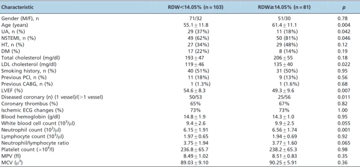
the cut-off RDW value ($14.0%vs,14.0%), we found that patients with higher RDW values were older and had higher LDL-C and cTnI values, a lower LVEF, a greater total number of diseased coronary arteries and lower MCH values than the patients with lower RDW values did (Table 3).
In the correlation analyses, the RDW was positively correlated with LDL-C (r = 0.21;p= 0.012) and cTnI (r = 0.19; p= 0.045) levels (Figure 1). Conversely, the RDW was
negatively correlated with the LVEF (r = -0.26; p= 0.0032).
Additionally, the RDW was significantly correlated with the WBC count (r = 0.18; p= 0.041) and the total number of
diseased coronary arteries (r = 0.25;p= 0.003). In the
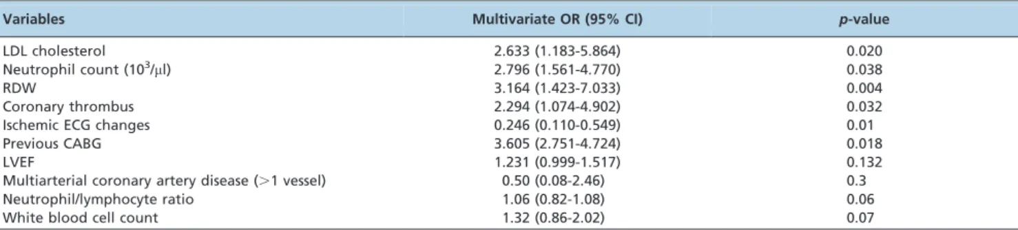
multi-variate analysis, multiarterial coronary disease, the WBC count, the neutrophil/lymphocyte ratio, ischemic ECG changes, the RDW, the neutrophil count and a history of CABG were independently correlated with NSTEMI in the patients admitted to the hospital with chest pain (Table 4). Regarding the diagnostic accuracy of the RDW, we calculated the composite score for sensitivity and specificity (i.e., the area under the curve (AUC)), as determined by the ROC curve analysis for the prediction of NSTEMI by the RDW. Overall, the AUC was 0.649 (95% confidence interval: 0.546-0.753; p,0.01), suggesting a modest model for the prediction of NSTEMI using the RDW. Using a cut-off value of 14.05%, the sensitivity and specificity of the RDW were 73% and 56%, respectively (Figure 2).
& DISCUSSION
The principal finding of our study was that the RDW might predict increased myocardial injury in patients with NSTE-ACS. In particular, in the study group, the degree of myocardial dysfunction (based on the LVEF) and the levels
of inflammatory mediators in the blood were associated with RDW values. Our study especially demonstrates an important relationship between RDW and increased cTnI levels in patients with NSTE-ACS.
The RDW has been reported to be a predictor of coronary heart disease events in different cardiovascular conditions and of all-cause mortality (9,10). In a study by Cavusoglu et al., the RDW was also found to be a strong independent predictor of all-cause mortality in the ACS subset of patients based on a multivariate analysis (11). In a study by Sandip et al. (12), a greater CAD risk category was associated with a linear increase in the RDW value, suggesting that the RDW is a potent predictor of CAD risk. In another study, Tonelli et al. (13) reported that among patients with CAD and without heart failure, mortality rates were significantly increased in patients with elevated RDW values compared with patients with RDW values within the normal range.
Chronic subclinical inflammation appears to be a poten-tial pathophysiologic mechanism underlying the association between the RDW and CAD and other cardiovascular events (14). Inflammation leads to anisocytosis due to the release of immature red blood cells into the peripheral circulation. The association between inflammation and an increased RDW is supported by findings of increased levels of CRP, interleukin-6 and soluble tumor necrosis factor receptors 1 and 2 in patients with elevated RDW values (15,16). In our study, we found that indicators of inflamma-tion such as the total leukocyte count and the neutrophil count were significantly correlated with higher RDW values. In addition to these markers, the neutrophil/ lymphocyte ratio and thrombus formation in diseased coronaries were significantly associated with higher RDW values.
Another mechanism suggested to underlie the association between the RDW and an increased frequency of CAD events is oxidative stress. High oxidative stress has been shown to be associated with an increased risk of cardiovas-cular events (17). Additionally, high oxidative stress reduces red blood cell survival, causes anisocytosis and promotes the release of premature red blood cells into the peripheral circulation (18).
In a study by Lippi et al. (19), researchers investigated the role of the RDW in patients with chest pain suggestive of ACS. These researchers reported that the combined mea-surement of cardiac troponin and the RDW at admission increased the already impressive sensitivity of cardiac troponin from 94% to 99% in diagnosing ACS. In another
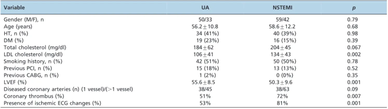
Table 1 -Baseline characteristics of the study population, stratified according to the final diagnosis of the study patients.
Variable UA NSTEMI p
Gender (M/F), n 50/33 59/42 0.79
Age (years) 56.2¡10.8 58.6¡12.2 0.68
HT, n (%) 34 (41%) 40 (39%) 0.98
DM (%) 19 (23%) 16 (15%) 0.39
Total cholesterol (mg/dl) 184¡62 204¡45 0.067
LDL cholesterol (mg/dl) 106¡41 134¡43 0.002
Smoking history, n (%) 42 (51%) 50 (50%) 0.78
Previous PCI, n (%) 15 (18%) 13 (13%) 0.52
Previous CABG, n (%) 1 (2%) 0 (0%) 0.35
LVEF (%) 55.6¡8.5 50.3¡9.6 0.001
Diseased coronary arteries (n) (1 vessel)/(.1 vessel) 38/45 38/63 0.09
Coronary thrombus (%) 51% 72% 0.007
Presence of ischemic ECG changes (%) 53% 81% 0.001
Table 2 -Blood parameters of the study population, stratified according to cardiac troponin I elevation.
Variable UA NSTEMI p Blood hemoglobin (g/dl) 14.4¡0.9 14.7¡2.6 0.95 White blood cell count (103/
ml) 9.1¡2.8 9.8¡2.4 0.055 Neutrophil count (103/
ml) 5.88¡1.92 6.8¡1.79 0.001 Lymphocyte count (103/
ml) 1.98¡0.65 1.95¡0.67 0.92 Neutrophil/lymphocyte ratio 3.25¡1.32 3.95¡1.91 0.032 Platelet count (6109/l) 229.9¡49.5 239.8¡70.4 0.98
MPV (fl) 8.41¡0.94 8.53¡0.95 0.35
MCV (m3) 89.4¡3.6 88.6¡8.9 0.36
Table 3 -Baseline characteristics of the entire cohort, stratified by the upper tertile of the baseline RDW values ($14.05%vs,14.05%).
Characteristic RDW,14.05% (n = 103) RDW$14.05% (n = 81) p
Gender (M/F), n 71/32 51/30 0.78
Age (years) 55.1¡11.8 61.4¡11.1 0.004
UA, n (%) 29 (37%) 11 (18%) 0.042
NSTEMI, n (%) 49 (62%) 50 (81%) 0.046
HT, n (%) 27 (34%) 29 (48%) 0.12
DM (%) 17 (22%) 8 (14%) 0.19
Total cholesterol (mg/dl) 193¡47 206¡55 0.18
LDL cholesterol (mg/dl) 119¡46 135¡40 0.022
Smoking history, n (%) 40 (51%) 31 (50%) 0.95
Previous PCI, n (%) 11 (18%) 9 (13%) 0.56
Previous CABG, n (%) 1 (1.3%) 1 (1.6%) 0.68
LVEF (%) 54.6¡8.3 49.3¡9.6 0.007
Diseased coronary (n) (1 vessel)/(.1 vessel) 50/53 25/56 0.011
Coronary thrombus (%) 65% 67% 0.82
Ischemic ECG changes (%) 73% 73% 1.00
Blood hemoglobin (g/dl) 14.8¡1.9 14.3¡1.0 0.95
White blood cell count (103/
ml) 9.4¡2.6 9.9¡2.5 0.055
Neutrophil count (103/
ml) 6.15¡1.91 6.56¡1.74 0.001
Lymphocyte count (103/
ml) 1.97¡0.65 1.94¡0.69 0.92
Neutrophil/lymphocyte ratio 3.75¡1.94 3.77¡1.60 0.065
Platelet count (6109/l) 236.8¡65.7 238.2¡65.3 0.98
MPV (fl) 8.49¡1.02 8.51¡0.83 0.35
MCV (m3) 89.03¡9.10 90.25¡5.91 0.36
study, by Cemin et al. (20), investigators studied the relationship between red blood cells, platelet morphology and AMI and also assessed whether they could supplement the role of traditional cardiac biomarkers in the early identification of patients with AMI. The researchers found that the RDW predicted AMI with statistical significance only in female patients. In our study, we found that among the clinical variables, ischemic ECG findings, LDL-C levels, coronary thrombus and a history of CABG were significant predictors of cardiac troponin increase in the patients with NSTE-ACS. Among the hematologic parameters, the WBC count and the RDW were significant predictors of increased cardiac troponin levels. The RDW is thus a simple and economical laboratory measurement that has relatively good diagnostic accuracy in predicting NSTEMI in patients with NSTE-ACS. Considering the fact that the specific
kinetics of troponin in the injured myocardium limit its usefulness within 2-4 h of symptom onset, baseline RDW measurement seems helpful for predicting myocardial injury at an earlier time point. We suggest that the RDW should be considered along with conventional cardiac markers for the prediction of NSTEMI in patients with NSTE-ACS and should serve as a guide for making appropriate treatment decisions.
Limitations
The main limitations of our study were the small number of study groups, the single-center design and inclusion limited to patients admitted to the intensive care unit. Because our study thus represents a very specific popula-tion, the results cannot be generalized. Increased RDW values are noted in clinical settings such as hemolysis;
Table 4 -Independent predictors of NSTEMI in a multivariate logistic regression analysis.
Variables Multivariate OR (95% CI) p-value
LDL cholesterol 2.633 (1.183-5.864) 0.020
Neutrophil count (103/
ml) 2.796 (1.561-4.770) 0.038
RDW 3.164 (1.423-7.033) 0.004
Coronary thrombus 2.294 (1.074-4.902) 0.032
Ischemic ECG changes 0.246 (0.110-0.549) 0.01
Previous CABG 3.605 (2.751-4.724) 0.018
LVEF 1.231 (0.999-1.517) 0.132
Multiarterial coronary artery disease (.1 vessel) 0.50 (0.08-2.46) 0.3
Neutrophil/lymphocyte ratio 1.06 (0.82-1.08) 0.06
White blood cell count 1.32 (0.86-2.02) 0.07
CI: confidence interval; OR: odds ratio.
Figure 2 -Receiver operating characteristic (ROC) curve analysis for prediction of NSTEMI by red blood cell distribution width (RDW).
increased red cell destruction after blood transfusion; and ineffective red cell production, such as during iron, vitamin B12, or folate deficiency. The RDW can also be increased in clinical conditions such as pregnancy, thrombotic thrombo-cytopenic purpura and inflammatory diseases (21-23). Only Hb levels were measured in the present study, whereas parameters such as iron, vitamin B12 and folate were not assessed. None of our patients received blood transfusion and no pregnant women or individuals with inflammatory diseases, thrombotic thrombocytopenic purpura, or malnu-trition were included.
A greater baseline RDW value is associated with myocardial injury; therefore, the RDW could be considered as an additional marker for evaluating patients with NSTE-ACS. RDW is also a widely available marker that, in contrast to other novel markers of cardiovascular risk, can be obtained at no additional cost.
& AUTHOR CONTRIBUTIONS
Tenekecioglu E was responsible for the collection of study data and manuscript writing and revision. Yilmaz M was responsible for manuscript writing and revision. Yontar OC was responsible for the statistical analyses and manuscript revision. Peker T was responsible for the collection of study data and statistical analyses. Karaagac K, Bekler A were responsible for the collection of study data and review of the literature. Ozluk OA was responsible for manuscript writing and revision. Agca FV, Senturk M, Aslan B performed the coronary angiography and collected the study data.
& REFERENCES
1. Hamm CW, Goldmann BU, Heeschen C, Kreymann G, Berger J, Meinertz T. Emergency room triage of patients with acute chest pain by means of rapid testing for cardiac troponin T or troponin I. N Engl J Med. 1997;337(23):1648-53, http://dx.doi.org/10.1056/NEJM199712043372302. 2. Antman EM, Tanasijevic MJ, Thompson B, Schactman M, McCabe CH,
Cannon CP, et al. Cardiac-specific troponin I levels to predict the risk of mortality in patients with acute coronary syndromes. N Engl J Med. 1996;335(18):1342-9.
3. James SK, Lindahl B, Siegbahn A, Stridsberg M, Venge P, Armstrong P, et al. N-terminal pro-brain natriuretic peptide and other risk markers for the separate prediction of mortality and subsequent myocardial infarction in patients with unstable coronary artery disease: a Global Utilization of Strategies To Open occluded arteries (GUSTO)-IV substudy. Circulation. 2003;108(3):275-81, http://dx.doi.org/10.1161/ 01.CIR.0000079170.10579.DC.
4. Apple FS, Wu AH. Myocardial infarction redefined: role of cardiac troponin testing. Clin Chem. 2001;47(3):37.
5. Dabbah S, Hammerman H, Markiewicz W, Aronson D. Relation between red cell distribution width and clinical outcomes after acute myocardial infarction. Am J Cardiol. 2010;105(3):312-7, http://dx.doi.org/10.1016/j. amjcard.2009.09.027.
6. Patel KV, Ferrucci L, Ershler WB, Longo DL, Guralnik JM. Red blood cell distribution width and the risk of death in middle-aged and older adults. Arch Intern Med. 2009;169(5):515-23, http://dx.doi.org/10.1001/ archinternmed.2009.11.
7. Azab B, Torbey E, Hatoum H, Singh J, Khoueiry G, Bachir R, et al. Usefulness of red cell distribution width in predicting all-cause long-term mortality after non-ST-elevation myocardial infarction. Cardiology. 2011;119(2):72-80, http://dx.doi.org/10.1159/000329920.
8. Tamhane UU, Aneja S, Montgomery D, Rogers EK, Eagle KA, Gurm HS. Association between admission neutrophil to lymphocyte ratio and outcomes in patients with acute coronary syndrome. Am J Cardiol. 2008;102(6):653-7, http://dx.doi.org/10.1016/j.amjcard.2008.05.006. 9. Centers for Disease Control and Prevention (CDC), National Center for
Health Statistics. National Health and Nutrition Examination Survey data. Available at: www.cdc.gov/nchs/nhanes.htm. Accessed on March 15, 2010.
10. Helfand M, Buckley DI, Freeman M, Fu R, Rogers K, Fleming C, et al. Emerging risk factors for coronary heart disease: a summary of systematic reviews conducted for the U.S. Preventive Services Task Force. Ann Intern Med. 2009;151(7):496-507, http://dx.doi.org/10.7326/ 0003-4819-151-7-200910060-00010.
11. Cavusoglu E, Chopra V, Gupta A, Battala VR, Poludasu S, Eng C, et al. Relation between red blood cell distribution width (RDW) and all-cause mortality at two years in an unselected population referred for coronary angiography. Int J Cardiol. 2010;141(2):141-6.
12. Zalawadiya SK, Veeranna V, Niraj A, Pradhan J, Afonso L. Red cell distribution width and risk of coronary heart disease events. Am J Cardiol. 2010;106(7):988-93, http://dx.doi.org/10.1016/j.amjcard.2010. 06.006.
13. Tonelli M, Sacks F, Arnold M, Moye L, Davis B, Pfeffer M. Relation between red blood cell distribution width and cardiovascular event rate in people with coronary disease. Circulation. 2008;117(2):163-8, http:// dx.doi.org/10.1161/CIRCULATIONAHA.107.727545.
14. Libby P, Ridker PM, Maseri A. Inflammation and atherosclerosis. Circulation. 2002;105(9):1135-43, http://dx.doi.org/10.1161/hc0902.104353. 15. Lippi G, Targher G, Montagnana M, Salvagno GL, Zoppini G, Guidi GC. Relation between red blood cell distribution width and inflammatory biomarkers in a large cohort of unselected outpatients. Arch Pathol Lab Med. 2009;133(4):628-32.
16. Forhecz Z, Gombos T, Borgulya G, Pozsonyi Z, Prohaszka Z, Janoskuti L. Red cell distribution width in heart failure: prediction of clinical events and relationship with markers of ineffective erythropoiesis, inflamma-tion, renal funcinflamma-tion, and nutritional state. Am Heart J. 2009;158(4):659-66, http://dx.doi.org/10.1016/j.ahj.2009.07.024.
17. Berliner JA, Navab M, Fogelman AM, Frank JS, Demer LL, Edwards PA, et al. Atherosclerosis: basic mechanisms. Oxidation, inflammation, and genetics. Circulation. 1995;91(9):2488-96.
18. Ghaffari S. Oxidative stress in the regulation of normal and neoplastic hematopoiesis. Antioxid Redox Signal. 2008;10(11):1923-40, http://dx. doi.org/10.1089/ars.2008.2142.
19. Lippi G, Filippozzi L, Montagnana M, Salvagno GL, Franchini M, Guidi GC, et al. Clinical usefulness of measuring red blood cell distribution width on admission in patients with acute coronary syndromes. Clin Chem Lab Med. 2009;47(3):353-7.
20. Cemin R, Donazzan L, Lippi G, Clari F, Daves M. Blood cells characteristics as determinants of acute myocardial infarction. Clin Chem Lab Med. 2011;49(7):1231-6. 2.
21. Clarke K, Sagunarthy R, Kansal S. RDW as an additional marker in inflammatory bowel disease/undifferentiated colitis. Dig Dis Sci. 2008; 53(9):2521-3, http://dx.doi.org/10.1007/s10620-007-0176-8.
22. Shehata HA, Ali MM, Evans-Jones JC, Upton GJ, Manyonda IT. Red cell distribution width (RDW) changes in pregnancy. Int J Gynaecol Obstet. 1998;62(1):43-6.