CRP Levels are Higher in Patients with ST Elevation Than Non-ST
Elevation Acute Coronary Syndrome
Syed Shahid Habib, Mohammad Ibrahim Kurdi, Zohair Al Aseri, Mohammad Owais Suriya
Department of Physiology, College of Medicine & KKUH, King Saud University, Department of Cardiology KKUH, King Saud University, Department of Emergency Medicine KKUH, King Saud University, Riyadh
Mailing address: Syed Shahid Habib • King Saud University - Riyadh - 11461 E-mail: [email protected]
Manuscript received April 18, 2009; revised manuscript received November 07, 2009; accepted December 16, 2009.
Abstract
Background: There is intense interest in the use of high-sensitivity C-reactive protein (hsCRP) for risk assessment. Elevated hsCRP concentrations early in acute coronary syndrome (ACS), prior to the tissue necrosis, may be a surrogate marker for cardiovascular co-morbidities.
Objective: Therefore we aimed to study different follow up measurements of hsCRP levels in acute coronary syndrome patients and to compare the difference between non-ST elevation myocardial infarction (NSTEMI) and ST myocardial infarction (STEMI) patients.
Methods: This is an observational study. Of the 89 patients recruited 60 patients had acute myocardial infarction (AMI). Three serial hsCRP levels at baseline on admission to hospital before 12 hours of symptom onset, peak levels at 36-48 hours and follow up levels after 4-6 weeks were analyzed and compared between non-ST elevation AMI and ST elevation AMI.
Results: STEMI patients had significantly higher BMI compared to NSTEMI patients. Creatine kinase myocardial bound (CKMB) and Aspartate aminotransferase (AST) levels were significantly higher in STEMI patients compared to NSTEMI patients (p<0.05). CRP levels at baseline and at follow up did not significantly differ between the two groups (p= 0.2152, p=0.4686 respectively). There was a significant difference regarding peak CRP levels between the two groups, as STEMI patients had significantly higher peak CRP levels compared to NSTEMI patients (p=0.0464).
Conclusion: STEMI patients have significantly higher peak CRP levels compared to NSTEMI patients. These data suggest that inflammatory processes play an independent role in the pathogenesis of myocardial infarction. Thus, CRP assessment may assist in risk stratification after myocardial infarction. (Arq Bras Cardiol 2011; 96(1): 13-17)
Keywords: C-reactive protein; acute myocardial infarction; acute coronary syndrome; inflammation.
Introduction
A large body of evidence suggests that inflammation plays a key role in the pathogenesis of atherosclerosis. The chronic inflammatory process can develop into an acute clinical event by the induction of plaque rupture, leading to acute coronary syndromes1. More than 20 large prospective trials
have shown that the inflammatory biomarker high-sensitivity C-reactive protein (hsCRP) is an independent predictor of future cardiovascular events, in addition to predicting the risk of incident hypertension and diabetes2.
In acute coronary syndromes, plaque rupture is induced by the inflammatory process in the atherosclerotic tissue. The pathogenesis of atherosclerosis is influenced by inflammatory mechanisms and different plasmatic markers of inflammation
have been studied. CRP has been the most extensively studied. Initially it was suggested that CRP was a by-stander3 marker
of inflammation, but subsequent works demonstrated that it was a risk marker in both acute coronary syndromes (ACS) and in patients with myocardial ischemia4,5.
CRP levels increase after acute myocardial infarction (AMI) but their changes in the process of an acute ischemic attack has been studied mainly in patients with non-ST elevation AMI6,7. Therefore, it is interesting to discuss the value of
follow-up measurements of hs-CRP in patients with coronary artery disease (CAD).
Therefore we aimed to study the differences in hsCRP levels in patients with two clinical forms of ACS of non-ST elevation myocardial infarction compared to ST myocardial infarction.
Patients and methods
December 2007. Consecutive eligible patients with either STEMI or NSTEMI who were admitted at King Khalid University Hospital were recruited. Of the 89 patients recruited, 60 had evidence of AMI on the basis of the aforementioned criteria. The other 29 subjects were used as a control group. Of these, 11 manifested signs of unstable angina, 8 had chronic ischemic heart disease, and 10 had non-ischemic diseases. The hs-CRP concentration of these patients with an acute coronary syndrome (ACS) was measured on admission to hospital before 12 hours of symptom onset, peak levels at 36-48 hours and follow up levels after 4-6 weeks.
This project was supported by College of Medicine Research Center (CMRC). The study protocol was approved by the Research Ethics Committee of CMRC. The individuals selected were informed about the details of the study and consent was obtained. Inclusion criteria included patients of any sex with both non-ST elevation acute coronary syndrome and ST elevation acute coronary syndrome. The diagnosis of myocardial infarction required the presence of at least 2
of these criteria (1) A history of characteristic prolonged ( ≥
30 min) pain or discomfort (2) Creatine kinase (CK) elevation
exceeding twice the upper limit of normal (or CK-MB ≥ 50%
of total CK). Presence of new Q waves or new abnormal ST-T features8.
Patients with STEMI were required to have: (1) continuous chest pain upon presentation, refractory to nitrates, and lasting
≥ 30 min; (2) ST-segment elevation of ≥ 0.2 mV in ≥ 2 contiguous precordial leads, or ≥ 0.1 mV in ≥ 2 contiguous
limb leads, or new (or presumably new) left bundle branch block on admission electrocardiogram; (3) presentation within the first 12 h from index pain. Patients with NSTEMI were required to have angina-like chest pain at rest in the last 24 h
lasting ≥ 5 min, with associated ST-segment depression of ≥ 0.1 mV in ≥ 2 contiguous leads upon presentation9.
Patient with (1) angina of secondary etiology, (2) recent surgery, (3) active infection, or chronic inflammatory diseases (thyroid disorders, acute infections, stroke, diabetic ketoacidosis, non-ketotic hyperosmolar diabetes, rheumatic diseases, chronic liver diseases, renal disorders, cancer and sepsis), (4) significant hepatic or renal dysfunction, and (5) malignancy, were not included as well as (5) individuals with body temperature of >37.8° C at admission, (6) those who had suffered a coronary or cerebral event in that same period, those with complete left bundle block, those with pacemaker rhythm, and those with serious aortic valve disease, obstructive hypertrophic cardiomyopathy, and subjects who were critically ill or with ongoing or recent (< 1 month) infectious diseases (7) patients with surgical procedures in last 3 months were excluded. We followed the guidelines of the American Heart Association for measurement, evaluation and expression of hsCRP10.
Fasting venous blood samples were analyzed for lipid levels, comprising total cholesterol (TC), Triglycerides (TG), Low density Lipoprotein (LDL) and High density lipoprotein (HDL). TC, TG, LDL and HDL were analyzed by an enzymatic colorimetric method. The equipment used was a Dimension autoanalyzer (USA) and the kits were also provided by the same company. Levels of hsCRP and Lp(a) were measured by turbidimetric assays with commercial kits (Quantex Lp(a)
supplied by BIOKIT, S.A., Barcelona, Spain) on a Hitachi 911 equipment (ROCHE diagnostics, USA). The kit had a working range from 0.10 to 20.0 mg/l for hsCRP. One important attribute of C-reactive protein is its stability over time and the availability of automated assay techniques. Besides, the new assays are very sensitive and provide measurement of C-reactive protein at levels substantially below those levels measured by other traditional methods. For Lp(a) the Limit of Quantification (LOQ) was 1.3 mg/dl and the Limit of Detection (LOD) was 0.4 mg/dl. The autoanalyzer used was Hitachi 911, manufactured by ROCHE diagnostics, USA.
Statistical analysis
The data were analyzed by the computer software program Statistical Package for Social Sciences (SPSS version 10, Chicago). Descriptive characteristics and the lipid profile of the study patients were calculated as Mean ± SD (Standard Deviation) or SEM (Standard error of mean) for continuous variables. Analysis of variance was used to assess differences in age, blood pressure, TC, LDL, HDL, TG and BMI. Data on hs-CRP, Lp(a) and cardiac enzymes, because of their extreme skewness, were analyzed by non-parametric Mann-Whitney U test and Wilcoxon (Kruskal-Wallis) test when comparing two or three groups, respectively. A p value of < 0.05 was considered statistically significant.
Results
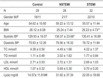
Clinical characteristics, lipid profile, Lp(a) and hsCRP levels of STEMI and NSTEMI patients are shown in table 1. There were non-significant differences regarding age and blood pressure levels between Control and CAD subjects. TC and LDL levels did not significantly differ between the two groups. Lp(a) levels were significantly higher in both STEMI and NSTEMI patients compared to control subjects, but the difference was non-significant between the two groups.
STEMI patients had significantly higher BMI compared to NSTEMI patients. We also observed significantly higher levels of hs-CRP in STEMI (1.59 ± 1.47) compared to NSTEMI (0.75 ± 0.99) patients (p = 0.0472).
Table 2 shows the differences in cardiac enzymes between the two groups. It was observed that Creatine kinase myocardial bound (CKMB) fraction and Aspartate aminotransferase (AST) levels were significantly higher in STEMI patients compared to NSTEMI patients (p<0.05).
CRP levels at baseline and at follow up did not significantly differ between the two groups (p= 0.2152, p=0.4686 respectively). There was a significant difference in peak CRP levels between the two groups, as it was significantly higher in STEMI patients when compared to NSTEMI patients (p = 0.0464) [Figure 1].
Discussion
Table 2 -Peak cardiac enzyme levels in ACS patients with STEMI compared to NSTEMI
Cardiac enzymes
IU/l NSTEMI STEMI
Troponin T 0.95 ± 1.51 2.61 ± 2.76
CKMB 111.75 ± 44.33 205.39 ± 152.15*
AST 38.00 ± 31.13 95.00 ± 72.66*
LDH 188.38 ± 92.63 309.00 ± 213.02
Creatine kinase myocardial bound (CKMB), Aspartate aminotransferase (AST), Lactate dehydrogenase (LDH). Differences were studied by Mann-Whitney -test. Data is expressed as Mean ± SD. *p<0.05 versus NSTEMI.
Table 1 - Clinical characteristics of ACS patients with STEMI compared to NSTEMI
Control NSTEMI STEMI
N 29 28 32
Gender M/F 18/11 21/7 22/10
Age 54.62 ± 10.60 59.22 ± 13.12 55.57 ± 11.44
BMI 26.12 ± 6.08 25.24 ± 7.44 29.23 ± 4.73**
Systolic BP 129.93 ± 19.07 136.37 ± 23.68* 130.41 ± 16.88
Diastolic BP 75.83 ± 12.26 79.56 ± 18.32 76.72 ± 11.84
TC mmol/l 4.38 ± 0.50 4.49 ± 1.66 4.22 ± 1.37
TG mmol/l 1.11 ± 0.49 2.02 ± 1.62 1.77 ± 0.84
LDL mmol/l 2.71 ± 0.53 2.72 ± 1.31 2.69 ± 1.03
HDL mmol/l 1.07 ± 0.32 0.69 ± 0.30 0.70 ± 0.20
Lp(a) mg/dl 14.57± 11.81## 31.92 ± 37.34 22.05 ± 18.66
Body mass index (BMI), Systolic blood pressure (SBP), Diastolic blood pressure (DBP), Total cholesterol (TC), Triglycerides (TG), Low-density lipoprotein (LDL) and high-density lipoprotein (HDL) and lipoprotein(a) [Lp(a)]. Differences were studied by Kruskal-Wallis -test for Lp(a) and ANOVA for the other parameters. Data is expressed as Mean ± SD; *p<0.05 versus STEMI & Control; **p<0.01 versus NSTEMI & Control; ## p<0.01 versus NSTEMI & STEMI.
A similarly designed study in ACS patients reported that although CRP levels on admission were similar in all groups, the pattern of CRP release and peak levels observed was clearly different in STEMI versus NSTEMI patients. Peak CRP level was 67 (36-112) mg/l in the STEMI group, 29 (20-87) mg/l in the NSTEMI group and 18 (12-36) mg/l in the unstable angina group. In our study, the difference in CRP levels was significant between STEMI and NSTEMI patients at peak levels only, but the difference was non-significant in the baseline and follow up readings. This suggests that it might be influenced by the degree of early myocardial tissue necrosis. Therefore, this variation in CRP kinetics should be taken into consideration when designing future studies12.
Figure 1 -Comparison of mean CRP levels at baseline, peak and at 4-6 weeks of follow up in all ACS, NSTEMI and STEMI patients. Differences were studied by Mann-Whitney -test.
Baseline Peak Follow up
All ACS 13.9 18.6 8.8
NSTEMI 10.6 13.5 10.1
STEMI 15.9 22.9 7.8
Brunetti et al13 reported that plasma CRP concentrations
showed a different release curve with Q-wave AMI in comparison with non Q-wave AMI and patients with UA. CRP peak concentrations did not correlate with ejection fraction and angiographic findings, but correlated with the incidence of major adverse cardiac events (MACE). The higher increase in CRP levels during Q-wave MI than in non Q-wave MI seems to be linked to the extension of myocardial damage, rather than to the pre-existing inflammation13.
The higher the maximum CRP recorded, the more severe the infarction suffered, the greater the likelihood of ventricular remodeling, the lower the ejection fraction, and the greater the risk of heart failure, heart rupture, and death14.
The results of the present study expand upon previous reports that demonstrated non-significant differences in CRP levels at baseline in patients with acute coronary syndromes, which tended to be higher in successive samples15.
Following an AMI, fibrinogen, CRP, and IL-6 levels are reported to be significantly higher in patients with complications, both as in-hospital and follow-up prognostic indicators16,17.
The level of C-reactive protein (CRP) can be used to identify patients with the most complicated coronary lesions and the greatest degree of intracoronary thrombosis, but it can also help identify patients with apparently non-complex lesions that are susceptible to rupture - a problem that would lead to patient instability18-20.
In a study by Jahn et al21, most events during a 3-year
observation period occurred in patients with follow-up levels of hs-CRP > 60% of the initial level. Therefore, it was hypothesized that a repeated measurement of hs-CRP levels in CAD patients could help to discriminate those at high risk of further events21.
CRP measurement has a lot of advantages. Firstly, it is a stable compound and secondly, it can be measured at any time of the day without regards to the biological clock. In contrast to results of cytokine measurements, such as IL-6, no circadian variation appears to exist for hs-CRP. Thus, clinical testing for hs-CRP can be accomplished without regard for the time of day22.
There is an intracardiac inflammatory response in ACS that appears to be the result of the evolution of myocardial necrosis
as shown by higher CRP, TNFα, IL-6 and Troponin T levels in patients with major adverse cardiac events, when compared to those without MACE23,24. This suggests that the systemic
inflammatory response may be the result of the evolution of myocardial infarction, thus showing a higher peak level in transmural infarction.
Further studies are needed to elucidate the inflammatory process in ACS, which may lead to novel therapeutic approaches and better application of currently available therapies. The present study can help us better understand the significance of CRP values, improve pharmacological therapies, and improve the design of research projects, assessing the prognostic significance of CRP levels over the ACS spectrum.
Conclusion
PeakCRP levels are significantly higher in STEMI patients, when compared to NSTEMI patients. These data suggest that inflammatory processes play an independent role in the pathogenesis of myocardial infarction. Thus, the measurement of CRP levels may assist in risk stratification after myocardial infarction.
Acknowledgement
The authors are thankful to Mr Mujeebul Haq for help in data collection and Mrs Ester for technical support.
Potential Conflict of Interest
No potential conflict of interest relevant to this article was reported.
Sources of Funding
This study was funded by College of medicine Research Center of kind Sand University.
Study Association
This study is not associated with any post-graduation program.
References
1. Libby P, Ridker PM, Maseri A. Inflammation and atherosclerosis. Circulation. 2002; 105 (9): 1135-43.
2. Ridker PM. C-reactive protein and the prediction of cardiovascular events among those at intermediate risk: moving an inflammatory hypothesis toward consensus. J Am Coll Cardiol. 2007; 49 (21): 2129-38.
3. Blake GJ, Ridker PM. Inflammatory bio-markers and cardiovascular risk prediction. J Intern Med. 2002; 252 (4): 283-94.
4. Zebrack JS, Anderson JL, Maycock CA, Horne BD, Bair TL, Muhlstein JB. Usefulness of high-sensitivity C-reactive protein in predicting long-term risk of death or acute myocardial infarction in patients with unstable or stable angina pectoris or acute myocardial infarction. Am J Cardiol. 2002; 89 (2): 145-9.
5. Topol EJ. A guide to therapeutic decision-making in patients with non-ST-segment elevation acute coronary syndromes. J Am Coll Cardiol. 2003; 41 (4 Suppl.): S123-9.
6. De Winter RJ, Bholasingh R, Lijmer JG, Koster RW, Gorgels JP, Schouten Y, et al. Independent prognostic value of C-reactive protein and troponin I in patients with unstable angina or non-Q-wave myocardial infarction. Cardiovasc Res. 1999; 42 (1): 240-5.
7. Ercan E, Tengiz I, Duman C, Onbasili OA, Baris N. Effect of tirofiban on C-reactive protein in non-ST-elevation myocardial infarction. Am Heart J. 2004; 147 (1): 54-7.
redefined–a consensus document of The Joint European Society of Cardiology/American College of Cardiology Committee for the redefinition of myocardial infarction. J Am Coll Cardiol. 2000; 36 (3): 959-69.
9. Braunwald E. Unstable angina: a classification. Circulation. 1989; 80 (2): 410-4.
10. Pearson TA, Mensah GA, Alexander RW, Anderson JL, Cannon RO 3rd, Criqui M, et al. Markers of inflammation and cardiovascular disease: application to clinical and public health practice. Circulation. 2003; 107 (3): 499-511.
11. Bursi F, Weston SA, Killian JM, Gabriel SE, Jacobsen SJ, Roger VL. C-reactive protein and heart failure after myocardial infarction in the community. Am J Med. 2007; 120 (7): 616-22.
12. Sánchez PL, Rodríguez MV, Villacorta E, Albarrán C, Cruz I, Moreiras JM, et al. Kinetics of C-reactive protein release in different forms of acute coronary syndrome. Rev Esp Cardiol. 2006; 59 (5): 441-7.
13. Brunetti ND, Troccoli R, Correale M, Pellegrino PL, Di Biase M. C-reactive protein in patients with acute coronary syndrome: correlation with diagnosis, myocardial damage, ejection fraction and angiographic findings. Int J Cardiol. 2006; 109 (2): 248-56.
14. Pietila KO, Harmoinen AP, Jokiniitty J, Pasternack AI. Serum C reactive protein concentration in acute myocardial infarction and its relationship to mortality during 24 months of follow-up in patients under thrombolytic treatment. Eur Heart J. 1996; 17 (9): 1345-9.
15. Auer J, Berent R, Lassnig E, Eber B. C-reactive protein and coronary artery disease. Jpn Heart J. 2002; 43 (6): 607-19.
16. Ziakas A, Gavrilidis S, Giannoglou G, Souliou E, Gemitzis K, Kalampalika D, et al. In-hospital and long-term prognostic value of fibrinogen, CRP, and IL-6 levels in patients with acute myocardial infarction treated with thrombolysis.
Angiology. 2006; 57 (3): 283-93.
17. Lindahl B, Toss H, Siegbahn A, Venge P, Wallentin L, for the FRISC Study Group. Markers of myocardial damage and inflammation in relation to long-term mortality in unstable coronary artery disease. N Engl J Med. 2000; 343 (16): 1139-47.
18. Zouridakis E, Avanzas P, Arroyo-Espliguero R, Fredericks S, Kaski JC. Markers of inflammation and rapid coronary artery disease progression in patients with stable angina pectoris. Circulation. 2004; 110 (13): 1747-53.
19. Arroyo-Espliguero R, Avanzas P, Cosín-Sales J, Aldama G, Pizzi C, Kaski JC. C-reactive protein elevation and disease activity in patients with coronary artery disease. Eur Heart J. 2004; 25 (5): 401-8.
20. Zairis MN, Lyras AG, Bibis GP, Patsourakos NG, Makrygiannis SS, Kardoulas AD, et al. Association of inflammatory biomarkers and cardiac troponin I with multifocal activation of coronary artery tree in the setting of non-ST-elevation acute myocardial infarction. Atherosclerosis. 2005; 182 (1): 161-7.
21. Jahn J, Hellmann I, Maas M, Giannitsis E, Dalhoff K, Katus HA. Time-dependent changes of hs-CRP serum concentration in patients with non-ST elevation acute coronary syndrome. Herz. 2004; 29 (8): 795-801.
22. Ewart HKM, Ridker PM, Rifai N, Price R, Dinges DF, Mullington JM. Absence of diurnal variation of C-reactive protein levels in healthy human subjects. Clin Chem. 2001; 47 (3): 426-30.
23. Cusack MR, Marber MS, Lambiase PD, Bucknall CA, Redwood SR. Systemic inflammation in unstable angina is the result of myocardial necrosis. J Am Coll Cardiol. 2002; 39 (12): 1917-23.