Dement Neuropsychol 2013 September;7(3):312-315
Case Report
312 Dissociation of depression from apathy Costa RQM, et al.
Dissociation of depression from
apathy in traumatic brain injury
A case report
Raquel Quimas Molina da Costa1, Fábio Henrique de Gobbi Porto2, Rogério Paysano Marrocos3
ABSTRACT. Although not evident clinically, lesions to the prefrontal cortex cause great social and functional impairment to patients. The anterior cingulate cortex is intimately involved with motivational behavior and after injury to this area the onset of an apathetic state can be observed. This paper describes the case of a patient with traumatic brain injury to the prefrontal lobe presenting with a depressive syndrome associated with apathetic symptoms. After appropriate treatment for depression, intense apathy was revealed, an irreversible sequelae of the traumatic brain injury, constituting the main barrier to the patient’s return of lifestyle and independence.
Key words: apathy, depression, traumatic brain injury.
DISSOCIAÇÃO DE DEPRESSÃO E APATIA EM LESÃO ENCEFÁLICA TRAUMÁTICA: UM RELATO DE CASO.
RESUMO. Apesar de não se destacarem ao olhar clínico, as lesões do córtex pré-frontal acarretam grande prejuízo funcional e social aos pacientes. O córtex do cíngulo anterior está intimamente envolvido com o comportamento motivacional e o que se observa após um dano a esta região é a instalação de um estado apático. Este trabalho visa relatar o caso de um paciente com lesão traumática do lobo pré-frontal que apresentou uma síndrome depressiva associada a sintomas apáticos. Após tratamento adequado da depressão foi possível perceber intensa apatia, sequela irreversível do traumatismo crânio encefálico, que se constituiu como principal obstáculo ao retorno do estilo de vida e independência do paciente.
Palavras-chave: apatia, depressão, lesão encefálica traumática.
INTRODUCTION
L
esions of the prefrontal cortex are often not easily recognized without a thorough neurological, cognitive and neuropsychiatric examination. Frequently, more evident ele-mentary neurologic deicit may mask the be-havioral symptoms. Although not evident in a casual encounter, lesions of the prefrontal cortex can cause great social and functional impairment in afected subjects, as is often reported by them and their relatives.1he prefrontal cortex can be subdivided into three major areas, each of which is pre-dominantly related to distinct cognitive func-tions. he orbitofrontal cortex (OFC) is asso-ciated with the regulation of social behaviors
and social cognition. Damage to its structures is associated with changes in personality, re-sulting primarily in disinhibition and disre-gard for social rules.1 he dorsolateral cortex
(DLC) is the region related to executive func-tions such as task organization, planning, hypothesis generation and decision making while the anterior cingulate cortex (ACC) is intimately involved with motivational behav-ior where damage to this region may result in lack of initiative, motivation and volition, with the onset of an apathetic state.1
Apathy has been deined in diferent ways in the literature, but lack of motivation can be identiied as the main feature in most of these
1MD. Universidade Federal do Estado do Rio de Janeiro (Unirio). Escola de Medicina e Cirurgia (EMC). Hospital Universitário Gaffrée e Guinle (HUGG). Departamento
de Medicina Especializada. Disciplina de Psiquiatria. Serviço de Psiquiatria.2MD. Behavioral and Cognitive Neurology Unit, Department of Neurology, and Cognitive
Disorders Reference Center (CEREDIC). Hospital das Clínicas of the University of São Paulo, São Paulo, SP, Brazil. 3MD, MSc. Universidade Federal do Estado do
Rio de Janeiro (Unirio). Hospital Universitário Gaffrée e Guinle (HUGG).
Raquel Quimas Molina da Costa. Rua Real Grandeza 108 / sala 314 – 22281-034 Rio de Janeiro RJ – Brazil. E-mail: [email protected] Disclosure: The authors report no conflicts of interest.
Dement Neuropsychol 2013 September;7(3):312-315
313 Costa RQM, et al. Dissociation of depression from apathy
deinitions.2 Motivation is understood as the direction,
intensity and persistence of goal-directed behaviors.3
Apathy may have several dimensions, such as motor apathy, causing akinetic mutism, cognitive apathy, de-creasing the curiosity and interest in learning, afective apathy, with reduced facial expression, emotional apa-thy, with reduced social interest and afection, and mo-tivational apathy causing decreased initiative. Lesions in the ACC impact motivational brain processing, the linking of external stimuli to the needs of the internal milieu.4 Bilateral damage to the ACC causes akinetic
mutism, in which patients are deeply impaired, rarely move and eat only when fed by others, the so-called apa-thetic abulic state.5 Unilateral injury causes less severe
apathetic abulic syndrome, but may present as transient akinetic mutism.5
Mood changes are also frequently seen in cases with prefrontal lesions6 and major depression is considered
a common sequela in traumatic brain injury. Depres-sion is associated with worse functional and cognitive recovery.7,8
Herein, we reported a case of a patient with traumat-ic injury to the prefrontal cortex, causing a dysexecutive syndrome, depressive symptoms and apathy. In this case, apathy was only evidenced by a thorough neuro-psychiatric and cognitive evaluation and the application of dedicated scales. We emphasize the diferences be-tween apathy and depression, review the neuroanatomy of regions linked to apathy and discuss the management of apathetic patients.
CASE REPORT
A 37-year-old right-handed man, without previous psy-chiatric history was evaluated in the outpatient neu-ropsychiatric unit. His history began eight years prior when he was struck by a train, resulting in a traumatic brain injury and multiple skull fractures. He was diag-nosed with left-side epidural hematoma and submit-ted to frontotemporal craniotomy for drainage. He re-mained in a coma for 32 days, slowly recovering his level of consciousness at the time.
In the irst interview, eight years after the acci-dent, he showed depressed mood and spoke little. He cried easily and reported unsatisfactory sleep. His wife, who only met him after the accident, said that he did not like to go out, had no interest in friends, ate only when someone prepared his food for him and could go all day long without eating. He watched television but was only interested in soccer matches. He had little initiative, spent many hours lying down “thinking” (as he described) and had no interest in sex. his pattern
of behavior intermittently changed to outbursts of ag-gression, stubbornness and irritability. he wife stated, however, that he used to be afectionate with her and their daughter.
In the following appointment, his brother reported that before the accident he was “playful”, had many friends, liked to hang out with them and was very cheer-ful. He loved playing football, was very afectionate and described by others as “a sweet person”. He noted that after the accident he had become cold emotionally, fat, lacked energy, was very aggressive and could not control his emotions, once even threatening his father. During the examination he was lucid and oriented, his language abilities were preserved and his general neurological exam was normal. Magnetic resonance image showed extensive injury to the left prefrontal DLC and ACC (Figure 1).
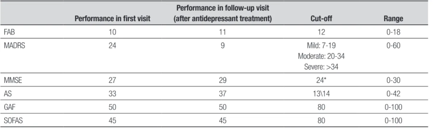
He was submitted to neurocognitive and functional evaluation and his performance is shown in Table 1.9-15
Performance on the Stroop Test (ST), Trail Making Test (TMT) and Wisconsin Card Sorting Test (WCST) was abnormal. He was treated for the depressive syndrome with nortriptyline 25 mg, twice a day. After 10 weeks of treatment for depression, he reported feeling better regarding sadness, had stopped crying and was sleeping well. He still had no interest in sex and only ate when his wife prepared his meal. His wife said that he did not in-ish daily tasks which he had started and could not do two
A
B
C
D
Dement Neuropsychol 2013 September;7(3):312-315
314 Dissociation of depression from apathy Costa RQM, et al.
things at the same time. She also said that he was less aggressive, but kept lying still for many hours, “without doing anything”. When asked about it, he didn’t show any critical judgment of his current condition. After 2 months of treatment, his wife was able to ind him a job in a local bus company. She reported that she needed to accompany him to work every day and this was the only way he was able to keep his job. He is still being followed and has remained stable ever since .
DISCUSSION
his case report shows the consequences of an injury to the prefrontal cortex causing apathy and depression si-multaneously. Although similar, the two entities are not the same.
Major depression is characterized by depressed mood, loss of interest or pleasure in nearly all activi-ties and symptoms such as sleep disturbances, appetite changes, decreased psychomotor activity, decreased en-ergy, diiculty concentrating and thoughts of worthless-ness or guilt.7,16 Some studies in the 1990s correlated a
higher incidence of depressive symptoms with lesions in the left frontal pole and in some cases, a signiicant cor-relation between the severity of the symptoms and the distance from the anterior edge of the cerebral lesion to the frontal lobe.1,8
Apathy is more closely linked to the “lack of moti-vation” concept.2 Motivation is understood as the
di-rection, intensity and persistence of goal-directed be-haviors.3 It can be evidenced by a reduction of directed
behavior, lack of energy, efort and need for external or-ders to carry tasks out. Concomitantly, changes in goal-direct cognition, for example, lack of interest in learning new things and loss of concern about personal problems also often occur. Behavioral abnormalities such as lack
of emotional reaction (both positive and negative) and the lat afect also occurs. Apathy is a clinical manifes-tation that can be measured by some dedicated scales such as the Apathy Scale proposed by Stekenstein and validated in Portuguese.2,5,9
he symptomatology related to apathy can be mis-diagnosed as depression. Sometimes, depression may cause some apathetic symptoms, but the two conditions are nosologically distinct. he importance of separating these syndromes hinges on the fact that depression and apathy have diferent types of management and prog-nosis. Depression has a better response to treatment, as seen in this patient. In fact, once treated, the mood disorder presented by the patient improved yet the apa-thetic symptoms became more evident and remained chronic. In this case, apathy was the main complaint re-ported by his family and his biggest obstacle to return-ing to an independent and workreturn-ing life.
In this patient, there was also great impairment in executive functions due to the extensive lesion to the DLC. A dysexecutive syndrome was noted through his low performance on the WCST, FAB, ST and TMT. Dur-Table 1. Patient performance before and after antidepressant treatment.
Performance in first visit
Performance in follow-up visit
(after antidepressant treatment) Cut-off Range
FAB 10 11 12 0-18
MADRS 24 9 Mild: 7-19
Moderate: 20-34 Severe: >34
0-60
MMSE 27 29 24* 0-30
AS 33 37 13\14 0-42
GAF 50 50 80 0-100
SOFAS 45 45 80 0-100
AS: Apathy scale9; FAB: Frontal assessment battery10; GAF: Global Assessment of Functioning11; MADRS: Montgomery and Asberg depression rating scale12; MMSE: Mini-mental statement examination13,14;
SOFAS: Social and Occupational Functioning Assessment Scale15. *According to educational level.
Table 2. Patient performance on executive function tests:
Test Patient Normative values
DSF 4 6 to 7
DSB 3 4 to 5
TMT A 37” (mean=35.8; SD =11.9)
TMT B 5’43’’ (mean=81.2; SD = 38.5)
ST 1 19.9’ (mean=12.56; SD=1.89)
ST 2 18.6’ (mean=16.16; SD=3.46)
ST 3 21.3’ (mean=31.32; SD=8.22)
Dement Neuropsychol 2013 September;7(3):312-315
315 Costa RQM, et al. Dissociation of depression from apathy
ing the interview, speciic complaints were identiied, such as “he does not inish the tasks he started”, “cannot do two things at the same time”, which are characteris-tic diiculties of dysexecutive syndrome.17
Due to the extensive prefrontal lesion, there were also behavior changes, such as aggressiveness and ex-plosive temper, which are typically related to the OFC. As traumatic brain injuries are often not selective to any prefrontal region, widespread damage usually occurs, generating a “blend” of frontal symptoms. Further-more, it is important to remember the presence of brain circuits between the prefrontal cortices, which further complicates the occurrence of a clinical presentation with symptoms unique to one or another region.1
A Japanese study published in 2011 evaluated de-pressive and apathetic symptoms in patients with traumatic brain injury and correlated them with the Functional Independence Measurement. he authors found that apathy was more strongly associated with a negative impact on the recovery of these patients than depressive symptoms.7 his patient has a lesion in the
left anterior pole of the frontal lobe, a site often related to the onset of depressive symptoms compared with le-sions in other areas or even in the right frontal pole.18,19
hese patients ind it very diicult to keep or even get a new job, because of the dysexecutive syndrome, lack of motivation and interest and also the diiculty in relating normally with colleagues.20 Sometimes this
is only possible with the continued efort of the family,
as seen in this case. he great social impairment and functional disability of the injuries involved are evident through relatively low GAF and SOFAS.
he appropriate investigation of cognitive impair-ment and patient behavior is the irst step to identify their limitations and guide a possible rehabilitation. Identifying these deicits is important for the family and the patient to understand these changes and know what to expect in the future. It is essential to provide adequate treatment for those manifestations that may show improvement with drug intervention and to iden-tify possible comorbid psychiatric disorders in order to take the necessary therapeutic approach. Often the damage is perceived months or years after the trauma but can cause long-term disability and major functional impairment.21
However, it is curious to note that these changes were not obvious or readily related to the trauma, as would be the case in a motor, aphasic or visual-perceptu-al lesion. he sequelae related to prefrontvisual-perceptu-al damage are diicult to measure, often requiring several question-naires, scales and tests to be able to draw a parallel be-tween the anatomical lesion and functional impairment and consequently allow devising of a treatment and/or rehabilitation plan tailored to these. he introduction of drug therapy in this case exempliies that depres-sive symptoms are potentially treatable and therefore reversible. he same is not true however for apathetic symptoms.
REFERENCES
1. Cummings JL, Miller BL. The Human Frontal Lobes. 2nd ed., Guilford Press; 2007.
2. Starkstein SE, Leentjens FG. The nosological position of apathy in clini-cal practice. J Neurol Neurosurg Psychiatry 2008;79:1088-1092. 3. Marin R. Apathy: a neuropsychiatric syndrome. J Neurop sychiatry Clin
Neurosci 1991;3:243-254.
4. Mesulam MM. From sensation to cognition. Brain 1998;121:1013-1052.
5. Guimarães HC, Paes P, Fialho A. Brazilian caregiver version of the Apa-thy Scale. Dement Neuropsychol 2009;3:321-326.
6. Jorge ER, Robrinson G, Moser D. Major depression following traumatic brain injury. Arch Gen Psychiatry 2004;61:42-50.
7. Hama S, Yamashita H, Yamawaki S, Kurisu K. Post-stroke depression and apathy: Interactions between functional recovery, lesion location, and emotional response. Psychogeriatrics 2011;11:68-76.
8. Schwartzbold M, Diaz A, Martins E. Psychiatric Disorders and Trau-matic Brain Injury. Neuropsychiatr Dis Treat 2008;4:797-816. 9. Starkstein SE, Mayberg HS, Preziosi TJ, et al. Reliability, validity and
clinical correlate of apathy in Parkinsons’s disease. J Neuropsychiatry Clin Neurosci 1992;4:134-139.
10. Dubois B, Slachevsky A, Litvan I, Pillon B. The FAB: a Frontal Assess-ment Battery at bedside. Neurology 2000;55:1621-1626.
11. Hall RC. Global assessment of functioning. A modified scale. Psycho-somatics 1995;36:267-275.
12. Hamilton M. A rating scale for depression. J Neurol Neurosurg Psychia-try 1960;23:56-62.
13. Brucki SMD, Nitrini R, Caramelli P, Bertolucci PHF, Okamoto IH. Sug-estões para o uso do mini-exame do estado mental no Brasil. Arq Neu-ropsiquiatr 2003;61:777-781.
14. Folstein MF, Folstein SE, McHugh PR. Mini-Mental State: a practical method for grading the cognitive state of patients for clinician. J Psychi-atr Res 1975;12:189-198.
15. American Psychiatric Association. 2000. Diagnostic and statistical man-ual of mental disorders (DSM-IV-TR). 4th ed. Washington, DC: Ameri-can Psychiatric Association.
16. Jorge M. Manual Diagnóstico e Estatístico de Transtornos Mentais. 4th ed., Artmed; 2003.
17. Hanna-Pladdy B. Dysexecutive syndromes in neurologic disease. J Neurol Phys Therapy 2007;31:119-127.
18. Robinson RG, Szetel B. Mood change following left hemispheric brain injury. Ann Neurol 1981;9:447-453.
19. Lipsey R, Robinson R, Pearlson G. Mood change following bilateral hemisphere brain injury. Br J Psychiatry 1983:266-273.
20. Nicholl J, LaFrance WC. Neuropsychiatric sequelae of traumatic brain injury. Semin Neurol 2009;29:247-255.
![Figure 1. Brain MRI. [A, B] Axial FLAIR. [C, D] coronal T2WI. Reveals a large area of encephalomalacia and surrounding gliosis involving left frontal lobe](https://thumb-eu.123doks.com/thumbv2/123dok_br/15188245.527006/2.892.484.829.683.1037/figure-coronal-reveals-encephalomalacia-surrounding-gliosis-involving-frontal.webp)