Arq Neuropsiquiatr 2008;66(3-B):758-760
758
Clinical / Scientiic note
NEUROENDOSCOPIC TREATMENT OF QUADRIGEMINAL
ARACHNOID CYST IN A TWO-YEAR-OLD CHILD
João Paulo C. de Almeida
1, Saul Quinino
2, Igor Vilela Faquini
2, Danilo Otávio de A. Silva
2,
Lucas Alverne F. de Albuquerque
1, Jean Carlos de Araújo Mendes
2, Hildo Azevedo-Filho
3TRATAMENTO NEUROENDOSCÓPICO DE CISTO ARACNÓIDE QUADRIGEMINAL EM CRIANÇA DE DOIS ANOS DE IDADE
Department of Neurosurgery, Hospital da Restauração, Recife PE, Brazil: 1Medical School, Federal University of Ceara, Fortaleza CE, Brazil; 2
Depart-ment of Neurosurgery, Hospital da Restauração, Recife PE, Brazil; 3Professor and Chairman, Department of Neurosurgery, Hospital da Restauração,
Recife PE, Brazil.
Received 29 May 2008, received in inal form 21 July 2008. Accepted 28 July 2008.
Dr. João Paulo Cavalcante de Almeida – Rua Paulo Morais 130 - 60175-175 Fortaleza CE - Brasil. E-mail: [email protected] Arachnoid cysts of the posterior fossa represent a rare
group of central nervous system lesions. Quadrigeminal cistern cysts, speciically, are unusual lesions, with only 79 cases described in the English literature1-8. Classical
treat-ment of such lesions consists of craniectomy and fenes-tration of the lesion or cystoperitoneal shunting. Neuro-endoscopy represents a new effective minimally invasive approach for such lesions.
We report the case of a two year old boy who present-ed a quadrigeminal cyst arachnoid cyst which was success-fully treated by neuroendoscopy in our department.
CASE
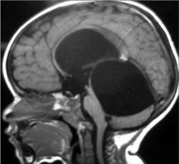
A two-year-old boy, presented to our department with one month history of daily headaches, vomiting and gait instabili-ty. There was no report of recent associated infections, previ-ous history of any classical childhood viral infection or alter-ations of psychomotor development. The exam at admission revealed a hypoactive child, obvious macrocrania and gait in-stability associated with lower limbs hypertonia. There was no papilloedema. There were no cranial nerves or sensitive altera-tions. Analysis of the cephalic perimeter growth curve showed mild upward deviation since the initial months of life. Computed tomography (CT) scan demonstrated supratentorial hydroceph-alus associated with a cystic lesion in the posterior fossa. Mag-netic resonance image (MRI) demonstarted enlargement of the supratentorial ventricular system secondary to a large quadri-geminal cistern arachnoid cyst compressing the brainstem, cere-bellum, aqueduct of Sylvius and fourth ventricle (Fig 1). A ventri-culo-peritoneal shunt was inserted at this time, with regression of the symptoms.
Seven months later, the patient returned to our department with history of new episodes of headache and vomiting. A new MRI revealed persistence of the arachnoid cyst. It was decided to perform endoscopic fenestration of the cyst and third ven-triculostomy for treatment of the hydrocephalus.
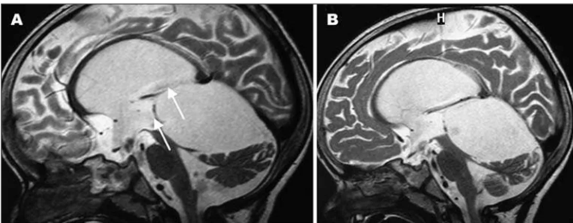
Informed consent was obtained from the family and the pa-tient underwent the surgical procedure. A paramedian incision was performed overlying the right coronal suture, 2.5 cm off the midline. Then a 14-mm burr hole was created and the dura-mater opened. After navigating through the right lateral ventricle and third ventricle, the arachnoid cyst was reached and cysto-ven-tricle shunting was realized. This was followed by a third ventric-ulostomy. There were no complications during the surgery. The patient presented no symptoms at time of discharge (Fig 2A).
At one year follow-up, the patient was asymptomatic. Neu-rological examination was unremarkable (Fig 2B).
DISCUSSION
Arq Neuropsiquiatr 2008;66(3-B)
759
Quadrigeminal arachnoid cyst Almeida et al.
cysts are all synonyms for quadrigeminal cistern arachnoid cyst1. Quadrigeminal cistern is a rare location for an
arach-noid cyst: only 53 cases were reported in the literature until 19992. Review of articles published in the last years
demonstrates that another 26 cases have been reported1,3-8.
Typical clinical presentation of a quadrigeminal cistern cyst includes headache, vomiting, visual complaints, and gait ataxia9. These symptoms are primarily caused by
pres-sure against the tectal plate, which can also compress the aqueduct of Sylvius and lead to hydrocephalus10.
Cerebel-lar and brainstem compression occasionally occur, as well10.
Quadrigeminal cysts appear as midline, supracerebel-lar, infratentorial cysts that abuts the quadrigeminal cistern on CT and MRI imaging with no enhancement and present-ing the same attenuation values as the cerebrospinal luid (CSF)11. The enlarging cyst projects downwards and
back-ward to lie over the superior surface of the cerebellum. The aqueduct and fourth ventricle are displaced down-wards and fordown-wards and tectal compression occurs2. The
third ventricle is distorted and anteriorly displaced. The aqueduct is stretched causing obstructive hydrocephalus.
Traditionally, arachnoid cysts of the quadrigeminal cis-tern have been treated in two ways, via craniotomy and fenestration and via cystoperitoneal shunting12. Although
open surgery allows arachnoid cysts to be exposed under direct vision and completely resected, it inevitably requires craniotomy. As many as one-third of those with arachnoid cysts and concomitant hydrocephalus who undergo open surgery require additional shunt placement for recurrent hydrocephalus13. Shunt placement without fenestration
of the cyst is an alternative treatment, but occlusion, dis-connection, or infection of the shunt system remains a potential threat to patients undergoing these procedures3.
In a review of the literature, Ruge et al. demonstrated the outcomes and complications of 48 quadrigeminal cis-tern arachnoid cysts treated by traditional techniques9.
Two patients treated with shunt died and three remained unchanged. Out of 23 patients treated with craniotomy and fenestration alone, one died and three remained with some degree of impairment (the outcome is unknown in 10 patients of this group); two of ive patients who were treated by craniotomy plus fenestration and shunt contin-ued to present some impairment after the procedures.
With the extent of the indications to ventricular en-doscopy, the neuroendoscopic approach to quadrigemi-nal cysts has gained an increasing rate of acceptance. Many authors have shown that limited openings of the cyst walls might be suficient to reestablish a functional CSF circulation and cyst size reduction. In this context, when compared to open surgery, neuroendoscopy has the main advantage to allow cysts to open into the ven-tricular system through a minimally invasive approach8.
Besides, endoscopic cystostomy associated with the third ventriculostomy presents a higher success rate than only endoscopic cystostomy for treatment of quadrigeminal cistern arachnoid cysts8.
Gangemi et al., studying 36 quadrigeminal cysts dem-onstrated a rate of cured or improved patients similar either treated by endoscopic fenestration or traditional surgery, respectively 87.5% (14/16) and 85% (17/20)14.
Tam-burrini et al., in a recent published series of 26 intra and paraventricular cysts, described the treatment of 11 quad-rigeminal cysts8. Six patients were initially treated only
by cistoventriculostomy and one patient was submitted to a second endoscopic cyst marsupialization. The other ive patients were initially treated by cistoventriculosto-my and endoscopic third ventriculostocistoventriculosto-my and one case required a second endoscopic cyst marsupialization. The nine symptomatic patients of this group had their symp-toms resolved after the procedure. Reduction of cyst size was obtained in all eleven patients. In this study there was no mortality or operative morbidity described8.
Arq Neuropsiquiatr 2008;66(3-B)
760
Quadrigeminal arachnoid cyst Almeida et al.
Neuroendoscopy, however, is not a risk-free proce-dure. The risks of neuroendoscopy include hemorrhage, with the inherent dificulty of controlling the bleeding, infection, and raised intracranial pressure from too much irrigation without proper venting13,15.
Endoscopic treatment appears as new, safe and secure alternative to open surgery and shunting in the manage-ment of quadrigeminal cistern arachnoid cyst. Results in the literature are comparable to the traditional methods with the beneit of low incidence of complications. Al-though, as a relatively new method, it is necessary a care-ful follow-up in order to provide evidence of long term eficiency of the endoscopic treatment for this uncom-mon lesion.
REFERENCES
1. Topsakal C, Kaplan M, Erol F, Cetin H, Ozercan I. Unusual arachnoid cyst of the quadrigeminal cistern in an adult presenting with apneic spells and normal pressure hydrocephalus: case report. Neurol Med Chir (Tokyo) 2002;42:44-50.
2. Hayashi N, Endo S, Tsukamoto E, Hohnoki S, Masuoka T, Takaku A. Endoscopic ventriculocystocisternostomy of a quadrigeminal cistern arachnoid cyst: case report. J Neurosurg 1999;90:1125-1128.
3. Inamasu J, Ohira T, Nakamura Y, et al. Endoscopic ventriculo-cysto-my for non-communicating hydrocephalus secondary to quadrigemi-nal cistern arachnoid cyst. Acta Neurol Scand 2003;107:67-71.
4. Rapana A, Bellotti A, Iaccarino C, Pascale M, Schonauer M. Intracra-nial pressure patterns after endoscopic third ventriculostomy: prelim-inary experience. Acta Neurochir (Wien) 2004;146:1309-1315. 5. Hayashi N, Hamada H, Umemura K, Kurosaki K, Kurimoto M, Endo
S. [Selection of surgical approach for quadrigeminal cistern arachnoid cyst]. No Shinkei Geka 2005;33:457-465.
6. Mitsos AP, Samelis AL, Panteleakou-Sameli HM, Kottas GD. Arachnoid cyst of quadrigeminal cistern presenting as trigeminal neuralgia.Acta Neurochir (Wien) 2006;148:93-94.
7. Kara B, Kayserili H, Imer M, Caliskan M, Ozmen M. Quadrigeminal cistern arachnoid cyst in a patient with Kabuki syndrome. Pediatr Neurol 2006; 34:478-480.
8. Tamburrini G, D’Angelo L, Paternoster G, Massimi L, Caldarelli M, Di Rocco C. Endoscopic management of intra and paraventricular CSF cysts. Childs Nerv Syst 2007;23:645-651.
9. Ruge JR, Johnson RF, Bauer J. Burr hole neuroendoscopic fenestration of quadrigeminal cistern arachnoid cyst: technical: case report. Neuro-surgery 1996;38:830-837.
10. Pagni CA, Canavero S, Vinci V. Left trochlear nerve palsy, unique symp-tom of an arachnoid cyst of the quadrigeminal plate: case report. Acta Neurochir (Wien) 1990;105:147-149.
11. Di Rocco C, Caldarelli M, Trapani G. Infratentorial arachnoid cysts in children. Childs Brain 1981;8:119-133.
12. Raffel C, McComb JG. To shunt or to fenestrate: which is the best sur-gical treatment for arachnoid cysts in pediatric patients? Neurosurgery 1988;23:338-342.
13. Fewel ME, Levy ML, McComb JG. Surgical treatment of 95 children with 102 intracranial arachnoid cysts. Pediatr Neurosurg 1996;25:165-173. 14. Gangemi M, Maiuri F, Colella G, Magro F. Endoscopic treatment of
quadrigeminal cistern arachnoid cysts. Minim Invasive Neurosurg 2005;48:289-292.