and Shock
Diego R. Barbosa Pereira, Fabio Augusto Pinton, Luiz Alberto Benvenuti
Instituto do Coração (InCor) HC-FMUSP, São Paulo, BrazilA 40-year-old man sought medical attention because of intense abdominal pain accompanied by profuse sweating (Nov. 2005).
He reported hypertension diagnosed at age of 35 and made use of atenolol 100 mg and aspirin 100 mg on a daily basis.
The tests taken at the time of diagnosis of hypertension (Oct. 2000) included echocardiogram revealing ascending aortic diameters of 42 mm; left atrium, 19 mm; right ventricle, 26 mm; left ventricle (diastole/systole), 59/33 mm; left ventricular ejection fraction was 75% and septal and left ventricular posterior wall thickness was 7 mm. We made the following echocardiographic diagnoses: dilated ascending aorta, bicuspid aortic valve without signs of stenosis and mild insufficiency.
Electrocardiogram (Aug. 2000) showed sinus rhythm and intraventricular stimulus conduction disorder (Fig. 1).
New echocardiographic assessment (in august 2004) revealed aortic diameters of 50 mm, left atrium, 42 mm; right ventricle, 20 mm; left ventricle (diastole/systole), 60/40 mm, ejection fraction, 70%. Septal and posterior wall 15 mm and 14 mm, respectively. Thus, there was left ventricular hypertrophy and ascending aorta ectasia. No valvopathy was found.
He had been asymptomatic for 10 months had, when short-duration palpitations appeared. There were no complaints of dyspnea, chest pain or syncope. The patient denied rheumatic history.
Physical examination (Jul 12) 2005; first visit to the hospital) revealed heart rate of 80 bpm; blood pressure, 160/80 mm Hg; presence of arterial pulsation in the neck; lung semiology revealed no changes; heart examination revealed stroke in the sixth intercostal space out of the left midclavicular line, extending two fingertips; heart sounds were normal and there was systolic murmur +/ 4+ in the aortic area and diastolic murmur aspiration +/ 4+ at the left sternal border. Abdominal
examination was normal and there was lower limb edema; neurological examination revealed left hemiparesis.
Chest radiography showed cardiomegaly at the expense of increased left ventricle +++/ 4+
Laboratory tests revealed 14.3 g/dL hemoglobin; hematocrit 44%; 7700 leukocytes/mm³; creatinine 0.7 mg/dL; potassium 4.4 mEq/L and sodium 139 mEq/L.
One month after that visit, he sought medical attention for palpitations that had begun four days before; atrial fibrillation was detected and warfarin and digoxin were added to the medications used. About four months after the first visit, the patient sought emergency medical attention for severe abdominal pain accompanied by profuse sweating. We carried out clinical and laboratory evaluation and the hypothesis of acute cholecystitis was ruled out.
Tomography with contrast (Nov. 18 2005) revealed moderate pleural effusion on the right and discrete on the left; enlarged liver, dilated portal and liver veins. The gallbladder was normal. The kidneys were normal and there were no changes in the aorta.
Ultrasound examination of the bladder was normal. A CT scan revealed right parietal cortical and subcortical hypoattenuation zone with effacement of adjacent sulci, compatible with old cerebral infarction. Additionally, there was opacification of the ethmoid sinuses.
Laboratory tests (Nov. 18 2005) revealed 13.9 g/dL hemoglobin; hematocrit 43%; leukocytes 23.380/mm³ without left deviation; platelets 135 000/mm³; prothrombin time (INR) 3.96; ratio of partial activated thromboplastin time 1,62; urea 74 mg/dL; creatinine 1.2 mg/dL; sodium 144 mEq/L; potassium 4.5 mEq/L; glucose 26 mg/dL; lactate 52 mg/dL; amylase 33 U/L; alkaline phosphatase 213 U/L; AST 2794 U/L; ALT 797 U/L; total bilirubin 4.1 mg/dL; direct bilirubin 2.9 mg/ dL; creatine phosphokinase 285 U/L, MB fraction of creatine phosphokinase 8.2 mg/dL; arterial pH 7.38; PaO2 97.7 mmHg;
PaCO2 24.6 mm Hg; O2 saturation 97.3%; blood bicarbonate 13.4 mEq/L; base excess (-) 9.4 mEq/L; albumin 2.9 g/dL and globulin 2.6 g/dL.
Diagnoses of decompensated heart failure with hepatic ischemia were made and the patient was transferred to the Hospital (InCor).
Physical examination (early morning of 19 November 2005) revealed patient sedated with endotracheal intubation and mechanical ventilation, pulse of 160 beats per minute, blood pressure 120 mm Hg x 70; decreased breath sounds at both
Mailing address: Vera D. Aiello •
InCor – Av. Dr. Enéas de Carvalho Aguiar, 44 – 05403-000 – São Paulo, SP E-mail: [email protected]
Keywords
Hypertension; aortic valve insufficiency; cardiomegaly; heart failure.
Editor: Alfredo José Mansur ([email protected])
hemithoraxes, heart sounds were arrhythmic, and there was systolic murmur ++/6 in the aortic area.
ECG (November 19 2005) revealed atrial fibrillation with ventricular rate averaged at 150 bpm, QRS duration 95 ms, low voltage QRS complex in the frontal plane, intraventricular stimulus conduction disorder and ventricular repolarization abnormalities (Fig. 2). There was reversal of atrial fibrillation to sinus rhythm a few hours later and low voltage in the frontal plane complex and ventricular repolarization abnormalities (Fig. 3).
Transthoracic echocardiogram (Nov. 19 2005), revealed aorta with 47 mm diameter; left atrium, 45 mm; left ventricle diastolic diameter of 51 mm. Left ventricle showed marked diffuse hypokinesia; right ventricle was dilated and markedly hypokinetic. There were also biatrial enlargement and mild aortic regurgitation, and dilatation of the sinus of Valsalva and discreet pericardial effusion.
Transesophageal echocardiogram (Nov. 19 2005) revealed dilated left ventricle with moderate dysfunction;
moderate right ventricular dysfunction; moderate aortic regurgitation, atrial septal defect with left to right flow and akinesis of the inferoposterior wall and signs suggestive of a fistula from the aorta to the right ventricle.
Chest radiography (Nov. 19 2005) revealed cardiomegaly +++/4+ and pulmonary congestion.
New laboratory tests (Nov. 19 2005) revealed hemoglobin 12.6 g/dL; hematocrit 40%; 19.300/mm³ leukocytes (63% neutrophils, 29% lymphocytes and 8% monocytes); platelets 146.000/mm3; urea 106 mg/dL, creatinine 1.2 mg/dL;
amylase 55 U/L, gamma GT 36 U/L; AST 868 U/L; ALT 514 U/L. Urine test revealed mild hematuria 75.000/mL and leukocyturia 75.000/mL. Blood cultures for aerobic and anaerobic germs were negative.
The patient developed shock and need for vasoactive drugs, and hyperthermia (39°C). Infective endocarditis was clinically suspected and oxacillin and ceftriaxone were administered. The patient went into shock and presented cardiac arrest in asystole. With resuscitation, ventricular
Figure 2 – ECG. Atrial ibrillation, rhythm conduction disorder, left ventricular hypertrophy.
fibrillation became unresponsive and the patient died (November 19 at 09:50 p.m. 2005).
Clinical aspects
Male patient, 40 years old, with a history of longstanding hypertension and echocardiography (ECHO) showing enlarged left atrium, impaired left ventricular hypertrophy (LVH) and size of aortic root dilation in the last four years. According to the latest reports found in the emergency service, the patient became symptomatic in the last 10 months, starting with palpitations and left hemiparesis, which can be explained by the presence of thromboembolic events due to paroxysmal atrial fibrillation (AF) or dilated cardiomyopathy because the chest radiograph revealed cardiomegaly at the expense of increased left ventricle (LV).
Shortly thereafter, this arrhythmia was confirmed after performing an electrocardiogram (ECG) in the emergency department.
The complaint that led to his admission was composed mainly of abdominal pain and profuse sweating, which normally characterizes syndromic diagnosis of acute abdomen. In addition to considering the most common causes such as cholecystitis, appendicitis, diverticulitis, and pyelonephritis in a patient with atrial fibrillation, heart failure and known aortic disease, we cannot avoid thinking of vascular acute abdomen.
Abdominal ultrasound, however, ruled out any change in the aorta, kidneys and gallbladder.
Laboratory tests showed more than 60x increase of transaminases, especially AST, as well as important evidence of hepatocytic failure, since the glycogen stores were depleted and the synthesis of albumin, bilirubin and clotting factors, were impaired. The causes of acute liver failure should include: hepatotoxic drugs, acute alcoholic hepatitis, viruses (hepatitis A, B and D), autoimmune diseases and ischemia1.
In this case, there are no reports of alcohol consumption. Besides, increase of transaminases, with slightly enlarged canalicular enzymes, make this diagnosis less likely.
Although there is no report of results of serological tests for hepatitis, the predominance of AST over ALT is not common in acute viral hepatitis. There is also no history of excessive use of acetaminophen or a history consistent with autoimmune disease.
Arterial blood gas analysis shows metabolic acidosis, respiratory alkalosis, with large consumption of bicarbonate and increased lactate, thus representing a state of poor tissue perfusion.
At this point, the hypothesis of intestinal ischemia should be considered as it falls within the patient’s clinical and laboratory profile. Some thrombus from the heart chambers could occlude vascular structures representing abdominal perfusion, such as hepatic or mesenteric artery. The literature shows that the main causes of intestinal ischemia are cardioembolic, and in patients with atrial fibrillation and dilated cardiomyopathy2. Arterial thrombosis occurs in
chronic artery diseases, with significant atherosclerosis, and
affects the proximal arteries, whereas occlusion by emboli occurs in more distal portions of the vessel. Mesenteric ischemia is associated with vasospasm secondary to drug use (cocaine and ergot) or low flow states, where there is loss of autoregulation dysfunction, such as in cardiogenic shock3. The main findings on physical examination are:
abdominal distension, absence of peritoneal irritation and decreased bowel sounds, i.e., the disproportion between the pain reported by the patients with physical findings of the abdomen. Markers of intestinal necrosis, such as elevation of CPK, LDH, hemoconcentration, hypernatremia, increased lactate are commonly found. CT angiography of the abdomen is a good test to confirm the diagnosis with a sensitivity of up to 90%4.
CT with contrast does not describe arterial vascular changes, although there are no reports of CT in the arterial phase. Pleural effusion especially on the right, and enlarged liver with major dilation of the hepatic portal veins. The Budd Chiari5 syndrome (thrombosis of suprahepatic veins)
could justify the presence of ischemic hepatitis and hepatic veins dilation described above, but would not explain the presence of pleural effusion which, as it is greater on the right, directing us to a condition which affects the heart and liver, reflecting in hepatic structures such as acute right heart failure.
One day after admission to the emergency room, there was significant clinical deterioration with respiratory failure and endotracheal intubation. A new ECG showed low voltage complexes in the frontal place and ventricular repolarization, which prompted another ECO. This, in turn, showed no significant pericardial effusion and brought right ventricle (RV) markedly hypokinetic, significant LV hypokinesis especially in the postero-inferior wall, and moderate aortic regurgitation and dilatation of the sinus of Valsalva. Soon after that, transesophageal echocardiography confirmed the transthoracic findings and also showed septal flow from left to right and signs suggestive of a fistula from the aorta to the RV.
Based on these new findings, the hypothesis of a ruptured sinus of Valsalva aneurysm becomes possible.
The sinus of Valsalva aneurysm is a rare congenital d i s e a s e , a c c o u n t i n g f o r a b o u t 0 . 1 % o f c a r d i a c malformations, and occurs mainly in eastern men6,7. It
follows from the weakening of the aortic wall, resulting from interruption of the vascular middle layer. It is located in the LV — Aorta transition, especially in the topography of the right sinus of Valsalva. Concurrent findings are: bicuspid aortic valve, ventricular septal defect, subaortic membrane and aortic coarctation.
If rupture does not occur, the aneurysm usually causes no symptoms, although there may be complications such as obstruction of RV outflow, coronary compression, conduction system abnormalities, stenosis or tricuspid and aortic regurgitation, thromboembolic events and infective endocarditis8.
hepatitis often happens. Infective endocarditis and stroke are additional complications. On examination, a continuous murmur that increases during diastole draws attention, when the fistula opens into the RV, and thrill along the right sternal edge. Surgical treatment is mandatory, with the closure of the aneurysm through suture or patching, and a direct suture or suture through prosthesis from the aorta to the heart. If this is not enough, the aortic valve should also be replaced9.
The patient presented clinical worsening rapidly. The patient had fever, leukocytosis and hematuria, which suggests that the cause of death was an association between septic shock and cardiogenic shock, and the focus of infection was infective endocarditis. (Dr. Diego R. Barbosa Pereira; Dr. Fabio Augusto Pinton)
Diagnostic hypotheses
Mesenteric ischemia.
Ruptured abdominal aortic aneurysm. (Dr. Diego R. Barbosa Pereira; Dr. Fabio Augusto Pinton)
Autopsy
The heart was enlarged and weighed 622 g. There was hypertrophy and dilation of the four heart chambers,
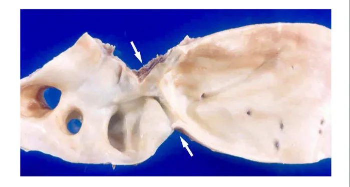
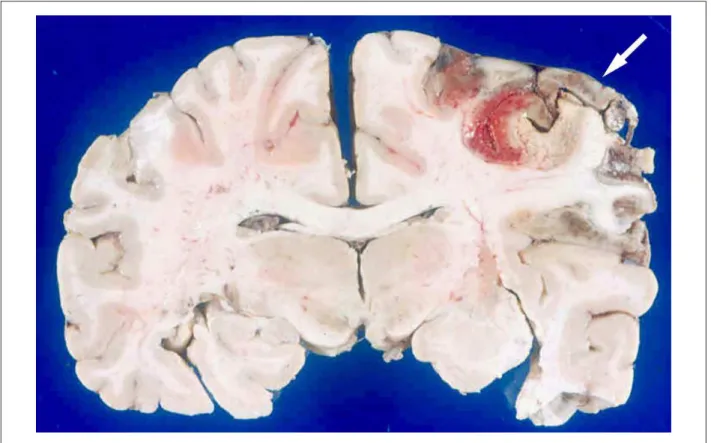
bicuspid, presenting fibrous thickening and fenestrations along the left free edge of the semilunar leaflets in the region of the lunula, and collapse of the right lunate, which also had a circular hole in the lunula measuring 0.5 cm; there was no thrombus, calcification or vegetation (Fig. 4). The right coronary sinus of Valsalva presented aneurysm formation measuring 3.8 x 2.0 cm (Fig. 4), which continued with multibosselated saccular structure, measuring 1.8 x 1.5 cm, projected into the right atrium, in its basal region near the tricuspid valve annulus (Fig 5). There were no perforations, lacerations or thrombi of the structures described above. The examination of the aorta showed severe coarctation of the aortic arch at the isthmus, pervious to 0.8 cm (Fig. 6). The brain weighed 1516 g and presented an extensive recent infarction, cortical and subcortical, confirmed by histological examination, located on the right parietal region (Fig. 7). There was no significant atherosclerosis of the aorta, coronary arteries or the arteries of the circle of Willis. The lungs showed chronic passive congestion and areas of alveolar edema. There was also widespread visceral congestion and centrilobular hemorrhagic necrosis of the liver. (Dr. Luiz Alberto Benvenuti)
Anatomical-pathological diagnoses
Bicuspid aortic valve failure; coarctation of the aortic arch; aneurysm of the right coronary sinus of Valsalva
Figure 4 – Macroscopic view of the bicuspid aortic valve and aortic root. The left semilunar lealet has fenestrations and freeboard thickening, with median raphe (*);
Figure 5 – View of the right heart chambers, in the atrioventricular transition. Aneurysm of the right sinus of Valsalva extends with full multibosselated saccular structure, protruding into the right atrium, at the level of the tricuspid valve annulus (arrow).
Figure 7 – Extensive recent infarction of the right parietal region of the brain (arrow).
with congestive heart failure; recent cerebral infarction, probably of embolic nature (cause of death). (Dr. Luiz Alberto Benvenuti)
Comments
Patient who was diagnosed with hypertension at 35 years of age and who developed aortic valve failure (bicuspid valve), progressive heart failure and cardiomegaly. In the terminal phase, the patient had fever, and suspected infective endocarditis. In addition to bicuspid aortic valve failure, the necropsy revealed coarctation of the aortic arch and aneurysm of the right coronary sinus of Valsalva. Death was due to progressive heart failure, with no infection.
Coarctation of the aortic arch is one of the secondary causes of hypertension and should always be investigated in young patients, as in our case. Correct diagnosis is very important because blood pressure tends to normalize after correction of the aortic defect10. In turn, bicuspid
which may be traditionally associated with coarctation of the aortic arch, but also several other malformations11.
However, triad bicuspid aortic valve, coarctation of the aortic arch and sinus of Valsalva aneurysm, as presented by the patient in question is considered rare12. A ruptured
sinus of Valsalva aneurysm, with the establishment of a fistula between the aorta and the right atrium or right ventricle is the cause of heart failure, which may cause the patient’s death13. However, in this case, the aneurysm
was intact and the death was due to progressive heart failure secondary to aortic valve insufficiency, probably worsened by the theft of blood flow during the diastole filling the aneurysm.
Treatment of aorta coarctation and sinus of Valsalva aneurysm is surgical and may occur at once or in two steps. Hybrid treatment has been recently described, with surgical closure of the aneurysm and subsequent percutaneous treatment of coarctation, using stent14. (Dr.
References
1. Dufour DR. Evaluation of liver function and injury. In: Henry JB (ed). Clinical diagnosis and management by laboratory methods. 20th ed. Philadelphia: WB Saunders; 2001. p. 264-80.
2. Sreenarasimhaiah, J. Diagnosis and management of intestinal ischemic disorders. BMJ. 2003;326(7403):1372-6.
3. Olderburg WA, Lau LL, Rodenberg TJ, Edmonds HJ, Burger CD. Acute mesenteric ischemia: a clinical review. Arch Intern Med. 2004;164(10):1054-62.
4. Kailidou E, Pikoulis E, Katsiva V, Papaconstantinou I, Athanassopoulou A, Gougoudi E, et al. Acute segmental intestinal ischemia: diagnosis with spiral computed tomography. JBR-BTR. 2006;89(2):72-6.
5. Horton JD, San Miguel FL, Membreno F, Wright F, Paima J, Foster P, et al. Budd-Chiari syndrome: illustrated review of current management. Liver Int. 2008;28(4):455-66.
6. Valdes-Cruz LM. Echocardiographic diagnosis of congenital heart disease. Philadelphia: Lippincott-Raven; 1999. p. 372-5.
7. Tanabe T, Yokota A, Sugie S. Surgical treatment of aneurysms of the sinus of Valsalva. Ann Thorac Surg. 1979;27(2):133-6.
8. Dong C, Wu QY, Tang Y. Ruptured sinus of valsalva aneurysm: a Beijing experience. Ann Thorac Surg. 2002;74(5):1621-4.
9. Shah RP, Ding ZP, Ng AS, Quek SS. A ten-year review of ruptured sinus of valsalva: clinico-pathological and echo-Doppler features. Singapore Med J. 2001;42(10):473-6.
10. Maia MM, Aiello VD, Barbero-Marcial M, Ebaid M. Coarctation of the aorta corrected during childhood: clinical aspects during follow-up. Arq Bras Cardiol. 2000;74(2):167-80.
11. Yuan SM, Jing H. The bicuspid aortic valve and related disorders. Sao Paulo Med J. 2010;128(5):296-301.
12. Hakami A, Stiller B, Hetzer R. Unruptured congenital aneurysm of the left sinus of Valsalva in an adult with complex left heart malformations. Heart. 2003;89(1):e3.
13. Mujanovic E, Kabil E, Bergsland J, Stanimirovic-Mujanovic S, Caluk J. Ruptured aneurysm of the noncoronary sinus of valsalva into the right atrium. Med Arch. 2010;64(5):307-8.