Alexandra Maria Gomes da Silva Gonçalves
Life after Transcatheter Aortic Valve Implantation:
a Clinical and Echocardiographic Appraisal
Dissertação de candidatura ao grau de Doutor Europeu apresentada à
Faculdade de Medicina da Universidade do Porto
Art.º 48º, § 3º - “A Faculdade não responde pelas doutrinas expendidas na dissertação.”
Corpo Catedrático da Faculdade de Medicina do Porto
PROFESSORES CATEDRÁTICOS EFECTIVOS
Doutor Manuel Alberto Coimbra Sobrinho Simões
Doutor Jorge Manuel Mergulhão Castro Tavares
Doutora Maria Amélia Duarte Ferreira
Doutor José Agostinho Marques Lopes
Doutor Patrício Manuel Vieira Araújo Soares Silva
Doutor Daniel Filipe Lima Moura
Doutor Alberto Manuel Barros da Silva
Doutor José Manuel Lopes Teixeira Amarante
Doutor José Henrique Dias Pinto de Barros
Doutora Maria Fátima Machado Henriques Carneiro
Doutora Isabel Maria Amorim Pereira Ramos
Doutora Deolinda Maria Valente Alves Lima Teixeira
Doutora Maria Dulce Cordeiro Madeira
Doutor Altamiro Manuel Rodrigues Costa Pereira
Doutor Rui Manuel Almeida Mota Cardoso
Doutor António Carlos Freitas Ribeiro Saraiva
Doutor José Carlos Neves da Cunha Areias
Doutor Manuel Jesus Falcão Pestana Vasconcelos
Doutor João Francisco Montenegro Andrade Lima Bernardes
Doutora Maria Leonor Martins Soares David
Doutor Rui Manuel Lopes Nunes
Doutor José Eduardo Torres Eckenroth Guimarães
Doutor Francisco Fernando Rocha Gonçalves
Doutor José Manuel Pereira Dias de Castro Lopes
PROFESSORES JUBILADOS OU APOSENTADOS
Doutor Abel Costa Sampaio da Costa Tavares
Doutor Abel Vitorino Trigo Cabral
Doutor Alexandre Alberto Guerra Sousa Pinto
Doutor Amandio Gomes Sampaio Tavares
Doutor António Augusto Lopes Vaz
Doutor António Carvalho Almeida Coimbra
Doutor António Fernandes da Fonseca
Doutor António Fernandes Oliveira Barbosa Ribeiro Braga
Doutor António Germano Pina Silva Leal
Doutor António José Pacheco Palha
Doutor António Luis Tomé da Rocha Ribeiro
Doutor António Manuel Sampaio de Araujo Teixeira
Doutor Belmiro dos Santos Patricio
Doutor Candido Alves Hipólito Reis
Doutor Carlos Rodrigo Magalhães Ramalhão
Doutor Cassiano Pena de Abreu e Lima
Doutor Daniel Santos Pinto Serrão
Doutor Eduardo Jorge Cunha Rodrigues Pereira
Doutor Fernando de Carvalho Cerqueira Magro Ferreira
Doutor Fernando Tavarela Veloso
Doutor Francisco de Sousa Lé
Doutor Henrique José Ferreira Gonçalves Lecour de Menezes
Doutor José Augusto Fleming Torrinha
Doutor José Carvalho de Oliveira
Doutor José Fernando Barros Castro Correia
Doutor José Luis Medina Vieira
Doutor José Manuel Costa Mesquita Guimarães
Doutor Levi Eugenio Ribeiro Guerra
Doutor Luis Alberto Martins Gomes de Almeida
Doutor Manuel Augusto Cardoso de Oliveira
Doutor Manuel Machado Rodrigues Gomes
Doutor Manuel Maria Paula Barbosa
Doutor Maria da Conceição Fernandes Marques Magalhães
Doutor Maria Isabel Amorim de Azevedo
Doutor Mário José Cerqueira Gomes Braga
Doutor Serafim Correia Pinto Guimarães
Doutor Valdemar Miguel Botelho dos Santos Cardoso
Doutor Walter Friedrich Alfred Osswald
Ao abrigo do Decreto–Lei nº 388/70 fazem parte desta dissertação
as seguintes publicações:
I. Gonçalves A, Zamorano J. Chapter 10 - Imaging for TAVI. Valvular Medicine Textbook 2011, Springer
US; in press
II. Goncalves A, Marcos-Alberca P, Zamorano JL. Echocardiography: guidance during valve
implanta-tion. EuroIntervention 2010;6 Suppl G:G14-9.
III. Goncalves A, Marcos-Alberca P, Almeria C, Feltes G, Rodriguez E, Hernandez-Antolin RA, Garcia
E, Maroto L, Fernandez Perez C, Silva Cardoso JC, Macaya C, Zamorano JL. Acute left ventricle diastolic
function improvement after transcatheter aortic valve implantation. Eur J Echocardiogr 2011;12:790-7.
IV. Goncalves A, Almeria C, Marcos-Alberca P, Feltes G, Hernandez-Antolin R, Rodriguez E, Silva
Cardoso JC, Macaya C, Zamorano JL. Three-Dimensional Echocardiography in Paravalvular Aortic
Regurgitation Assessment after Transcatheter Aortic Valve Implantation. J Am Soc Echocardiogr
2012;25:47-55.
V. Goncalves A, Marcos-Alberca P, Almeria C, Feltes G, Hernandez-Antolin RA, Rodriguez E, Rodrigo
JL, Cobiella J, Maroto L, Silva Cardoso JC, Macaya C, Zamorano JL. Quality of life improvement at
mid-term follow-up after transcatheter aortic valve implantation. Int J Cardiol 2011. [Epub ahead of print]
This research was performed in the Cardiovascular Institute, University Clinic San Carlos, Madrid, under
supervision of Professor José Luis Zamorano and co-supervision of Professor José Carlos Silva Cardoso.
To my family
To Professor José Zamorano
“A dream is just a dream. A goal is a dream with a plan and a deadline.”
CONTENTS
Part I
Introduction ... 1
Part II
Aims ... 9
Part III
Papers ... 13
1.
Imaging for TAVI ... 15
2.
Echocardiography: guidance during valve implantation ... 29
3.
Acute left ventricle diastolic function improvement after transcatheter
aortic valve implantation ... 37
4.
Three-Dimensional Echocardiography in Paravalvular
Aortic Regurgitation Assessment after Transcatheter Aortic Valve Implantation ... 47
5.
Quality of life improvement at midterm follow-up after transcatheter
aortic valve implantation ... 59
Attachment:
EAE/ASE recommendations for the use of echocardiography in
new transcatheter interventions for valvular heart disease ... 67
Part IV
Conclusions ... 97
Part V
Abstract ... 101
1.
Abstract ... 103
2.
Resumo ... 105
INTRODUCTION
Aortic stenosis (AS) is the most frequent native valve disease of the elderly in western industrialized
coun-tries.
1Once symptomatic it is responsible for serious disability, restrictions in the normal daily living and reduced
life expectancy.
2Until recently, the only effective treatment was surgical aortic valve replacement.
3-4However,
surgery is associated with higher morbidity and mortality rates in the elderly compared with younger patients.
5It is estimated that about 31% of patients with severe AS, older than 75 years are not referred to surgery, or
are refused by the cardiac surgeon because of co-morbidities and perceived high surgical risk.
6Transcatheter
aortic valve implantation (TAVI) techniques have emerged as an alternative treatment for this group of
pa-tients.
7-9After the first human TAVI procedure, performed by Alain Cribier in April 2002
10the number of
patients has increased exponentially and more than 40 000 patients had undergone TAVI worldwide.
11Good
procedural success and favourable clinical outcomes at short and midterm follow-up have been reported.
12-15Nevertheless, TAVI is a demanding procedure that requires a multidisciplinary team approach and still faces
important safety issues, such as paravalvular leaks, vascular complications, stroke or conduction disorders. An
additional challenge is the lack of operator direct visualization. Consequently, this technique greatly depends
on imaging, especially on echocardiography, which plays an essential role in identifying patients suitable for the
intervention, in providing intra-procedural monitoring and as primary modality for post-procedure follow-up.
16During peri-procedural guidance 2D and 3D transesophageal echocardiography (TEE) can contribute for
bal-loon and prosthesis positioning, to confirm prosthesis normal functioning and to rapidly detect complications.
17Moreover, TEE guidance represents a unique opportunity to firstly assess left ventricle physiology and
behavior following afterload release. Most patients referred for TAVI are elderly and present symptomatic AS
with heart failure in NYHA class III-IV, in spite of preserved left ventricle (LV) systolic function.
18-19Immediate
almost complete afterload normalization and significant clinical improvement at short-term follow-up have
been reported.
19-21At medium-term, LV mass reduction and improvement in tissue Doppler velocities have
also been described.
22In view of that we performed the first study evaluating the acute hemodynamic effects
of TAVI in diastolic performance, immediately after aortic valvuloplasty and prosthesis deployment.
23Aortic regurgitation is the most common complication after TAVI.
14,8It may occur as a consequence of
result, in spite of low incidence of prosthesis-patient mismatch,
20some degree of residual aortic
regurgita-tion (AR) is common, mainly at the paravalvular locaregurgita-tion.
12, 25The degree of AR appears to be minor in most
patients, however in some it may be responsible for left ventricular remodelling and moderate/severe AR
was shown to be a strong predictor of increased mortality and poor treatment response at midterm
follow-up.
26-27Therefore, accurate evaluation and detailed description of AR are essential for proper identification
of patients at risk and for appropriate follow-up, but up to now this remained challenging, as no systematic
methodology had been proposed. Bearing these concerns in mind, we developed a new method for accurate
description of paravalvular AR, based on three-dimensional (3D) transthoracic echocardiography evaluation,
suitable for accurate identification of moderate AR and proper 3D echocardiographic follow-up.
28Patients referred for TAVI are elderly and their expectations for the treatment are beyond procedural
success and favourable clinical outcomes.
19, 29Quality of life improvement is also a major concern for this
elderly patient’s profile.
7Clinical benefit and neurohormonal activation reduction has been reported at short
term after TAVI,
30but the midterm results of quality of life were scarce and the reports conflicting.
31-32Further than mortality and hemodynamic valve performance, quality of life assessment is crucial for the
evaluation of procedure efficiency and to guide clinical decision-making. Therefore, we looked into changes
in the quality of life of patients undergoing TAVI at a midterm follow-up.
Finally, considering the new demands placed by TAVI, a partnership between the European Association
of Echocardiography and the American Society of Echocardiography, developed the recommendations for
the use of echocardiography in new transcatheter interventions, providing an actual reference for
echocar-diographers participating in new transcatheter treatments for patients with valvular heart disease.
16Setting
This study included consecutive patients referred for TAVI at Hospital Clinico San Carlos, Madrid, from
April 2009 to April 2010. All patients had severe aortic valve stenosis (aortic valve area ≤1 cm
2) and were
References
1. Nkomo VT, Gardin JM, Skelton TN, Gottdiener JS, Scott CG, Enriquez-Sarano M. Burden of valvular heart diseases: A
popula-tion-based study. Lancet. 2006;368:1005-1011
2. Bouma BJ, van Den Brink RB, van Der Meulen JH, Verheul HA, Cheriex EC, Hamer HP, Dekker E, Lie KI, Tijssen JG. To operate
or not on elderly patients with aortic stenosis: The decision and its consequences. Heart. 1999;82:143-148
3. Vahanian A, Baumgartner H, Bax J, Butchart E, Dion R, Filippatos G, Flachskampf F, Hall R, Iung B, Kasprzak J, Nataf P, Tornos P,
Torracca L, Wenink A. Guidelines on the management of valvular heart disease: The task force on the management of valvular heart disease of the european society of cardiology. Eur Heart J. 2007;28:230-268
4. Bonow RO, Carabello BA, Chatterjee K, de Leon AC, Jr., Faxon DP, Freed MD, Gaasch WH, Lytle BW, Nishimura RA, O’Gara
PT, O’Rourke RA, Otto CM, Shah PM, Shanewise JS, Smith SC, Jr., Jacobs AK, Adams CD, Anderson JL, Antman EM, Fuster V, Halperin JL, Hiratzka LF, Hunt SA, Nishimura R, Page RL, Riegel B. Acc/aha 2006 guidelines for the management of patients with valvular heart disease: A report of the american college of cardiology/american heart association task force on practice guide-lines (writing committee to revise the 1998 guideguide-lines for the management of patients with valvular heart disease) developed in collaboration with the society of cardiovascular anesthesiologists endorsed by the society for cardiovascular angiography and interventions and the society of thoracic surgeons. J Am Coll Cardiol. 2006;48:e1-148
5. Alexander KP, Anstrom KJ, Muhlbaier LH, Grosswald RD, Smith PK, Jones RH, Peterson ED. Outcomes of cardiac surgery in
patients > or = 80 years: Results from the national cardiovascular network. J Am Coll Cardiol. 2000;35:731-738
6. Iung B, Baron G, Butchart EG, Delahaye F, Gohlke-Barwolf C, Levang OW, Tornos P, Vanoverschelde JL, Vermeer F, Boersma E,
Ravaud P, Vahanian A. A prospective survey of patients with valvular heart disease in europe: The euro heart survey on valvular heart disease. Eur Heart J. 2003;24:1231-1243
7. Vahanian A, Alfieri O, Al-Attar N, Antunes M, Bax J, Cormier B, Cribier A, De Jaegere P, Fournial G, Kappetein AP, Kovac J,
Ludgate S, Maisano F, Moat N, Mohr F, Nataf P, Pierard L, Pomar JL, Schofer J, Tornos P, Tuzcu M, van Hout B, Von Segesser LK, Walther T. Transcatheter valve implantation for patients with aortic stenosis: A position statement from the european as-sociation of cardio-thoracic surgery (eacts) and the european society of cardiology (esc), in collaboration with the european association of percutaneous cardiovascular interventions (eapci). Eur Heart J. 2008;29:1463-1470
8. Smith CR, Leon MB, Mack MJ, Miller DC, Moses JW, Svensson LG, Tuzcu EM, Webb JG, Fontana GP, Makkar RR, Williams M,
Dewey T, Kapadia S, Babaliaros V, Thourani VH, Corso P, Pichard AD, Bavaria JE, Herrmann HC, Akin JJ, Anderson WN, Wang D, Pocock SJ. Transcatheter versus surgical aortic-valve replacement in high-risk patients. N Engl J Med. 2011;364:2187-2198
9. Leon MB, Smith CR, Mack M, Miller DC, Moses JW, Svensson LG, Tuzcu EM, Webb JG, Fontana GP, Makkar RR, Brown DL,
Block PC, Guyton RA, Pichard AD, Bavaria JE, Herrmann HC, Douglas PS, Petersen JL, Akin JJ, Anderson WN, Wang D, Pocock S. Transcatheter aortic-valve implantation for aortic stenosis in patients who cannot undergo surgery. N Engl J Med. 2010;363:1597-1607
10. Cribier A, Eltchaninoff H, Bash A, Borenstein N, Tron C, Bauer F, Derumeaux G, Anselme F, Laborde F, Leon MB. Percutane-ous transcatheter implantation of an aortic valve prosthesis for calcific aortic stenosis: First human case description. Circulation. 2002;106:3006-3008
11. Rodes-Cabau J. Transcatheter aortic valve implantation: Current and future approaches. Nat Rev Cardiol. 2011;9:15-29 12. Grube E, Schuler G, Buellesfeld L, Gerckens U, Linke A, Wenaweser P, Sauren B, Mohr F-W, Walther T, Zickmann B, Iversen S,
Felderhoff T, Cartier R, Bonan R. Percutaneous aortic valve replacement for severe aortic stenosis in high-risk patients using the second- and current third-generation self-expanding corevalve prosthesis: Device success and 30-day clinical outcome. Journal of
13. Thomas M, Schymik G, Walther T, Himbert D, Lefevre T, Treede H, Eggebrecht H, Rubino P, Michev I, Lange R, Anderson WN, Wendler O. Thirty-day results of the sapien aortic bioprosthesis european outcome (source) registry: A european registry of transcatheter aortic valve implantation using the edwards sapien valve. Circulation. 2010;122:62-69
14. Rodes-Cabau J, Webb JG, Cheung A, Ye J, Dumont E, Feindel CM, Osten M, Natarajan MK, Velianou JL, Martucci G, DeVarennes B, Chisholm R, Peterson MD, Lichtenstein SV, Nietlispach F, Doyle D, DeLarochelliere R, Teoh K, Chu V, Dancea A, Lachapelle K, Cheema A, Latter D, Horlick E. Transcatheter aortic valve implantation for the treatment of severe symptomatic aortic stenosis in patients at very high or prohibitive surgical risk: Acute and late outcomes of the multicenter canadian experience. J Am Coll
Cardiol. 2010;55:1080-1090
15. Bleiziffer S, Mazzitelli D, Opitz A, Hettich I, Ruge H, Piazza N, Lange R. Beyond the short-term: Clinical outcome and valve per-formance 2 years after transcatheter aortic valve implantation in 227 patients. J Thorac Cardiovasc Surg. 2011
16. Zamorano JL, Badano LP, Bruce C, Chan KL, Goncalves A, Hahn RT, Keane MG, La Canna G, Monaghan MJ, Nihoyannopoulos P, Silvestry FE, Vanoverschelde JL, Gillam LD. Eae/ase recommendations for the use of echocardiography in new transcatheter interventions for valvular heart disease. Eur Heart J. 2011;32:2189-2214
17. Goncalves A, Marcos-Alberca P, Zamorano JL. Echocardiography: Guidance during valve implantation. EuroIntervention. 2010;6 Suppl G:G14-19
18. Zajarias A, Cribier AG. Outcomes and safety of percutaneous aortic valve replacement. J Am Coll Cardiol. 2009;53:1829-1836 19. Grube E, Schuler G, Buellesfeld L, Gerckens U, Linke A, Wenaweser P, Sauren B, Mohr FW, Walther T, Zickmann B, Iversen S,
Felderhoff T, Cartier R, Bonan R. Percutaneous aortic valve replacement for severe aortic stenosis in high-risk patients using the second- and current third-generation self-expanding corevalve prosthesis: Device success and 30-day clinical outcome. J Am Coll
Cardiol. 2007;50:69-76
20. Clavel MA, Webb JG, Pibarot P, Altwegg L, Dumont E, Thompson C, De Larochelliere R, Doyle D, Masson JB, Bergeron S, Ber-trand OF, Rodes-Cabau J. Comparison of the hemodynamic performance of percutaneous and surgical bioprostheses for the treatment of severe aortic stenosis. J Am Coll Cardiol. 2009;53:1883-1891
21. Grube E, Buellesfeld L, Mueller R, Sauren B, Zickmann B, Nair D, Beucher H, Felderhoff T, Iversen S, Gerckens U. Progress and current status of percutaneous aortic valve replacement: Results of three device generations of the corevalve revalving system.
Circ Cardiovasc Interv. 2008;1:167-175
22. Gotzmann M, Lindstaedt M, Bojara W, Mugge A, Germing A. Hemodynamic results and changes in myocardial function after transcatheter aortic valve implantation. Am Heart J. 2010;159:926-932
23. Goncalves A, Marcos-Alberca P, Almeria C, Feltes G, Rodriguez E, Hernandez-Antolin RA, Garcia E, Maroto L, Fernandez Perez C, Silva Cardoso JC, Macaya C, Zamorano JL. Acute left ventricle diastolic function improvement after transcatheter aortic valve implantation. Eur J Echocardiogr. 2011;12:790-797
24. Détaint D, Lepage L, Himbert D, Brochet E, Messika-Zeitoun D, Iung B, Vahanian A. Determinants of significant paravalvular regurgitation after transcatheter aortic valve implantation: Impact of device and annulus discongruence. JACC: Cardiovascular
Interventions. 2009;2:821-827
25. Rodes-Cabau J, Dumont E, De LaRochelliere R, Doyle D, Lemieux J, Bergeron S, Clavel MA, Villeneuve J, Raby K, Bertrand OF, Pibarot P. Feasibility and initial results of percutaneous aortic valve implantation including selection of the transfemoral or trans-apical approach in patients with severe aortic stenosis. Am J Cardiol. 2008;102:1240-1246
27. Gotzmann M, Pljakic A, Bojara W, Lindstaedt M, Ewers A, Germing A, Mugge A. Transcatheter aortic valve implantation in patients with severe symptomatic aortic valve stenosis-predictors of mortality and poor treatment response. Am Heart J. 2011;162:238-245 e231
28. Goncalves A, Almeria C, Marcos-Alberca P, Feltes G, Hernandez-Antolin R, Rodriguez E, Silva Cardoso JC, Macaya C, Zamo-rano JL. Three-dimensional echocardiography in paravalvular aortic regurgitation assessment after transcatheter aortic valve implantation. J Am Soc Echocardiogr. 2011
29. Piazza N, Grube E, Gerckens U, den Heijer P, Linke A, Luha O, Ramondo A, Ussia G, Wenaweser P, Windecker S, Laborde JC, de Jaegere P, Serruys PW. Procedural and 30-day outcomes following transcatheter aortic valve implantation using the third gen-eration (18 fr) corevalve revalving system: Results from the multicentre, expanded evaluation registry 1-year following ce mark approval. EuroIntervention. 2008;4:242-249
30. Gotzmann M, Hehen T, Germing A, Lindstaedt M, Yazar A, Laczkovics A, Mumme A, Mugge A, Bojara W. Short-term effects of transcatheter aortic valve implantation on neurohormonal activation, quality of life and 6-minute walk test in severe and symp-tomatic aortic stenosis. Heart. 2010;96:1102-1106
31. Krane M, Deutsch MA, Bleiziffer S, Schneider L, Ruge H, Mazzitelli D, Schreiber C, Brockmann G, Voss B, Bauernschmitt R, Lange R. Quality of life among patients undergoing transcatheter aortic valve implantation. Am Heart J. 2010;160:451-457
AIMS
This research sought to improve the understanding of transcatheter aortic valve implantation (TAVI),
focusing on the importance of the information provided by echocardiography. Additionally the effects of
TAVI on quality of life were explored. The aims were:
1. To describe the role of imaging in TAVI, from patient‘s selection to follow-up;
2. To describe the role of echocardiography during TAVI, in terms of procedure guidance and
assess-ment of complications;
3. To determine the acute hemodynamic effects of TAVI in left ventricle diastolic performance,
immedi-ately after aortic valvuloplasty and prosthesis deployment;
4. To illustrate an accurate methodology for paravalvular aortic regurgitation evaluation by
echocardiography;
5. To evaluate quality of life results at midterm follow-up after TAVI;
CHAPTER 1
CHAPTER 1 – IMAGING FOR TAVI
Alexandra Gonçalves, MD,
1,2José Luis Zamorano, MD, PhD, FESC
1Authors Affiliation:
1Cardiovascular Institute, Hospital Clínico San Carlos, Madrid, Spain 2 Hospital S. João/ University of Porto Medical School, Porto, Portugal
Gonçalves A, Zamorano J. Chapter 10 - Imaging for TAVI. Valvular Medicine Textbook 2011, Springer US; in press
Introduction
Transcatheter aortic valve implantation (TAVI) is a recent technique for the treatment of patients with
severe symptomatic aortic stenosis (AS), who are at high risk for conventional aortic valve replacement or
considered inoperable.
1It is a challenging procedure that requires a multidisciplinary team approach, involving
interventional cardiologists, cardiac and vascular surgeons, anesthesiologists and imaging specialists. Currently
the Edwards SAPIEN
TMand CoreValve
TMvalve are the prosthesis approved for transcatheter aortic stenosis
treatment, both with good clinical and hemodynamic results at short and midterm follow-up.
23-4Each valve
has specific characteristics and different aortic anatomic requirements, in consequence, imaging plays an
essential task for proper patient‘s selection, decision on procedure access route and also for procedure
guid-ance and patients’ follow-up.
Pre-procedure evaluation
At the time of patients’ evaluation for TAVI the assessment of the anatomy of the aortic valve, aorta,
and peripheral vasculature will determine the feasibility of the procedure, its best access approach and the
prosthesis kind and size.
The Edwards SAPIEN
TMvalve is a cylindrical stainless steel balloon-expandable stent with three
sym-metric leaflets, made of bovine pericardium mounted inside (Figure 10.1 A). The stent also has a
polyethyl-ene terephthalate fabric skirt that decreases paravalvular leaks and it may be deployed via transfemoral or
transapical route. The CoreValve
TMReValving system is a prosthesis made of porcine pericardial tissue sewn
to form a trileaflet valve mounted within an asymmetrical self-expanding nitinol frame (Figure 10.1 B). The
lower portion of the frame affixes the valve to the left ventricle outflow tract (LVOT), the mid-portion has a
constrained waist that must be deployed at the level of the sinuses of Valsalva and coronary ostia and the
up-per section is designed to fix and stabilize the prosthesis in the ascending aorta. The Corevalve
TMis designed
for arterial access (femoral or subclavian), although there are case reports of deployment using a transapical
route.
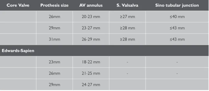
5Both valves are currently available in three sizes, as presented in Table 1.
Core Valve
Prothesis size
AV annulus
S. Valsalva
Sino tubular junction
26mm
20-23 mm
≥27 mm
≤40 mm
29mm
23-27 mm
≥28 mm
≤43 mm
31mm
26-29 mm
≥28 mm
≤43 mm
Edwards-Sapien
23mm
18-22 mm
-
-26mm
21-25 mm
-
-29mm
24-27 mm
Table 1: Aortic anatomical requirements of contemporary transcatheter aortic valve prosthesis
TRANSTHORACIC ECHOCARDIOGRAPHY
The transthoracic echocardiography (TTE) evaluation provides anatomic and hemodynamic information.
It establishes the presence of severe AS, which is defined by an aortic valve area of ≤1 cm
2(<0.6 cm
2/m
2) or a
mean aortic valve gradient of ≥40 mmHg.
67The left and right ventricular dimensions, morphology and function
are also evaluated by TTE. The presence of LV thrombus or a haemodynamically significant LVOT obstruction,
due to basal septal hypertrophy should be excluded, as they represent contraindications for the procedure.
Subaortic septal bulge may also create an obstacle to proper seating of the aortic prosthesis.
8The existence
of a patch in the LV as well as significant pericardial calcification is a contraindication for TAVI using the
valves should be evaluated and described.
TTE is the first assessment of the aortic annular dimension and anatomic characteristics of the aortic
valve. It can be used to describe the number of cusps, mobility, thickness and calcification. The presence of a
bicuspid aortic valve is still a contra-indication for TAVI because of the risk of spontaneous aortic dissection
or incorrect deployment of the aortic prosthesis due to the elliptical valvular orifice. However, successful
TAVI in bicuspid AS has already been reported.
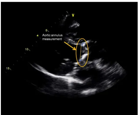
9As presented in Table 1 the aortic annulus dimension dictates the eligibility for TAVI and prosthesis
size, which is determinant to the procedural success. Using TTE the aortic annular dimension is measured
in systole, in a parasternal long-axis view, zoomed on the LVOT, at the point of insertion of the aortic valve
cusps, from tissue–blood interface to blood–tissue interface—trailing edge to leading edge (Figure 10.2).
102D Transesophageal echocardiography (TEE) and even 3D TEE evaluation may be necessary when TTE
measurements of the annulus are doubtful, particularly if they are near critical cut-offs for valve selection, or
if the calcification extends from the aortic valve onto the anterior mitral leaflet or the septum.
Figure 10.2 Transthoracic echocardiography parasternal long-axis view for measurement of aortic annular dimension.
TRANSESOPHAGEAL ECHOCARDIOGRAPHY
TEE is recommended prior to TAVI to a better evaluation of the aortic root anatomy, particularly if there
are any concerns about the assessment of the aortic annular size and the number of cusps.
The annular diameter at the level of the basal attachment of the aortic valve cusps, measured in systole
determines the size of the prosthesis, irrespective of the type of the valve inserted (Table 1). There is a
good correlation between TEE aortic annular measurements and TTE results; however TTE to some extent
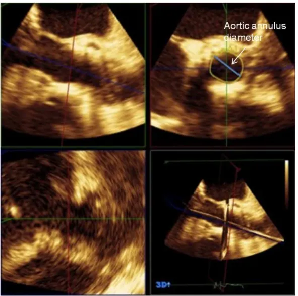
When measuring the annular diameter with 2D TEE, there is an assumption of annular circularity, which
may result in erroneous measurements in patients whose annuli are more oval shaped. This limitation can
be overcome using multiplanar tools of 3D TEE (Figure 10.3)
10or multidetector computed tomography
(MDCT). The later will be described in detail underneath in MDCT proper section. The final decision for the
appropriate valve size mainly considers the annulus diameter, but in cases of borderline size decisions, the
existence of large calcification in the native valve, may require a smaller prosthesis than the annular dimension
alone would advise. Up to now there is no gold standard imaging technique for annular sizing for TAVI, but
from the practical point of view, TTE complemented with TEE accomplishes good results in most patients.
Using short-axis views of the aortic valve the number of cusps can be precisely illustrated and its
opening classified as central or eccentric. It is relevant to describe the severity and eventual asymmetry of
calcification, as the differences in the tension–force across the valve may cause asymmetric deployment
of the prosthesis and increase the risk of compression of the coronary arteries. However TEE presents
constrains at the time of calcification evaluation, being MDCT the best method available.
12The distance
from the aortic annulus to the ostia of the coronary arteries should be measured, because patients with
cusps length larger than annular-ostial distances are at risk of ostial coronary occlusion at time of valve
deployment, as the native cusps will be crushed to the side. Only the right coronary annular-ostial distance
is possible to measure with 2D TEE, as the left coronary annular-ostial distance requires 3D TEE or MDCT.
Using TEE the characteristics of the ascending aorta, the aortic arch, and the descending thoracic aorta
should be considered as the presence of large atheromas may increase the risk of peri-procedural
emboliza-tion at the time of transfemoral approach.
MULTIDETECTOR COMPUTED TOMOGRAPHY (MDCT)
MDCT has a complementary role to echocardiography and angiography at the time of evaluation of
pa-tients before TAVI.
13It provides detailed anatomic visualization of the aortic root, thoraco-abdominal aorta
and the iliofemoral access. Nevertheless, MDCT imaging is associated with the administration of iodinated
contrast and exposure to ionizing radiation exposure, thus its use should be considered for individual patients
based on risk and benefit.
Three-dimensional (3D) derived MDCT measurements provide precise dimensions for best prosthesis
selection and sizing. Anatomically, the annulus is a crown-shaped 3-dimensional structure rather than a
circu-lar plane. However, 2D echocardiography and aortic angiography give the extent of a single diameter, making
the assumption of annular orifice circularity. In contrast, 3D MDCT reconstruction of the annulus, orthogonal
to the center-axis of the LVOT, allows the assessment of minimal and maximal diameters, circumference and
area measurements (Figure 10.4). Although the use of 3D MDCT measurements for best prosthesis sizing
may reduce the probability of error and present a more comprehensive definition of the annular size and
shape, the mean of the maximum and minimum diameter measurements is comparable to 2D TEE
measure-ment, remaining 2D TEE the most used technique.
11Moreover, the aortic valve can be assessed in detail, and its morphology precisely described, regarding
the number of cusps and the aortic valve area, measured by planimetry. Moreover, in comparison with
echo-cardiography, MDCT has the ability to provide precise description of localization and extent of aortic cusps
calcification. Relationships between aortic valve calcification and pos TAVI paravalvular aortic regurgitation
have been described, being paravalvular aortic regurgitation associated with a larger aortic annulus and with
more severe aortic root calcification.
12Furthermore, dense aortic leaflet calcification measured on contrast
MDCT discerned the need for an additional balloon post-dilatation, when using CoreValve
TM, for significant
paravalvular regurgitation reduction.
14Figure 10.5 Multidetector computed tomography measuring the distance between annulus and coronary ostia. Courtesy of Dr. Pedro Marcos-Alberca
At the time of pre-procedural evaluation, MDCT includes a complete assessment of coronary anatomy
with conventional coronary angiography, which generally is limited by the advanced calcified disease. However,
the relationship between leaflet height and distance between annulus and coronary ostia, can be accurately
measured, identifying patients at higher risk for coronary occlusion during the procedure, when the
annulus-coronary ostia distance is <11 mm (Figure 10.5).
junction and ascending and descending aorta, using centerline reconstructions. The occurrence of significant
aneurismal dilatation is considered a contraindication for the use of CoreValve (Table 1). Precise coaxial
align-ment of the prosthesis along the centerline of the aortic valve and aortic root is important during positioning
to decrease the likelihood of complications as prosthesis embolization. MDCT offers the assessment of the
aortic root in relation to the body axis and its pre-procedural angle prediction may decrease the number of
aortograms required during the procedure, therefore shortening procedure time and contrast usage.
15Finally, MDCT allows the assessment of peripheral vasculature, considering caliber, tortuosity and
calci-fication, being the minimum caliber of the common femoral artery dependent on the size of the prosthesis
valve chosen. Using the previous Edward Sapien
TMvalve it was necessary a common femoral artery with
a minimum of 8 mm of diameter, currently a minimum of 6 mm is required for both Edwards-XT
TMand
CoreValve
TM. Tortuosity and calcification are not prohibitive factors, but its combination is an adverse feature
for site complications and central embolization. In addition the location of the bifurcation of the femoral
artery and the degree of calcification have to be considered the time of deciding the feasibility of the
trans-femoral access or for choosing the alternative of subclavian or transapical approach
.
ANGIOGRAPHY
Previously to the procedure, aortography produces images of the coronary arteries, thoracic and
abdominal aorta. It allows the measurement of vessel diameters, tortuosity and excludes the presence of
aorta aneurism. Nevertheless, at time of measuring vessels calcification, angiography presents limitations,
being MDCT the best method.
Fluoroscopy is used for measurement of aortic annulus, at the time of definitive prosthesis sizing
deci-sion and to guide the procedure. The alignment of the three aortic sinuses in the same plane defines the
optimal plane and angiography projection for TAVI.
Peri-procedural echocardiography during transcatheter aortic valve
implantation
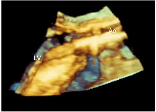
Peri-procedural 2D and 3D TEE can contribute for balloon and prosthesis positioning, to confirm
pros-thesis function immediately after implantation and to rapidly detect complications.
As in other intervention TEE guided procedures, the use of 3D, by its larger spacial resolution, presents
advantages over 2D. It allows a better visualization of the guide wire path through the LV and around the
mitral valve and subvalvular apparatus and permits a better evaluation of the prosthesis position on the
bal-loon, relative to the native valve annulus and surrounding structures (Figure 10.6).
the correct position of the valve and it is usually used in conjunction with fluoroscopy. The optimal position
for the Edwards SAPIEN
TMvalve is with the ventricular side of the prosthesis positioned 2–4 mm below the
annulus and for the CoreValve
TM, the ventricular edge of the prosthesis should be placed 5–10 mm below
the aortic valve annular plane. After the deployment it is important to confirm that all the prosthetic cusps are
moving well, that the valve stent has a circular configuration and to exclude significant valvular or paravalvular
aortic regurgitation. Mild aortic regurgitation through the prosthesis, until the guidewire is removed and at
the next few minutes after deployment is common. Small jets of paravalvular aortic regurgitation are frequent
and it may occur even in a successful procedure, however severe aortic regurgitation is a serious complication
and additional balloon inflation may be required.
Figure 10.6 3D Transesophageal echocardiography showing the catheter and the balloon through the native aortic valve. Ao- Aorta; LV- left ventricle.
ASSESSMENT OF COMPLICATIONS
Among the various complications assessable by TEE, aortic regurgitation is the most common (Figure
10.7).
2,4It may occur as a consequence of incomplete expansion, incorrect positioning, restricted cusp motion,
or inappropriate prosthetic size.
16An undersized prosthesis may result in paravalvular aortic regurgitation. On
the opposite, an oversized prosthesis has the risk of suboptimal stent expansion and central aortic
regurgita-tion. Other mechanisms eventually responsible for aortic regurgitation are the presence of severe asymmetric
calcification, causing paravalvular aortic leaks, or it may happen as a consequence of residual native aortic
valve leaflet tissue prolapsing into the prosthesis, interfering with cusp motion and coaptation. The aortic
regurgitation severity should be evaluated by the International recommended criteria.
10Furthermore, in result of prosthesis mismatch, or failed pacing capture, prosthetic embolism may occur.
If the embolization happens towards the aorta, it might be resolved through successful transcatheter
Figure 10.7 2D transesophageal echocardiography showing aortic prosthesis central regurgitation.
Other possible complications detectable by TEE are cardiac tamponade, secondary to wire perforation
of the left or right ventricle and anew LV dysfunction, which may be secondary to ostial occlusion by fragment
embolization or by an obstructive portion of the valve frame, sealing cuff or by native cusp.
18Moreover, sudden worsening of MR may occur due to right ventricular pacing, causing LV asynchrony,
or as a consequence of prosthetic misplacement, towards the left ventricle outflow tract, with pressure
exerted on the anterior mitral leaflet or by direct damage or distortion of the subvalvular apparatus.
19A tear
or ruptures of the aortic root have been also observed during the procedure after balloon valvuloplasty or
prosthesis deployment, especially in the presence of extensive annular calcification or prosthesis oversizing.
20TEE is not mandatory during TAVI, as it usually requires general anesthesia and the probe may also
partially obstruct the optimal fluoroscopic view, however it may be the main technique used for procedure
guidance, particularly in patients with limited native valve calcification,
10providing the best guidance and rapid
assessment of complications.
Post-implantation follow-up
The imaging follow-up evaluation of patients with transcatheter aortic valves is mainly based on TTE. It is
usually performed at the time of hospital discharge, at one month follow-up and at 6 or 12 months,
depend-ing on each institution protocol. The echocardiographic evaluation is mostly similar to surgically implanted
prostheses, as guided by previously published guidelines for prosthetic valves.
21≤25% suggests mild, 26–64% suggests moderate, and >65% suggests severe. These methods are limited in
the setting of paravalvular jets which are frequently eccentric and irregular in shape. The ASE/EAE guidelines
advise that for paravalvular jets, the proportion of the circumference of the sewing ring occupied by the jet
gives a semi-quantitative guide to severity: <10% of the sewing ring suggests mild, 10–20% suggests
moder-ate, and >20% suggests severe
21. However, this concept assumes jet continuity, which may not be the case for
transcatheter valves and therefore may overestimate the severity in presence of multiple small jets. For the
quantitative approach the width of the vena contracta is a robust estimate of regurgitant severity, but in the
setting of prostheses, portions of the sewing ring may not be imaged due to acoustic shadowing. In addition,
there has been no validation for adding the vena contracta widths of multiple jets as it may be encountered
post-TAVI. A new method using 3D TTE vena contracta planimetry has been recently described, showing an
accurate alternative for quantitative evaluation and moderate AR recognition of paravalvular aortic
regurgita-tion after TAVI.
22The quantitative methods for calculating regurgitant volume and effective regurgitant orifice
area that rely on the comparison of stroke volumes across the aortic valve and a nonregurgitant valve may be
also used for prosthetic valves evaluation.
21However, the final interpretation should follow the principle of
comprehensive evaluation and integrated approach.
At the time of effective orifice area calculation, it is essential that the pre-valvular velocity is recorded
proximal to the stent and the post-valvular velocity (typically recorded with continuous-wave Doppler) distal
to the stented valve. If the LVOT velocity used in calculations is erroneously recorded within the stent but
proximal to the cusps, the result will be an overestimation of valve area.
10Similarly the LVOT diameter should
be measured immediately proximal to the stent.
23In selected cases with suspicion of complications such as prosthesis malposition, MDCT by its higher
spatial resolution is an appropriate technique, providing accurate information on the position and deployment
of prosthesis.
Conclusion
References
1. Vahanian A, Alfieri O, Al-Attar N, Antunes M, Bax J, Cormier B, Cribier A, De Jaegere P, Fournial G, Kappetein AP, Kovac J,
Ludgate S, Maisano F, Moat N, Mohr F, Nataf P, Pierard L, Pomar JL, Schofer J, Tornos P, Tuzcu M, van Hout B, Von Segesser LK, Walther T. Transcatheter valve implantation for patients with aortic stenosis: A position statement from the european as-sociation of cardio-thoracic surgery (eacts) and the european society of cardiology (esc), in collaboration with the european association of percutaneous cardiovascular interventions (eapci). Eur Heart J. 2008;29:1463-1470
2. Rodes-Cabau J, Webb JG, Cheung A, Ye J, Dumont E, Feindel CM, Osten M, Natarajan MK, Velianou JL, Martucci G, DeVarennes
B, Chisholm R, Peterson MD, Lichtenstein SV, Nietlispach F, Doyle D, DeLarochelliere R, Teoh K, Chu V, Dancea A, Lachapelle K, Cheema A, Latter D, Horlick E. Transcatheter aortic valve implantation for the treatment of severe symptomatic aortic stenosis in patients at very high or prohibitive surgical risk: Acute and late outcomes of the multicenter canadian experience. J Am Coll
Cardiol. 2010;55:1080-1090
3. Leon MB, Smith CR, Mack M, Miller DC, Moses JW, Svensson LG, Tuzcu EM, Webb JG, Fontana GP, Makkar RR, Brown DL,
Block PC, Guyton RA, Pichard AD, Bavaria JE, Herrmann HC, Douglas PS, Petersen JL, Akin JJ, Anderson WN, Wang D, Pocock S. Transcatheter aortic-valve implantation for aortic stenosis in patients who cannot undergo surgery. N Engl J Med. 2010;363:1597-1607
4. Smith CR, Leon MB, Mack MJ, Miller DC, Moses JW, Svensson LG, Tuzcu EM, Webb JG, Fontana GP, Makkar RR, Williams M,
Dewey T, Kapadia S, Babaliaros V, Thourani VH, Corso P, Pichard AD, Bavaria JE, Herrmann HC, Akin JJ, Anderson WN, Wang D, Pocock SJ. Transcatheter versus surgical aortic-valve replacement in high-risk patients. N Engl J Med. 2011;364:2187-2198
5. Lange R, Schreiber C, Gotz W, Hettich I, Will A, Libera P, Laborde JC, Bauernschmitt R. First successful transapical aortic valve
implantation with the corevalve revalving system: A case report. Heart Surg Forum. 2007;10:E478-479
6. Bonow RO, Carabello BA, Chatterjee K, de Leon AC, Jr., Faxon DP, Freed MD, Gaasch WH, Lytle BW, Nishimura RA, O’Gara
PT, O’Rourke RA, Otto CM, Shah PM, Shanewise JS. 2008 focused update incorporated into the acc/aha 2006 guidelines for the management of patients with valvular heart disease: A report of the american college of cardiology/american heart association task force on practice guidelines (writing committee to revise the 1998 guidelines for the management of patients with valvular heart disease). Endorsed by the society of cardiovascular anesthesiologists, society for cardiovascular angiography and interven-tions, and society of thoracic surgeons. J Am Coll Cardiol. 2008;52:e1-142
7. Vahanian A, Baumgartner H, Bax J, Butchart E, Dion R, Filippatos G, Flachskampf F, Hall R, Iung B, Kasprzak J, Nataf P, Tornos P,
Torracca L, Wenink A. Guidelines on the management of valvular heart disease: The task force on the management of valvular heart disease of the european society of cardiology. Eur Heart J. 2007;28:230-268
8. Piazza N, de Jaegere P, Schultz C, Becker AE, Serruys PW, Anderson RH. Anatomy of the aortic valvar complex and its
implica-tions for transcatheter implantation of the aortic valve. Circ Cardiovasc Interv. 2008;1:74-81
9. Delgado V, Tops LF, Schuijf JD, van der Kley F, van de Veire NR, Schalij MJ, Bax JJ. Successful deployment of a transcatheter aortic
valve in bicuspid aortic stenosis: Role of imaging with multislice computed tomography. Circ Cardiovasc Imaging. 2009;2:e12-13 10. Zamorano JL, Badano LP, Bruce C, Chan KL, Goncalves A, Hahn RT, Keane MG, La Canna G, Monaghan MJ, Nihoyannopoulos
P, Silvestry FE, Vanoverschelde JL, Gillam LD. Eae/ase recommendations for the use of echocardiography in new transcatheter interventions for valvular heart disease. Eur Heart J. 2011;32:2189-2214
11. Messika-Zeitoun D, Serfaty JM, Brochet E, Ducrocq G, Lepage L, Detaint D, Hyafil F, Himbert D, Pasi N, Laissy JP, Iung B, Vaha-nian A. Multimodal assessment of the aortic annulus diameter: Implications for transcatheter aortic valve implantation. J Am Coll
12. Schultz CJ, Tzikas A, Moelker A, Rossi A, Nuis RJ, Geleijnse MM, van Mieghem N, Krestin GP, de Feyter P, Serruys PW, de Jae-gere PP. Correlates on msct of paravalvular aortic regurgitation after transcatheter aortic valve implantation using the medtronic corevalve prosthesis. Catheter Cardiovasc Interv. 2011;78:446-455
13. Schwartz JG, Neubauer AM, Fagan TE, Noordhoek NJ, Grass M, Carroll JD. Potential role of three-dimensional rotational angi-ography and c-arm ct for valvular repair and implantation. Int J Cardiovasc Imaging. 2011
14. Schultz C, Rossi A, van Mieghem N, van der Boon R, Papadopoulou SL, van Domburg R, Moelker A, Mollet N, Krestin G, van Geuns RJ, Nieman K, de Feyter P, Serruys PW, de Jaegere P. Aortic annulus dimensions and leaflet calcification from contrast msct predict the need for balloon post-dilatation after tavi with the medtronic corevalve prosthesis. EuroIntervention. 2011;7:564-572
15. Gurvitch R, Wood DA, Leipsic J, Tay E, Johnson M, Ye J, Nietlispach F, Wijesinghe N, Cheung A, Webb JG. Multislice computed tomography for prediction of optimal angiographic deployment projections during transcatheter aortic valve implantation. JACC
Cardiovasc Interv. 2010;3:1157-1165
16. Détaint D, Lepage L, Himbert D, Brochet E, Messika-Zeitoun D, Iung B, Vahanian A. Determinants of significant paravalvular regurgitation after transcatheter aortic valve implantation: Impact of device and annulus discongruence. JACC: Cardiovascular
Interventions. 2009;2:821-827
17. Tuzcu EM. Transcatheter aortic valve replacement malposition and embolization: Innovation brings solutions also new chal-lenges. Catheter Cardiovasc Interv. 2008;72:579-580
18. Webb JG, Chandavimol M, Thompson CR, Ricci DR, Carere RG, Munt BI, Buller CE, Pasupati S, Lichtenstein S. Percutaneous aortic valve implantation retrograde from the femoral artery. Circulation. 2006;113:842-850
19. Goncalves A, Marcos-Alberca P, Zamorano JL. Echocardiography: Guidance during valve implantation. EuroIntervention. 2010;6 Suppl G:G14-19
20. Masson JB, Kovac J, Schuler G, Ye J, Cheung A, Kapadia S, Tuzcu ME, Kodali S, Leon MB, Webb JG. Transcatheter aortic valve im-plantation: Review of the nature, management, and avoidance of procedural complications. JACC Cardiovasc Interv. 2009;2:811-820
21. Zoghbi WA, Chambers JB, Dumesnil JG, Foster E, Gottdiener JS, Grayburn PA, Khandheria BK, Levine RA, Marx GR, Miller FA, Jr., Nakatani S, Quinones MA, Rakowski H, Rodriguez LL, Swaminathan M, Waggoner AD, Weissman NJ, Zabalgoitia M. Rec-ommendations for evaluation of prosthetic valves with echocardiography and doppler ultrasound: A report from the american society of echocardiography’s guidelines and standards committee and the task force on prosthetic valves, developed in con-junction with the american college of cardiology cardiovascular imaging committee, cardiac imaging committee of the american heart association, the european association of echocardiography, a registered branch of the european society of cardiology, the japanese society of echocardiography and the canadian society of echocardiography, endorsed by the american college of cardiology foundation, american heart association, european association of echocardiography, a registered branch of the european society of cardiology, the japanese society of echocardiography, and canadian society of echocardiography. J Am Soc
Echocardiogr. 2009;22:975-1014; quiz 1082-1014
22. Goncalves A, Almeria C, Marcos-Alberca P, Feltes G, Hernandez-Antolin R, Rodriguez E, Silva Cardoso JC, Macaya C, Zamo-rano JL. Three-dimensional echocardiography in paravalvular aortic regurgitation assessment after transcatheter aortic valve implantation. J Am Soc Echocardiogr. 2011
23. Clavel MA, Rodes-Cabau J, Dumont E, Bagur R, Bergeron S, De Larochelliere R, Doyle D, Larose E, Dumesnil JG, Pibarot P. Validation and characterization of transcatheter aortic valve effective orifice area measured by doppler echocardiography. JACC
CHAPTER 2
Echocardiography: guidance during valve implantation
Alexandra Gonçalves, MD; Pedro Marcos-Alberca, MD, PhD; José Luis Zamorano*, MD, PhD, FESC
Cardiovascular Institute, Hospital Clínico San Carlos, Madrid, Spain
The authors have no conflict of interest to declare.
This paper also includes accompanying supplementary data published at the following website: www.eurointervention.com
G1
-Abstract
Transcatheter aortic valve implantation (TAVI) by percutaneous or transapical aproach has emerged as an
effective and less-invasive treatment for patients with severe symptomatic aortic valve stenosis and high
surgical risk. Echocardiography is a fundamental tool in patients’ selection for TAVI, for guiding the
intervention as well as evaluating the position, deployment and function of the prosthesis. This review describes the role of echocardiography during the intervention, in procedure guidance and in the
assessment of complications. KEYWORDS
Transcatheter aortic valve implantation, aortic valve stenosis, valvuloplasty, aortic prosthesis, three dimensional (3D) transesophageal echocardiography, procedure complications, transfemoral, transapical approach
Part I – Guidance for transcatheter aortic and mitral valve implantation
* Corresponding author: Cardiovascular Institute, University Clinic San Carlos, Plaza de Cristo Rey, 28040 Madrid, Spain E-mail: jlzamorano@vodafone.es
© Europa Edition 2010. All rights reserved.
EuroIntervention Supplement (2010) Vol. 6 (Supplement G) G0-G0
G2
-Echocardiography: guidance during valve implantation
Introduction
Transcatheter aortic valve implantation (TAVI) has recently emerged as an effective, less-invasive treatment for patients with severe
symptomatic aortic valve stenosis and high surgical risk, either in its
percutaneous or transapical approach.1Rates of success in device
implantation of around 95%, and procedure-related mortality rates between 5 and 18% have been reported.2-4
Currently, two different systems are available either through
transfemoral or transapical approach: the balloon-expandable
Edwards SAPIEN® prosthesis (Edwards Lifesciences, Irvine, CA, USA), a trileaflet symmetrical bovine pericardial valve mounted within
a stainless steel stent, and the self-expanding CoreValve ReValving®
system (Medtronic, Minneapolis, MN, USA). The CoreValve prosthesis consists of a trileaflet bioprosthetic porcine pericardial
tissue valve, which is sutured into a self-expanding nitinol frame with
an asymmetric shape. For the CoreValve, the sizes of the developed
delivery systems have been gradually reduced to 18 Fr, at the third generation, and recently, a new 18 Fr delivery system for the SAPIEN
valve has been approved, facilitating the vascular access and
deployment of the device. Presently, each system has two different
sizes available, compliant with annulus dimensions from 19 to 27 mm. Both systems have been extensively described elsewhere.5,6
Echocardiography is a fundamental tool in patient selection for TAVI,
for guiding the intervention and to evaluate the position, deployment
and function of the prosthesis. The procedure is usually performed under fluoroscopic and transesophageal echocardiographic (TEE)
imaging, but a fully echo-guided transapical aortic valve
implantation has already been reported.7,8This is an important step,
considering the risk of acute postoperative renal failure following extensive use of contrast medium, which might reach 28%,
according to recent reports.9
Transesophageal echocardiographic approach
before valvuloplasty and prosthesis implantation
Before starting the procedure, the echocardiographer has tocarefully describe the aortic valve anatomy and its anatomical
landmarks, distribution of aortic valve calcification, geometry of the
left ventricle outflow tract (LVOT) and its spatial relationships, the distance between coronary cusps insertion and coronary arteries
ostium and also an eventual ectopic calcification of the basal portion
of the anterior mitral leaflet and annulus. The aortic valve annulus
diameter is measured, from the insertion of the non-coronary cusp to the insertion of the right coronary cusp in a 135º view (Figure 1). In
the 45º view the orthogonal diameter of the aortic root is measured in
an upper plane of the coronary arteries arisen. The precise distance
of the coronary arteries to the annulus should be measured in order to minimise the risk of complications, namely myocardial ischaemia.
It is advisable that at the time of deployment, coronary ostia should
be minimally located 14 mm away from the leaflets insertion for the
CoreValve and 11 mm for the Edwards SAPIEN prosthesis. Left and right ventricular systolic function, regional wall motion
abnormalities, mitral or tricuspid regurgitation and thoracic aortic arch
atheroma should also be evaluated.10For the transapical aortic valve
implantation approach, the echocardiographer can use transthoracic echocardiography to point the left ventricle apex position.11
Although bi-dimensional (2D) echocardiography was the standard
technique in TEE, currently three dimensional (3D) TEE is highly
available and there is a common perception that it frequently
shortens the learning curve of the procedure, which might influence early outcome. Moreover, the 3D transesophageal probe (X7-2t®,
7 MHz, Philips Medical Systems, Eindhoven, The Netherlands) has
the capability of presenting two orthogonal bi-dimensional
simultaneous plane views (e.g., 45º short-axis and 135º) which provides additional information, especially during the intervention
(Figure 2). Throughout patient selection, and in the sensitive process
of aortic annulus measurement, 3D imaging improves the
assessment of valve anatomy and geometry of the LVOT (Figure 3), which was shown to be frequently elliptical instead of circular in
form.12,13Besides, the sectorial 3D image and amplified or zoom
mode 3D acquisition allow the visualisation of the entire guidewires,
catheters and prostheses throughout the process. This added value of 3D has become critical in the operating room, so fluoroscopy
needs to be in a postero-anterior projection during transapical 56. 57. 58. 59. 60. 61. 62. 63. 64. 65. 66. 67. 68. 69. 70. 71. 72. 73. 74. 75. 76. 77. 78. 79. 80. 81. 82. 83. 84. 85. 86. 87. 88. 89. 90. 91. 92. 93. 94. 95. 96. 97. 98. 99. 100. 101. 102. 103. 104. 105. 106. 107. 108. 109. 110.
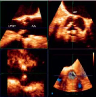
Figure 1. Two-dimensional TEE long axis view (123º) showing measurement of aortic valve annulus and sinotubular junction diameter. LV, left ventricle; AA, ascending aorta; LVOT, left ventricle outflow tract.
G3 -intervention. As a result, the calcified aortic annulus is positioned
over the spine and the echocardiographic support is the only imaging technique which visualises and can guide, as well, the exact placement of the prosthesis.8In the process of valve size selection, approximately 10% of oversizing is applied, based on these measurements. Although some oversizing is essential to avoid severe paravalvular leakage, however, in the presence of a rigid aortic root, too much oversizing entails a high risk of serious complications and should be avoided. In addition, it is wise to exclude patients with an annulus larger than the largest available prosthesis, in whom significant paravalvular regurgitation might be expected.14In spite of the experience of the operators, the lack of congruence between the annulus and the device is related to significant paravalvular aortic regurgitation, thus the process of precise aortic annulus measurement is essential for better outcomes.15
Recent studies have also highlighted the value of multislice computed tomography to evaluate aortic valve annulus morphology and size, however TEE remains the reference technique due to its reliability, safeness and non radiation exposure.16
Valvuloplasty and prosthesis implantation
Balloon valvuloplasty is performed with a balloon filled with 1:4 diluted contrast placed in the aortic valve, during rapid ventricular pacing. Echocardiography is used to watch for antegrade or retrograde slippage of the balloon throughout its inflation. Inappropriate motion of the balloon towards the ventricle during inflation may happen due to axial motion of the heart, or to a small sinotubular junction. Conversely, an upward shifting of the balloon towards the aorta may be caused by the presence of a prosthetic mitral valve.17Part I – Guidance for transcatheter aortic and mitral valve implantation
Figure 3. Post-processing of 3D volumetric acquisition using multiplanar reformatting or MPR showing valve anatomy and geometry of the LVOT. LVOT, left ventricle outflow tract; AA, ascending aorta; AV, aortic valve.
Using the Live 3D mode, the dilatation of the valve with the valvuloplasty balloon and the anatomic results are accurately displayed (Video 1). Following pre-dilation of the native aortic valve, the prosthesis is advanced and deployed within the aortic annulus. This is one of the most critical steps during the intervention, because of the possible misplacement and embolism of the device. After the valve is introduced into the annulus, the pusher is retrieved back into the delivery sheath.
A long-axis view of the aortic root with 2D TEE identifies the end of the delivery catheter through the aortic annulus and, joined with fluoroscopy imaging, allows observation of the initial position, deployment, and final placement of the prosthesis. However, the single plane of the 2D image has several constraints when looking for catheter alignment (Figure 2). Conversely, in spite of the relatively low temporal resolution of the 3D mode as compared to 2D TEE, using the Live 3D mode, the trajectory view is precisely identified (Figure 4). In addition, this discloses with detail the prosthesis deployment in relation to the aortic annulus (Video 2). Therefore, the 3D TEE approach provides relevant information to the interventional cardiologist during this decisive step, with an excellent visual agreement between the fluoroscopic and the 3D TEE images. In order to accomplish correct position in the deployment process, the prosthesis should be oriented coaxially with the long axis of the ascending aorta and perpendicularly to the aortic annulus, as showed in Figure 5. An oblique position may lead to valve misplacement, particularly in patients with a calcified aortic root and/or narrow sinotubular junction, in whom restriction of balloon inflation may occur with consequent downward displacement of the valve into the ventricle.17
One of the issues of confronting TEE during the procedure, is to identify, in one imaging plane, the exact location of the valve stent in relation to the deployment balloon and its ventricular and aortic rims. The stent is recognised as an echogenic rectangular structure seen in bristly profile to the balloon (Figure 5). The image interpretation is easier if the echocardiographer is aware of its length and if 3D TEE is used. As the prosthesis can move up to 2 to 4 mm towards the ascending aorta with balloon inflation, the optimal position is achieved when the ventricular edge of the stent is
G4
-Echocardiography: guidance during valve implantation
positioned approximately 2 to 4 mm below the aortic valvular
annular plane for the Edwards SAPIEN valve and 5–10 mm for the CoreValve ReValving system. The aortic rim of the stent should
cover the upper limit of the native aortic leaflets, and the valve
should be deployed when both echocardiographer and
interventionist agree that is in the best position.
During deployment, the native aortic leaflets are compressed
between the valve stent and the wall of the aortic root. To avoid an
upper positioning of the prosthesis in the aorta upon deployment
caused by left ventricular ejection flow, pacing is performed until the balloon is completely deflated.
After the permanent aortic prosthesis implantation, the correct
positioning is confirmed and aortic regurgitation evaluated
immediately after removal of the deployment catheter and guidewire. The aortic end of the valve stent should be below the
level of the coronary ostia and the ventricular end of the valve stent
should not interfere with anterior mitral leaflet function (Video 3).10
The prosthesis is expected to present a circular expansion, with native aortic leaflets perfectly contained and prosthesis leaflets
moving amply, without significant aortic regurgitation (Videos 4 and
5). Finally, the transgastric window is used for Doppler interrogation
of the aortic valve and to record the transaortic pressure gradient reduction and improvements in aortic valve area.
A
ssessment of results and detection of
complications
One of the most common complications is aortic regurgitation,
either perivalvular or central. It might be caused by severe
asymmetric calcification of the native aortic valve (Figure 6 and
Video 6), incomplete expansion of the device, incorrect positioning or inappropriate prosthesis size.15An excessively small size might
cause paravalvar aortic regurgitation (Figure 7). In contrast, a
prosthesis implantation too large for the aortic root can cause
suboptimal stent expansion, impaired leaflet mobility and central aortic regurgitation (Figure 8, Videos 7 and 8).
166. 167. 168. 169. 170. 171. 172. 173. 174. 175. 176. 177. 178. 179. 180. 181. 182. 183. 184. 185. 186. 187. 188. 189. 190. 191. 192. 193. 194. 195. 196. 197. 198. 199. 200. 201. 202. 203. 204. 205. 206. 207. 208. 209. 210. 211. 212. 213. 214. 215. 216. 217. 218. 219. 220.
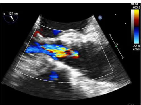
Figure 6. Two-dimensional transesophageal echocardiography long axis view showing moderate perivalvular regurgitation caused by asymmetric calcification. See Video 6 for viewing the entire sequence. LV, left ventricle; AA, ascending aorta; LVOT, left ventricle outflow tract.
Figure 7. Two-dimensional transesophageal echocardiography short axis view showing perivalvular regurgitation.
Figure 8. Two-dimensional transesophageal echocardiography long axis view showing central regurgitation. AA, ascending aorta; LVOT, left ventricle outflow tract.
Figure 5. Two-dimensional transesophageal echocardiography long axis view showing the prosthesis, oriented coaxial with the long axis of the ascending aorta and perpendicular to the aortic annulus. LV, left ventricle; AA, ascending aorta; LVOT, left ventricle outflow tract.
Sometimes, mild to moderate aortic paravalvular regurgitations are
seen due to minimal defects of sealing involving the posterior
commissure, mainly when leaflets highly calcified. As a consequence of the accommodation of the struts to these calcified
valves, frequently, after several minutes or several hours, the mild