NT-proBNP Levels in Patients with Non-ST-segment Elevation Acute
Coronary Syndrome
Luiz Ricardo A. Castro
1,2,3, Maria Clara N. Alencar, Márcia M. Barbosa
1,3,5, Maria do Carmo P. Nunes
1,2,5, José Ronaldo
Cardoso
4, Antonio Luiz P. Ribeiro
1,2Serviço de Cardiologia e Cirurgia Cardiovascular - Hospital das Clínicas1 - UFMG; Departamento de Clínica Médica - Faculdade de Medicina - UFMG2; Hospital Socor3; Laboratel4; Ecocenter Socor5, Belo Horizonte, Brazil
Abstract
Background: Non-ST-segment elevation acute coronary syndrome is associated with elevation of brain natriuretic peptide and markers of myocardial necrosis, although its relationship with the TIMI score and left ventricular function are largely unknown.
Objetive: To evaluate the correlation between plasma N-terminal pro-brain natriuretic peptide (NT-proBNP) and markers of myocardial necrosis [creatine phosphokinase muscle-brain fraction (CK-MB) and troponin I], TIMI risk score and left ventricular ejection fraction in patients with non-ST-segment elevation acute coronary syndrome.
Methods: Eighty-seven patients with non-ST-segment elevation acute coronary syndrome were divided into two groups: 37 (42.5%) with unstable angina and 50 (57.5%) with non-ST-segment elevation myocardial infarction.
Results: Left ventricular ejection fraction more than 40% was found in 86.2% of the total sample. Serum levels of NT-proBNP was higher in patients with non-ST elevation myocardial infarction than in those with unstable angina (p<0.001). Increased levels of NT-proBNP were associated with increases in troponin I (rs=0.425, p<0.001), peak CK-MB (rs=0.458, p<0.001) and low left ventricular ejection fraction (rs=-0.345, p=0.002); no correlation was found with the TIMI risk score (rs=0.082, p=0.44). Multivariate analysis revealed that left ventricular ejection fraction and troponin I levels were independently correlated with NT-proBNP levels (p=0.017 and p=0.002, respectively).
Conclusion: Increased levels of NT-proBNP in patients with non-ST-segment elevation acute coronary syndrome are not related exclusively to low left ventricular ejection fraction, but can also be caused by the presence of myocardial ischemia and necrosis. (Arq Bras Cardiol 2011;97(6):454-461)
Keywords: Acute coronary syndrome; natriuretic peptides; myocardial infarction; angina, unstable.
Mailing Address: Antônio Luiz Pinho Ribeiro •
Rua Campanha, 98/ l0l, Carmo – 30310770 – Belo Horizonte, MG, Brazil E-mail: [email protected], [email protected]
Manuscript received March 09, 2011; revised manuscript received May 12, 2011; accepted June 07, 2011.
itself, is known to increase BNP and NT-proBNP levels; as a result, the real role of high levels of natriuretic peptides as markers of ischemia and necrosis on the prognosis of NSTE-ACS remains uncertain.
The objective of this study was to assess plasma NT-proBNP levels in NSTE-ACS inpatients without heart failure, and to correlate these findings with other markers of myocardial necrosis (CK-MB and troponin I), TIMI risk score and left ventricular ejection fraction (LVEF).
Methods
Study Group
This observational, cross-sectional study was conducted at Hospital Socor, Belo Horizonte, MG, Brazil. Eighty-seven patients with suspicion of NSTE-ACS admitted to the intensive care unit (ICU) were consecutively selected and followed up until discharge. Inclusion criteria were age over 18 years, established diagnosis of NSTE-ACS, and admission within less than 72 hours after the onset of symptoms. Patients who refused or were unable to participate in the study, or who presented one of the following conditions,
Introduction
Determining the concentration of circulating natriuretic peptides is important in the diagnostic and prognostic evaluation of patients with acute coronary syndrome (ACS)1,2. In ACS patients with ST-segment elevation
myocardial infarction (STEMI), natriuretic peptides can be used to detect left ventricular dysfunction and are powerful independent predictors of death, heart failure and further myocardial infarctions1,3.
Several studies have consistently associated increased plasmatic levels of brain natriuretic peptides (BNP) or N-terminal pro-brain natriuretic peptide (NT-proBNP) with poor long-term prognosis in NSTE-ACS patients4-8. However,
were excluded: renal failure (serum creatinine > 2.5 mg/dL), preexisting valvular disease, hypertrophic cardiomyopathy, severe systemic hypertension (systolic blood pressure > 180 mmHg or diastolic blood pressure > 110 mmHg), cardiogenic shock, refractory ventricular arrhythmia, recent acute myocardial infarction (< four weeks prior to hospitalization), recent myocardial revascularization surgery (< four weeks), recent percutaneous coronary intervention (< two weeks), previous diagnosis or signs/symptoms of congestive heart failure, severe non-cardiovascular disease limitinglife expectancy, previous transplant of vital organs (lungs, liver, heart, and kidneys) or patients on transplant waiting lists. All patients included in the study signed an informed consent form. The study protocol was approved by the Research Ethics Committees of Universidade Federal de Minas Gerais and Hospital Socor, Belo Horizonte, Brazil.
Clinical and electrocardiographic data
Clinical data and information on previous conditions or interventions, risk factors for coronary artery disease (CAD) and medications in use were collected during each patient’s stay in the ICU. Electrocardiograms were performed to assess ST-segment and T-wave abnormalities.
Diagnosis of unstable angina was confirmed based on the presence of at least one of the three features: (i) typical and prolonged (> 20 minutes) chest pain at rest; (ii) new-onset angina of at least class III severity according to the Canadian Cardiovascular Society (CCS)9; and (iii)
increasing angina previously diagnosed and becoming more frequent, longer in duration or lower in threshold; any of these findings should be associated with absence of increased markers of myocardial necrosis (CK-MB and troponin I), with or without ST-segment or T-wave abnormalities on electrocardiogram10. NSTEMI was defined
as the presence prolonged chest pain (> 20 minutes) associated with increased CK-MB or troponin I levels (the presence of increased troponin levels only, without a CK-MB increase, was also considered diagnostic), with or without electrocardiographic abnormalities10. Electrocardiographic
findings were classified as T-wave abnormalities, ST-segment depression or transitory ST-segment elevation in at least in two contiguous leads10.
Treatment and laboratory routines followed the guidelines of the American College of Cardiology (ACC)/ American Heart Association (AHA) for the management of patients with unstable angina and NSTEMI10.
Laboratory data
CK-MB measurements were performed using the Cobas Mira Plus systemTM (Roche Diagnostics GmbH, Mannheim,
Germany), at baseline and at 8 and hours 16 after admission. CK-MB results were expressed in U/l (reference value: < 10U/l). Troponin I levels were measured using a chemiluminescence method on the Immulite® system (DPC, New Jersey, USA), usually on admission. The reference limit recommended by the manufacturer was < 1.0 ng/mL.
Plasma NT-proBNP concentration was determined by electrochemiluminescence using an Elecsysâ 2010/Modular
Analytics E170 immunoassay analyzer (Roche Diagnostics GmbH, Mannheim, Germany). Blood samples were collected in a tube without anticoagulants within 72 hours after the onset of symptoms, with the patient resting quietly while semirecumbent. Samples were centrifuged at 3,000 revolutions per minute during 10 minutes and the serum was extracted. NT-proBNP results were expressed in pg/ml.
The TIMI risk score (thrombolysis in myocardial infarction) was used for risk stratification, as previously described11.
Doppler Echocardiography
Doppler echocardiography was performed using a 5500 Phillips system (Phillips Medical Systems, Washington, USA). Two investigators performed and analyzed all the tests and both were blinded to patients’ clinical data. Both investigators evaluated the exams and disagreements were solved by consensus. The area-length method was used to determine ejection fraction12.
Statistical analysis
Sample size was calculated to find a correlation (r) of at least 0.3 between NT-proBNP levels and necrosis markers (CK-MB and troponin I), considering a significance level of 0.05 and a power of 0.80 in a two-sided test.
Qualitative variables were described in terms of frequency; quantitative variables were presented as measures of central tendency (mean and median) and dispersion (standard deviation and interquartile range, IQR). The distribution of continuous variables was tested for normality, and mathematical transformations were carried out when necessary. Comparisons between groups were carried out using Student’s t-test (variables with normal distribution) or nonparametric tests. The correlation between NT-proBNP, troponin I, CK-MB, TIMI risk score and LVEF was analyzed using Pearson’s or Spearman’s correlation coefficient. Statistical significance was set at 0.05.
A multivariate analysis was carried out to assess whether the relationship between the markers of myocardial necrosis and NT-proBNP levels was independent of ejection fraction. The partial correlation coefficient found between necrosis markers and NT-proBNP levels was evaluated using Pearson’s method, adjusted to ejection fraction. A multiple linear regression analysis was also performed to test for residual normality, where the natural logarithm (ln) NT-proBNP was the dependent variable, and LVEF and levels of necrosis markers were the independent variables.
Results
Clinical data
Clinical characteristics are shown in Table 1. Patients were divided into two groups according to the diagnosis: (i) unstable angina (troponin I < 1.0 ng/ml), 37 patients
(42.5%), and (ii) NSTEMI (troponin I ≥ 1.0 ng/ml), 50
61.1 (±11.4) years and it was similar in the two groups; there was a predominance of male patients (64.4%). Most patients were using aspirin (97.7%) and clopidogrel (95.4%). A great number of NSTEMI patients had been treated with glycoprotein IIb/IIIa inhibitors (n = 36, 72.0%), whereas only three (8.1%) of the group with unstable angina (p < 0.001) had received this medication. Fifty-two patients (60.5%), mostly from the NSTEMI group (n = 40, 76.9%), were submitted to coronary angiography during their stay at the ICU.
Events were rare among NSTEMI patients during hospital stay: one death due to cardiogenic shock, two patients with refractory angina, one with acute pulmonary edema, and one with supraventricular tachycardia; in the unstable angina group, only one patient presented acute atrial fibrillation.
Electrocardiographic data
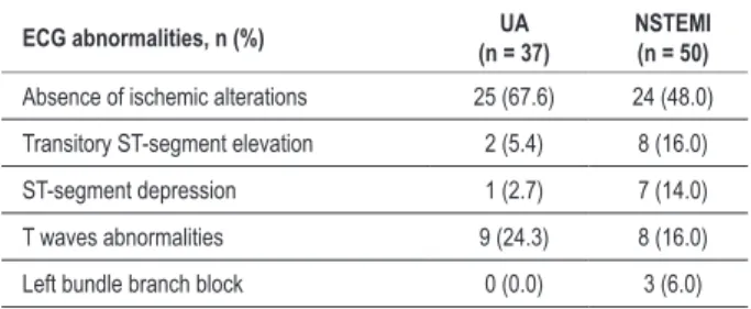
More than half of the patients (n = 49, 58.3%) did not present electrocardiographic ST-segment and T-wave abnormalities that could be associated with ischemia. Electrocardiographic abnormalities were found in 35 (41.7%) patients: ST-segment abnormalities (transitory depression or elevation) in 18 (21.4%), and T waves abnormalities in 17 (21.3%) (Table 2).
Laboratory data
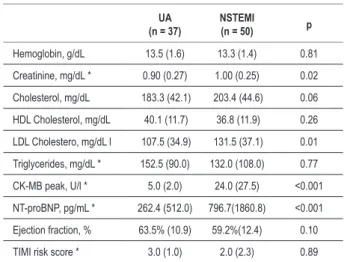
Laboratory results were similar in both groups. Median creatinine was 0.9mg/dL (IQR=0.3), the highest value being 2.0mg/dL (Table 3).
Median (IQR) levels of NT-proBNP were significantly higher in NSTEMI patients (796.7pg/ml, 1860.8) than in patients with unstable angina (262.4pg/ml, 512.0) (p < 0.001). TIMI risk score ranged from zero to five points, with a median of 3.0 (IQR=2.0). Its median and IQR values were similar in both groups: 3.0 (1.0) in the unstable angina group and 2.0 (2.3) among NSTEMI patients (p = 0.89).
Ejection Fraction
Mean LVEF in the 87 patients was 60.0% (±11.0), 63.5% (±10.9) in the unstable angina group and 59.2% (±12.4%) in the NSTEMI group (p=0.10) (Table 3). LVEF > 40% was found in 86.2% of the patients, with similar number of patients in both groups (p = 0.39).
NT-proBNP correlations
There was no evidence of correlation between NT-proBNP plasma levels and TIMI risk scores (rs=0.082, p = 0.44). However, a highly significant correlation was observed between NT-proBNP, peak CK-MB (rs=0.458, p < 0.001) (Figure 1), and troponin I levels (rs=0.425, p < 0.001). There was also a correlation between NT-proBNP levels and LVEF (rs=-0.345, p = 0.002) (Figure 2).
The correlation between NT-proBNP levels, peak CK-MB (r=0.387, p < 0.001) and troponin I levels (r=0.389, p < 0.001) persisted even after adjustment for LVEF. In the multiple linear regression analysis, both troponin I
levels (p=0.002) and LVEF (p = 0.017) correlated with NT-proBNP levels (r2=0.24, p < 0.001).
Discussion
the main finding of this study was the positive correlation between NT-proBNP levels and markers of myocardial necrosis in patients with NSTEMI, irrespective of LVEF. Our findings revealed a negative correlation between NT-proBNP levels and LVEF (rs=-0.345, p = 0.002), suggesting that the correlation between NT-proBNP and markers of myocardial necrosis was not caused by left ventricular dysfunction. In fact, 86.2% of our patients presented LVEF > 40%, and after a multiple linear regression analysis (r2=0.24, p < 0.001), both troponin I (p = 0.002) and
ejection fraction (p = 0.017) were significantly and independently associated with NT-proBNP levels. Bazzino et al13 hadalready described the existence of a correlation
between NT-proBNP and troponin T (r=0.23, p = 0.01), as well as a weak correlation with CK-MB (r=0.07, p < 0.001); however, those authors did not assess LVEF, a condition that, by itself, may provoke increases in natriuretic peptide levels. More recently, Palazzuoli et al14 have shown a progressive increase of BNP in stable
angina, unstable angina and NSTEMI, with preservation of systolic function.
Previous studies have shown that increased levels of BNP and NT-proBNP in NSTE-ACS patients are associated with increased mortality in long-term follow-up periods after hospital discharge and a higher risk for developing heart failure4,13,15-18. Omland et al19 conducted a study with
ACS patients (STEMI, NSTEMI and unstable angina) and found that NT-proBNP is a powerful indicator of long-term mortality. The authors used echocardiography to assess LVEF in all patients; however, the association between LVEF and NT-proBNP levels was not investigated in patients with NSTE-ACS. Omland et al19 aimed to investigate the
association between NT-proBNP levels and the occurrence of cardiovascular events later in life, while the investigation of this study was limited to patient’s stay at the hospital. In addition, our study excluded patients with a history of heart failure, to prevent this variable from interfering with serum NT-proBNP levels. As far as we know, this is the first study to simultaneously evaluate the correlation of NT-proBNP levels with markers of myocardial necrosis, TIMI risk score and LVEF in NSTE-ACS inpatients.
In our study, patient selection was based on objective diagnostic criteria, for unstable angina and for NSTEMI, and patients with non-ischemic chest pain were excluded10,20.
The study focused on patients with ischemia or acute myocardial damage who presented necrosis markers above reference values, regardless of the presence of electrocardiographic abnormalities. However, the use of these strict inclusion criteria has not been the rule in previous study4,5,21,22. In the present study, 41.7% of the
patients presented electrocardiographic ischemic changes indicative of unstable coronary disease23. This incidence
electrocardiographic abnormalities were not inclusion criteria in this study, in accordance with international guidelines4,10,22,23.
In all our patients, the time elapsed between onset of ischemic symptoms and NT-proBNP assessment was shorter than 72 hours; the exact interval, however, has not been assessed. The lack of a precise time is also present in many other studies, which may represent a further limitation for the comparison of results, considering the time-dependent changes that occur in natriuretic peptide levels in NSTE-ACS patients4,7,13,17,18.
It is known that renal failure and aging increase plasma NT-proBNP levels24. The mean age of our patients was
61.15 (11.38) years, which is similar to values reported in other studies21,22. Both NT-proBNP and BNP are influenced
by renal function, and NT-proBNP levels increase significantly when serum creatinine levels exceed 2.0mg/
dL14,25,26. However, in this study, no patient had creatinine
levels above 2.0mg/dL, suggesting that the increased levels of NT-proBNP in our patients were not associated with renal failure.
Risk stratification is essential in managing ACS and the TIMI risk score is a valuable tool in NSTE-ACS11. In our
study, TIMI risk score it did not correlate with plasma NT-proBNP levels (rs=0.082, p = 0.448). Bazzino et al13 found
that NT-proBNP had a more significant prognostic value than TIMI risk score and the ACC/AHA classification in predicting the risk of cardiovascular events in patients with NSTE-ACS. However, they did not report any association between NT-proBNP and TIMI risk score, which suggests that this study is the first to note the absence of such a correlation. In fact, our findings suggest that NT-proBNP and TIMI risk score reflect distinct aspects of NSTE-ACS. TIMI risk score is classically used in ACS as a risk marker
Table 1 - Characteristics of 87 patients with non-ST-segment elevation acute coronary syndrome according to the diagnosis
Characteristics Unstable angina
(n = 37)
NSTEMI
(n = 50) p
General features
Age, mean (Standard deviation) 62.7 (11.8) 60.0 (11.1) 0.28
Male, n (%) 20 (54.1) 36 (72.0) 0.08
Coming from hospital emergency, n(%) 34 (94.4) 44 (89.8) 0.57
Risk factors, n (%)
Smoking 10 (27.8) 14 (28.0) 0.98
Diabetes mellitus 12 (32.4) 7 (14.0) 0.04
Hypertension 32 (86.5) 30 (60.0) 0.007
Dyslipidemia 26 (70.3) 30 (62.5) 0.45
Family history for CAD 9 (25.0) 12 (24.5) 0.95
Clinical history, n (%)
Stable angina 7 (18.9) 11 (22.0) 0.72
Previous MI 11 (30.6) 8 (16.0) 0.10
Previous CABG 8 (21.6) 5 (10.0) 0.13
Previous PCI 14 (37.8) 6 (12.0) 0.005
Peripheral arterial disease 8 (21.6) 11 (22.0) 0.96
Medication upon admission, n (%)
AAS 20 (54.1) 9 (18.0) <0.001
Nitrates 8 (21.6) 1 (2.0) 0.003
Angiotensin-converting enzyme inhibitor 18 (48.6) 10 (20.0) 0.005
Beta-blocker 18 (48.6) 12 (12.0) 0.017
Statins 17 (45.9) 11 (22.0) 0.018
Hospital and ICU length of stay, median (IQI)
Hospital length of stay(days) 5.0 (2.0) 6.0 (3.0) 0.009
ICU length of stay(days) 2.0 (1.0) 3.0 (0.5) <0.001
for combined events of coronary artery bypass grafting, myocardial infarction and death in 30 days, while NT-proBNP may be related to ischemia only during the in-hospital period.
The demonstration of a significant increase in plasma NT-proBNP levels regardless of left ventricular dysfunction in NSTE-ACS patients has important pathophysiologicaland clinical consequences. In experimental models with rats, the induction of myocardial hypoxia has been shown to stimulate
the production of atrial and brain natriuretic peptides, as well as the rapid induction of ventricular BNP27,28. Studies in
patients submitted to percutaneous coronary interventions showed that a short-duration increase in natriuretic peptides correlated with the size of the ischemic area29,30.
Furthermore, a positive association was also observed between BNP gene expression in ventricular biopsies and plasma BNP levels in patients submitted to coronary artery bypass grafting31.Hence, both reversible myocardial ischemia
and myocardial necrosis may provoke increases in the levels of type B natriuretic peptides. More recently, it was suggested that increased BNP level can identify inducible ischemia as detected by standard noninvasive stress tests in patients with documented or suspected coronary artery disease32. This study adds information to the findings of these
studies by showing that, in NSTE-ACS patients, increased NT-proBNP levels are at least partly related to the degree of ischemic myocardial damage, which can have prognostic importance33.
Some limitations of this study should be stressed. The studied sample is relatively small and our results have to be confirmed in larger studies. Furthermore, blood samples were collected and the echocardiogram was performed any time between the admission and 72 hours after the
Tabela 2 - Electrocardiographic data upon admission in 87 patients with non-ST-segment elevation acute coronary syndrome
ECG abnormalities, n (%) UA
(n = 37)
NSTEMI (n = 50)
Absence of ischemic alterations 25 (67.6) 24 (48.0)
Transitory ST-segment elevation 2 (5.4) 8 (16.0)
ST-segment depression 1 (2.7) 7 (14.0)
T waves abnormalities 9 (24.3) 8 (16.0)
Left bundle branch block 0 (0.0) 3 (6.0)
p = 0.08 by 2 x K test; NSTEMI - non-ST-segment elevation myocardial infarction; UA - Unstable angina.
Figure 1 - Correlation between the NT-proBNP logarithm and the maximum peak of CK-MB in 87 patients with non-ST-segment elevation acute coronary syndrome (rs= 0.458, p < 0.001).
CK-MB peak
200 100
50 40 30 20 10
5 4 3 2 1
N
T
-p
ro
BN
P
20000
10000
5000 4000 3000 2000
1000
500 400 300 200
100
50 40 30 20
10
beginning of the symptoms. Since both ventricular function indexes and NT-pro BNP levels oscillate dynamically during the ACS, this relatively imprecise time window could be a cause of variability of the results. However, this same time window has been used in other studies4,17.
From a clinical point of view, our findings suggest that natriuretic peptide concentration in patients with NSTE-ACS can bring more relevant information than other biological markers (troponin I, CK-MB) because of their potential to better define the amount of injured myocardium. New imaging methods can define more accurately the area and location of ischemia or myocardial necrosis in the acute phase of NSTE-ACS. Studies using these methods should be done, so that the size of the ischemic/necrotic area can be analyzed in correlation with BNP and NT-proBNP levels.
Conclusion
In patients with NSTE-ACS, NT-proBNP correlated not only with LVEF, but also with CK-MB and troponin I levels. Increased levels of NT-proBNP in patients with
Table 3 - Tests during hospital stay in 87 patients with non-ST-segment elevation acute coronary syndrome
UA (n = 37)
NSTEMI
(n = 50) p
Hemoglobin, g/dL 13.5 (1.6) 13.3 (1.4) 0.81
Creatinine, mg/dL * 0.90 (0.27) 1.00 (0.25) 0.02
Cholesterol, mg/dL 183.3 (42.1) 203.4 (44.6) 0.06
HDL Cholesterol, mg/dL 40.1 (11.7) 36.8 (11.9) 0.26
LDL Cholestero, mg/dL l 107.5 (34.9) 131.5 (37.1) 0.01
Triglycerides, mg/dL * 152.5 (90.0) 132.0 (108.0) 0.77
CK-MB peak, U/l * 5.0 (2.0) 24.0 (27.5) <0.001
NT-proBNP, pg/mL * 262.4 (512.0) 796.7(1860.8) <0.001
Ejection fraction, % 63.5% (10.9) 59.2%(12.4) 0.10
TIMI risk score * 3.0 (1.0) 2.0 (2.3) 0.89
Data are mean (standard deviation), except * expressed by median (interquartile range); NSTEMI - non-ST-segment elevation myocardial infarction; UA - Unstable angina.
Ejection fraction %
90 80
70 60
50 40
30
N
T
-p
ro
BN
P
5000 4000 3000
2000
1000
500 400 300
200
100
50 40 30
20
10
References
1. Jernberg T, James S, Lindahl B, Johnston N, Stridsberg M, Venge P, et al. Natriuretic peptides in unstable coronary artery disease. Eur Heart J. 2004;25(17):1486-93.
2. Wiviott SD, de Lemos JA, Morrow DA. Pathophysiology, prognostic significance and clinical utility of B-type natriuretic peptide in acute coronary syndromes. Clin Chim Acta. 2004;346(2):119-28.
3. Richards AM, Nicholls MG, Espiner EA, Lainchbury JG, Troughton RW, Elliott J, et al. B-type natriuretic peptides and ejection fraction for prognosis after myocardial infarction. Circulation. 2003;107(22):2786-92.
4. de Lemos JA, Morrow DA, Bentley JH, Omland T, Sabatine MS, McCabe CH, et al. The prognostic value of B-type natriuretic peptide in patients with acute coronary syndromes. N Engl J Med. 2001;345(14):1014-21.
5. Jernberg T, Stridsberg M, Venge P, Lindahl B. N-terminal pro brain natriuretic peptide on admission for early risk stratification of patients with chest pain and no ST-segment elevation. J Am Coll Cardiol. 2002;40(3):437-45.
6. Omland T, de Lemos JA, Morrow DA, Antman EM, Cannon CP, Hall C, et al. Prognostic value of N-terminal pro-atrial and pro-brain natriuretic peptide in patients with acute coronary syndromes. Am J Cardiol. 2002;89(4):463-5.
7. James SK, Lindahl B, Siegbahn A, Stridsberg M, Venge P, Armstrong P, et al. N-terminal pro-brain natriuretic peptide and other risk markers for the separate prediction of mortality and subsequent myocardial infarction in patients with unstable coronary artery disease: a Global Utilization of Strategies To Open occluded arteries (GUSTO)-IV substudy. Circulation. 2003;108(3):275-81.
8. Galvani M, Ferrini D, Ottani F. Natriuretic peptides for risk stratification of patients with acute coronary syndromes. Eur J Heart Fail. 2004;6(3):327-33.
9. C a m p e a u L . L e t t e r : g r a d i n g o f a n g i n a p e c t o r i s . C i r c u l a t i o n . 1976;54(3):522-3.
10. Anderson JL, Adams CD, Antman EM, Bridges CR, Califf RM, Casey DE Jr, et al; ACC/AHA 2007 guidelines for the management of patients with unstable angina/non-ST-Elevation myocardial infarction: a report of the American College of Cardiology/American Heart Association Task Force on Practice Guidelines (Writing Committee to Revise the 2002 Guidelines for the Management of Patients With Unstable Angina/Non-ST-Elevation Myocardial Infarction) developed in collaboration with the American College of Emergency Physicians, the Society for Cardiovascular Angiography and Interventions, and the Society of Thoracic Surgeons endorsed by the American Association of Cardiovascular and Pulmonary Rehabilitation and the Society for Academic Emergency Medicine. J Am Coll Cardiol. 2007;50(7):e1-e157.
11. Antman EM, Cohen M, Bernink PJ, McCabe CH, Horacek T, Papuchis G, et al. The TIMI risk score for unstable angina/non-ST elevation MI: a method for prognostication and therapeutic decision making. JAMA. 2000;284(7):835-42.
12. Wahr DW, Wang YS, Schiller NB. Left ventricular volumes determined by two-dimensional echocardiography in a normal adult population. J Am Coll Cardiol. 1983;1(3):863-8.
13. Bazzino O, Fuselli JJ, Botto F, Perez De Arenaza D, Bahit C, Dadone J. Relative value of N-terminal probrain natriuretic peptide, TIMI risk score, ACC/AHA prognostic classification and other risk markers in patients with non-ST-elevation acute coronary syndromes. Eur Heart J. 2004;25(10):859-66.
14. Palazzuoli A, Deckers J, Calabro A, Campagna MS, Nuti R, Pastorelli M, et al. Brain natriuretic peptide and other risk markers for outcome assessment in patients with non-ST-elevation coronary syndromes and preserved systolic function. Am J Cardiol. 2006;98(10):1322-8.
15. Omland T, de Lemos JA, Morrow DA, Antman EM, Cannon CP, Hall C, et al. Prognostic value of N-terminal pro-atrial and pro-brain natriuretic peptide in patients with acute coronary syndromes. Am J Cardiol. 2002;89(4):463-5.
16. James SK, Lindahl B, Siegbahn A, Stridsberg M, Venge P, Armstrong P, et al. N-terminal pro-brain natriuretic peptide and other risk markers for the separate prediction of mortality and subsequent myocardial infarction in patients with unstable coronary artery disease: a Global Utilization of Strategies To Open occluded arteries (GUSTO)-IV substudy. Circulation. 2003;108(3):275-81.
17. Jernberg T, Lindahl B, Siegbahn A, Andren B, Frostfeldt G, Lagerqvist B, et al. N-terminal pro-brain natriuretic peptide in relation to inflammation, myocardial necrosis, and the effect of an invasive strategy in unstable coronary artery disease. J Am Coll Cardiol. 2003;42(11):1909-16.
18. Morrow DA, de Lemos JA, Sabatine MS, Murphy SA, Demopoulos LA, DiBattiste PM, et al. Evaluation of B-type natriuretic peptide for risk assessment in unstable angina/non-ST-elevation myocardial infarction: B-type natriuretic peptide and prognosis in TACTICS-TIMI 18. J Am Coll Cardiol. 2003;41(8):1264-72.
19. Omland T, Persson A, Ng L, O’Brien R, Karlsson T, Herlitz J, et al. N-terminal pro-B-type natriuretic peptide and long-term mortality in acute coronary syndromes. Circulation. 2002;106(23):2913-8.
20. Thygesen K, Alpert JS, White HD. Universal definition of myocardial infarction. Eur Heart J. 2007;28(20):2525-38.
21. Hasdai D, Behar S, Wallentin L, Danchin N, Gitt AK, Boersma E, et al. A prospective survey of the characteristics, treatments and outcomes of patients with acute coronary syndromes in Europe and the Mediterranean basin; the Euro Heart Survey of Acute Coronary Syndromes (Euro Heart Survey ACS). Eur Heart J. 2002;23(15):1190-201.
22. Fox KA, Goodman SG, Klein W, Brieger D, Steg PG, Dabbous O, et al. Management of acute coronary syndromes: variations in practice and outcome; findings from the Global Registry of Acute Coronary Events (GRACE). Eur Heart J. 2002 Aug;23(15):1177-89.
23. Bassand JP, Hamm CW, Ardissino D, Boersma E, Budaj A, Fernandez-Aviles F, et al. Guidelines for the diagnosis and treatment of non-ST- segment elevation acute coronary syndromes. Eur Heart J. 2007;28(13):1598-660.
24. Vanderheyden M, Bartunek J, Goethals M. Brain and other natriuretic peptides: molecular aspects. Eur J Heart Fail. 2004;6(3):261-8. NSTE-ACS are not related exclusively to low LVEF, but can
also be caused by the presence of myocardial ischemia and necrosis.
Potential Conflict of Interest
No potential conflict of interest relevant to this article was reported.
Sources of Funding
There were no external funding sources for this study. Study Association
25. Chenevier-Gobeaux C, Claessens YE, Voyer S, Desmoulins D, Ekindjian OG. Influence of renal function on N-terminal pro-brain natriuretic peptide (NT-proBNP) in patients admitted for dyspnoea in the Emergency Department: comparison with brain natriuretic peptide (BNP). Clin Chim Acta. 2005;361(1-2):167-75.
26. Vickery S, Price CP, John RI, Abbas NA, Webb MC, Kempson ME, et al. B-type natriuretic peptide (BNP) and amino-terminal proBNP in patients with CKD: relationship to renal function and left ventricular hypertrophy. Am J Kidney Dis. 2005;46(4):610-20.
27. Toth M, Vuorinen KH, Vuolteenaho O, Hassinen IE, Uusimaa PA, Leppaluoto J, et al. Hypoxia stimulates release of ANP and BNP from perfused rat ventricular myocardium. Am J Physiol. 1994;266(4 Pt 2):H1572-80.
28. Hama N, Itoh H, Shirakami G, Nakagawa O, Suga S, Ogawa Y, et al. Rapid ventricular induction of brain natriuretic peptide gene expression in experimental acute myocardial infarction. Circulation. 1995;92(6):1558-64.
29. Marumoto K, Hamada M, Hiwada K. Increased secretion of atrial and brain natriuretic peptides during acute myocardial ischaemia induced by dynamic exercise in patients with angina pectoris. Clin Sci (Lond). 1995;88(5):551-6.
30. Tateishi J, Masutani M, Ohyanagi M, Iwasaki T. Transient increase in plasma brain (B-type) natriuretic peptide after percutaneous transluminal coronary angioplasty. Clin Cardiol. 2000;23(10):776-80.
31. Goetze JP, Christoffersen C, Perko M, Arendrup H, Rehfeld JF, Kastrup J, et al. Increased cardiac BNP expression associated with myocardial ischemia. FASEB J. 2003;17(9):1105-7.
32. Nadir MA, Witham MD, Szwejkowski BR, Struthers AD. Meta-analysis of B-type natriuretic peptide’s ability to identify stress induced myocardial ischemia. Am J Cardiol. 2011;107(5):662-7.