Abstract
Background: Recent studies describe the participation of reactive oxygen and nitrogen species in hypertension. Objective: To identify the redox imbalance in the blood of hypertensive.
Methods: Superoxide dismutase (SOD), catalase (CAT), glutathione peroxidase (GPx), glutathione (GSH), vitamin C, transferrin, ceruloplasmin, malondialdehyde (MDA) and carbonyl group were quantified in the blood of 20 hypertensives
and 21 controls. The individuals had a Body Mass Index of ≥ 18.5 and ≤ 30 kg/m2, glycemia ≤ 100 mg/dL, serum
cholesterol ≤ 200 mg/dL, and were nonsmokers, non-pregnant and non-lactating women, non-users of alopurinol and probucol, with hypertensives on antihypertensive medication. All individuals underwent a preparatory period of 4 weeks without alcohol, vitamin supplements, dexamethasone and paracetamol.
Results: Reduced levels of CAT (p 0.013), GSH (p 0.003) and MDA (p 0.014), and high levels of GPx (p 0.001) and ceruloplasmin (p 0.015) were obtained in the hypertensive group compared with controls. A positive correlation between systolic pressure and MDA in hypertensive and diastolic pressure and CAT in controls was obtained.
Conclusion: The data obtained suggest that the hypertensives were in redox imbalance, despite the possibly attenuating effect of their antihypertensive medication. (Arq Bras Cardiol 2011; 97(2) : 141-147)
Keywords: Hypertension; biological markers; antioxidants; oxidative stress; enzymes.
Markers of Redox Imbalance in the Blood of Hypertensive Patients of
a Community in Northeastern Brazil
Sandra Mary Lima Vasconcelos
1,2,4, Marília Oliveira Fonseca Goulart
1,2, Maria Alayde Mendonça da Silva
1,5,
Vanusa Manfredini
6, Mara da Silveira Benfato
6, Luiza Antas Rabelo
1,3, Gilberto Fontes
1,3Universidade Federal de Alagoas-UFAL1, Instituto de Química e Biotecnologia-IQB2, Instituto de Ciências Biológicas e da Saúde-ICBS3, Faculdade de Nutrição-FANUT4, Faculdade de Medicina-FAMED5; Maceió, AL-Brasil. Universidade Federal do Rio Grande do Sul-UFRGS6; Porto Alegre, RS-Brazil.
Mailing address: Sandra Mary Lima Vasconcelos •
Av. Dr. Hamilton Falcão, 379 – Cond. Chácaras da Lagoa, quadra F, lote 13 - Santa Amélia - 57063-250 – Maceió, AL, Brazil
E-mail: [email protected]
Manuscript received June 10, 2010; revised manuscript received December 14, 2010; accepted February 08, 2011.
Introduction
Hypertension has been the object of many studies due to its high prevalence and high impact on morbidity and mortality: population-based surveys conducted in various Brazilian cities indicate its prevalence in 22.3% to 43.9%1.
Among the factors associated with the development of hypertension, an extremely important, complex and current one is the participation of reactive oxygen species (ROS) and reactive nitrogen species (RNS) in its pathogenesis. Hence, the oxidative hypothesis of hypertension is based on the fact that the vascular endothelium, the central organ within the scope of hypertension, is the site of a number of redox processes, mainly through the enzymes NAD(P)H oxidase, xanthine oxidase (XO), and endothelial nitric oxide synthase (eNOS), which act not only by increasing the production of superoxide radical anion (O2•) but also through mechanical forces that
stimulate O2• production. Overproduction of O
2
• favors
the reaction with nitric oxide (•NO) and forms peroxynitrite (ONOO—), a particularly harmful reactive intermediary, since
it is able to form hydroxyl radical (•OH) regardless of the presence of transition metal. Among other related phenomena, the diversion of •NO from its vasodilatory function promotes the growth of endothelial cells and vasoconstriction2-13.
Oxidative stress can be determined by means of redox balance biomarkers that are quantifiable in biological fluids14.
The main purpose of this study was to quantify some antioxidants and markers of oxidative damage in the blood of a group of hypertensives and controls.
Methods
Selection of study participants
Individuals
The participants of this study were selected from a sample of 433 out of the 803 hypertensives registered by the public health teams of the city of Flexeiras in 2005. They represented 53.92% of the hypertensives monitored by the local health service. All of the 433 hypertensives were evaluated from January to June 2005, based on anthropometric data (weight, height, waist circumference), clinical data (arterial pressure levels, diabetes diagnosis, use of medication), biochemical data (fasting glycemia, total cholesterol and triglycerides after 12 hours of fasting), and lifestyle (smoking, lack of exercising). Inclusion criteria were: (1) patients with hypertension1; (2) 40
to 60 years old; (3) not obese, with body mass index (BMI) ≤ 30 kg/m2 and ≥ 18.5 kg/m2; (4) with fasting glycemia of
≤ 100 mg/dL and serum cholesterol of ≤ 200 mg/dL; (5) non-menopausal, non-pregnant, non-lactating women not using contraceptives. Patients with diagnosis of diabetes mellitus, fasting glycemia of > 100 mg/dL, cholesterol > 200 mg/dL, users of alopurinol and probucol, and smokers were excluded.
A total of 63 non-hypertensive individuals (control group), volunteers, also residents of Flexeiras, signed the informed consent form and were subjected to the same selection criteria.
The protocols complied with the principles of the Declaration of Helsinki and the patients who met the selection criteria were included after reading and signing a written and informed consent form approved by the research ethics committee of the Federal University of Alagoas (UFAL), under process number 009991/2004-79, dated November 20, 2005.
Socioeconomic data
In addition to the aforementioned information, the individuals were surveyed in terms of social class, based on the Brazil Economic Classification criteria of the ABEP (Brazilian Association of Market Research Firms), per capita income and education level.
Preparation for blood collection
The individuals selected were instructed to stop using interfering medication such as vitamin and mineral supplements, paracetamol, dexamethasone and alcoholic beverages four weeks prior to drawing blood. During this period, the individuals were contacted systematically through home visits and phone calls to ensure that the preparatory phase was being followed.
Blood samples and analytical procedures
After 12 hours of fasting, blood samples were drawn and deposited into vacutainers in 3 aliquots of total blood: (1) 10 mL in heparin for analysis of antioxidant enzymes: superoxide dismutase (SOD), glutathione peroxidase (GPx) and catalase (CAT), glutathione (GSH), all of which in erythrocytes and carbonyl in plasma; (2) 5 mL in EDTA for hemogram; and (3) 5 mL, without anticoagulant, for uric acid, ceruloplasmin (CER), transferrin, malondialdehyde (MDA) and vitamin C readings in serum. Total blood was centrifuged at 3,000 rpm for 5 min. An aliquot of 500 µL of serum was stored in triplicate at a temperature of -20˚C, while one aliquot of 500 µL of serum,
one aliquot of 200 µL of plasma and four aliquots of 500 µL of plasma were stored in triplicate at a temperature of -80˚C.
SOD, CAT and GPx activity were determined, respectively, according to the RANDOX-Ransod enzyme kit, the Aebi method, and Paglia and Valentine; GSH according to the Akerboom and Sies method; carbonyl according to the method of Levine; and MDA and vitamin C by HPLC-UV as described by Katepe. CER and transferrin were analyzed with the Spinreact kit. All of the methods used were described by Vasconcelos14.
Statistical analysis
The statistical analysis initially involved the application of Kolmogorov-Smirnov’s test to check the Gaussian distribution. To compare the groups, Student’s t test, Pearson’s chi-square (χ2) and Fischer’s test were used for the variables with normal
distribution, and Mann-Whitney’s and Spearman’s test for the variables with asymmetric distribution. In all of the tests, p < 0.05 was adopted as statistically significant.
Results
General characteristics of the study population
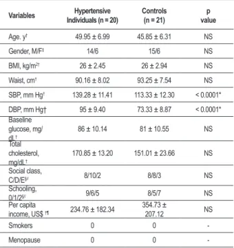
Out of the 433 hypertensives, 24 (5.5%) met the selection criteria and 20 (4.6%) completed the protocol. As for the control group, out of the 63 volunteers studied, 21 (33.33%) met the selection criteria and completed the protocol. Therefore, the study was conducted with 20 hypertensives and 21 controls, whose demographic, socioeconomic, anthropometric and biochemical characteristics are listed in Table 1.
Table 1 - General characteristics of controls and hypertensive individuals
Variables Individuals (n = 20)Hypertensive Controls(n = 21) valuep
Age. y† 49.95 ± 6.99 45.85 ± 6.31 NS
Gender, M/F‡ 14/6 15/6 NS
BMI, kg/m2† 26 ± 2.45 26 ± 2.94 NS
Waist, cm† 90.16 ± 8.02 93.25 ± 7.54 NS
SBP, mm Hg† 139.28 ± 11.41 113.33 ± 12.30 < 0.0001*
DBP, mm Hg† 95 ± 9.40 73.33 ± 8.87 < 0.0001*
Baseline glucose, mg/ dL†
86 ± 10.14 81 ± 10.55 NS
Total cholesterol, mg/dL†
170.85 ± 13.20 151.01 ± 23.66 NS
Social class,
C/D/E§// 8/10/2 8/8/3 NS
Schooling,
0/1/2§// 9/6/5 8/5/7 NS
Per capita
income, US$ †¶ 234.76 ± 182.34
354.73 ±
207.12 NS
Smokers 0 0
-Menopause 0 0
-*P values denote differences between hypertensive and control; † Student’s t test; ‡ Pearson’s χ2 test; § Fischer’s exact test; // CCEB criteria ; ¶ November 10. 2006.
Table 2- Antihypertensive drug treatment
Antihypertensive drug treatment
Medication use and dosage (mg)
Patients (n, %)
Diuretic HCT†, 25 mg 6 (30%)
ACE‡ inhibitor Captopril 25 mg 3 (15%)
Captopril 50 mg 2 (10%)
Diuretic + ACE inhibitor
HCT 25 mg + Captopril 25 mg 4 (20%)
HCT 50 mg + Captopril 50 mg 2 (10%)
Diuretic + ACE inhibitor + beta-blocker
HCT 25 mg + Captopril 25 mg
+ Atenolol 40 mg 1 (5%)
No drug 2 (10%)
Total patients 20 (100%)
†HCT- Hydrochlorothiazide; ‡ ACE - Angiotensin-Converting Enzyme.
Table 3 - Antioxidants and biomarkers of oxidative stress in the study population
Groups of Biomarkers
Study population and values obtained
(mean ± SD and median) p value
Hypertensives (H) (n = 20)
Controls(C) (n = 21)
Antioxidant enzymes
SOD (U/gHb) † 1,498.85 ± 575.04
1,470.00
1,649 ± 407.43
1,722.00 NS
CAT (KU/gHb)† 68.00 ± 32.83
67.24
102.08 ± 49.36
102.48 0.013*
GPx (U/gHb)‡ 20.51± 9.42
21.51
7.77 ± 9.60
2.00 0.0001*
Antioxidant of low molecular mass
GSH (mM)‡ 5.33 ± 2.87
4.80
8.96 ± 5.19
7.00 0.003*
Uric acid (mg/dL) ‡ 4.08 ± 1.33
3.80
3.49 ± 0.83
3.60 NS
Ascorbate (mmol/L) † 40.53 ± 12.93
37.63
34.58 ± 9.55
32.00 NS
Transport proteins Fe+2/+2 Cu+/+2
Transferrin (mg/dL) † 189.60 ± 32.96
188.00
194.14 ± 38.42
189.00 NS
Ceruloplasmin (mg dL) ‡ 38.60 ± 8.61
37.00
33.86 ± 3.97
33.00 0.015*
Biomarkers of oxidative damage
Malondialdehyde (mmol/L) ‡ 2.80 ± 4.49
1.42
8.51 ± 6.83
8.81 0.014*
Carbonyl (nmol/gPtna) ‡ 2.13 ± 1.66
1.71
1.91 ± 1.42
1.67 NS
*P values denote differences between hypertensive and controls; † Student’s t test; ‡ Mann-Whitney’s test. NS - not signiicant. Treatment with antihypertensive medication
The hypertensives were under antihypertensive therapy and regular users of antihypertensive medication (Table 2). In this group, 30% were users of diuretics, 25% used an ACE inhibitor, and 30% used an association of two medications.
Antioxidants and biomarkers of oxidative damage
Hypertensive individuals presented reduced blood levels of SOD, CAT, GSH, transferrin and MDA and high blood levels of GPx, ascorbate, CER and carbonyl compared to the controls. However, only the blood levels of CAT, GSH and MDA (reduced) and GPx and CER (increased) showed significant differences (Table 3).
Correlation among pressure levels and Biomarkers of Oxidative Damage
Pearson and Spearman correlation tests had revealed a positive correlation between SBP and MDA (r = 0.44 e p = 0.04) in the study population, and between PAD and CAT (r = 0.54 and p = 0.01) into the controls. Among the biomarkers, a negative correlation was found between MDA and GPx in both groups (r = -0.63 and p = 0.003 in the study population and r = -0.63 and p = 0.004 into the controls).
Discussion
have demonstrated an increase in the production of reactive species in patients with essential hypertension17-36 (Table 4).
There is a high production of O2•
in hypertension that would
lead to the formation of ONOO (by reaction of O
2• with
•NO), which occurs at a constant rate of 6.7 x 10⁹ mol L-1 s-1 16, while the dismutation reaction of O
2
• by SOD occurs at a
slower rate: 1.6 x 10⁹ mol L-1 s-1 4. Furthermore, SOD activity
is favored when the concentration of O2• is low and that of
SOD is high, which occurs only in physiological conditions3.
These aspects would explain, in part, the prooxidant phenomena of hypertension3,4,16. Additionally, the activity of
the antioxidant systems would be reduced, which would also favor the oxidative stress present in hypertension.
Clinical studies have demonstrated reduced antioxidant enzyme activity among hypertensives compared with controls17,18, but it is not yet clear if this is a cause or a
consequence of the hypertensive condition. Thus, oxidative stress in a chronic process such as hypertension could consume the reserves and impair antioxidant enzyme activity.
In view of the above, one would expect to see high levels of oxidative damage and low levels of antioxidants in hypertensives. However, in this study, the hypertensive group showed only lower levels of CAT and GSH antioxidants, higher levels of GPx and CER, and lower levels of the marker of oxidative damage, MDA, in comparison with the control group (Table 3). In fact, as far as antioxidant enzymes are concerned, studies of hypertensives have found low levels of SOD, CAT
Table 4 - Studies of biomarkers of oxidative stress in hypertension
Studies/
References n
Biomarkers Studied
Results Obtained H vs. Ca and Hb
19 H = 30
C = 164 SOD and GPx SOD and GPx decreased
a
34 H = 30C = 30 F2-ISO No difference
a
18 H = 66
C = 16
GSH, GSSG, GSH/GSSG SOD, CAT, GPx, MDA,
8-OxoGUA
GSH/GSSG and MDA augmenteda
SOD, CAT, GPx and GSH decreaseda
34
H = 38 C1 = 21 C2 = 17
GSH SOD, CAT, GPx, GST
Nitrate/Nitrite Carbonyl and MDA
Carbonyl and MDA augmenteda
SOD decreaseda
CAT and GPx similara
24 H = 89 GSH, GSSG,GSH/GSSG
SOD and MDA AO raised and MDA
b decreased
23
H1 = 70 H2 = 85 C = 40
F2ISO
Vitamin C and E, Uric Acid F2ISO decreased in H treated (H2)
a
17 H and C,
do not refer to n
GST, GPx. Asc., thiols
FRAP ( Fe 3+) GST, GPx, Asc., Thiols and FRAP decreased
a
37 H + ICC = 23
C = 50
SOD and CAT MDA
SOD and CAT augmenteda
MDA decreaseda
20 H = 39 FRAP FRAP raised H diureticb
36 H = 83
C = 50
F2ISO
CRP and TNFα F2ISO augmented
a
CRP and TNFα augmenteda
H – Hypertensive; C – Control; SOD - superoxide dismutase; GPx - glutathione peroxidase; F2ISO- F2-Isoprostane; GSH - glutathione reduced form; GSSG - glutathione oxidized form; MDA – malondialdehyde; CAT – catalase; 8-OxoGUA - 8-oxoguanina; GST - glutathione S-transferase; AO – antioxidant; Asc – Ascorbate;
FRAP - Ferric reducing ability of plasma; CRP- C reactive protein; TNFα -Tumor necrosis factor-alpha.
and GPx17-19 (Table 3) similarly to those of this study, except
GPx, which was inexplicably high for Ide et al20.
Catalase may be inhibited in the presence of inadequately removed O2•, generating ferroxycatalase which does not
decompose H2O2 rapidly10. This would explain not only
the low CAT levels but also the high GPx levels, possibly indicating a greater demand for this enzyme, since it is able to reduce ONOO efficiently, thus preventing macromolecular oxidation and protein nitration10. Moreover, although no
significant differences were found in SOD, one can consider that the reduced levels of this enzyme were probably due to the reaction of O2•and •NO, which is faster than the
reaction of O2•and SOD, along with the fact that it acts
more efficiently in physiological conditions (low concentration of O2•). Returning to CAT, one can consider that, with
O2•“deviated” to form ONOO, H2O2 production via SOD is
lower or remains at normal levels, not requiring extra activity. Moreover, as mentioned earlier, this enzyme can be inhibited in the presence of accumulated O2•and its action confined
to the cell’s peroxisomes.
Concerning drug therapy, the antihypertensive medications that act as antioxidants are: (1) ACE inhibitors and AT1 receptor blockers, which act indirectly by inhibiting the renin-angiotensin-aldosterone system (RAAS), an important source of ROS in the endothelium 3,4,7,8,11; (2) third generation
increasing the availability of nitric oxide in the endothelial cell and increasing the expression of MnSOD in vascular smooth muscle cells22.
In the hypertensive group, the use of an ACE inhibitor (captopril) by 60% of the patients (40% with 25 mg/day and 20% with 50 mg/day) requires additional comments. The antioxidant mechanism of this medication, though indirect and without a defined dose-response relationship, involves inhibition of the RAAS by inhibiting the action of angiotensin II (ANG II), which is the antioxidant link and a potent stimulus for ROS production in the endothelial cell, increasing the NADPH oxidase activity. In addition, ANG II also supra-regulates eNOS activity, which is accompanied by decoupling of the enzyme, reduction of •NO production and increase of superoxide production10. This effect was observed in a study
that found an inverse association between f2-isoprostane and number and type of antihypertensive medication: 46% of treated hypertensive patients were using ACE inhibitor and 26%, AT1 receptor blocker23. In addition, the antioxidant
action of beta-blockers and AT1 receptor antagonists was
also reported in a study of hypertensives treated with these drugs, whose SOD, CAT and GPx levels increased and 8-oxo-2’-deoxyguanosine and MDA levels decreased24. In contrast,
another study25 found increased total antioxidant capacity in
the plasma of hypertensives using thiazide diuretic, but did not find the same positive correlation with ACE inhibitor and beta-blocker, despite their antioxidant action.
The positive correlation evidenced between SBP and MDA in hypertensives is indicative of the association between HAS and oxidative stress, mainly after the negative relationship obtained between MDA and GPx in both groups.
Because glutathione acts together with GPx, the results were analyzed jointly, considering both of them fundamental in the defense against lipid peroxidation. Each unit of GPx acts by consuming 2 molecules of GSH, which is the most important intracellular antioxidant and is present in the cell predominantly in the reduced form (GSH), to the detriment of the oxidized form (GSSG). The GSH/GSSG > 1 ratio, which is vital for the cell, is maintained by an efficient recycling system of GSH from GSSG10.
In hypertension, low GSH levels and GSH/GSSG <1 ratio have both been found18. In this study, GSSG was not
measured. Our findings of low GSH levels are consistent with the literature and would be explained by oxidative stress, since ROS oxidize GSH to GSSG, leading to a drop in GSH, which is worsened by the conversion of GSH into GSSG in the process of peroxide detoxification by the action of GPx. As for uric acid and vitamin C, the higher levels observed in the hypertensive group were not statistically different, although it is known that hyperuricemia is associated with hypertension28 and the antioxidant activity of urate involves
different reactions (with R•, ROO•, ONOO, and ONO
2
•)10,14
acting cyclically, since it can be recovered by ascorbate, among others. It is considered a potent plasmatic antioxidant, once its concentration in plasma is tenfold higher than other antioxidants such as vitamins E and C13.
The transferrin levels were similar in the two groups, but the levels of ceruloplasmin, which is also a ferroxidase,
were higher in the hypertensive group (Table 3). This finding also represents an important factor of protection, in view of the activity of ceruloplasmin in transporting copper and oxidizing iron for capture by transferrin, i.e., it acts on the most important transition metals with respect to the ability to transfer electrons in their free form in biological systems. Other antioxidant mechanisms include O2• and H
2O2
sequestration, inhibition of the Fenton reaction, protecting the biological tissues from the damaging effects of iron decompartmentalization, inhibition of lipid oxidation and blocking of protein and DNA damages, which is verified by inhibition of carbonyl formation and protection of the cell against damage and lysis caused by ROS11,27. However,
under oxidative stress, ceruloplasmin may act as a prooxidant in the intravascular medium, since ONOOand H2O2 can
induce the dissociation of the free Cu2+ bond of the protein,
favoring its release into the intracellular medium, as well as diminishing its ferroxydase activity27. Indeed, several
studies have found a correlation between ceruloplasmin and cardiovascular disease, and some prospective studies and control cases have indicated it as a cardiovascular risk factor28. This is a biomarker whose prooxidant activity
seems to predominate in certain circumstances, including cardiovascular disease.
A decrease was observed in the phenomenon of lipid peroxidation (LP) among the hypertensives, since the presence of MDA (the most abundant reactive aldehyde of LP) in the serum of these individuals was lower than in that of the controls (Table 3). The presence of carbonyl groups at similar levels, in this case, may reinforce the assumption that, if the hypertensive group was under oxidative stress, ONOO would be the prevailing reactive species, since it is
a poor inducer of carbonyl proteins37. It is worth mentioning
that, in the choice of the marker of oxidative stress, the nature of the oxidative stress under study plays an extremely important role. However, the methodology available and the feasibility of applying analytical techniques are of equal importance, and were particularly determinant in the choices for this study.
Several studies have found a positive correlation between high levels of MDA and cardiovascular diseases such as acute myocardial infarction, congestive heart failure and hypertension29-31 in hypertensives without drug therapy18 and
in elderly hypertensives using antihypertensive medication32.
On the other hand, studies involving hypertensives who have never been treated33 and treated hypertensives23,34 found no
differences, among hypertensives and controls, in the levels of F2-isoprostane, another marker of lipid peroxidation, despite
the antioxidant action of antihypertensive drugs. However, another study found a positive correlation between this damage marker and inflammation markers in hypertensives not yet under drug therapy35. Patients with class II to class
IV congestive heart failure showed reduced levels of MDA36.
The results indicate the possible association and LP protection mechanisms, such as GPx and CER, which were found in high levels.
channel-blocking drugs and ECA inhibitors) produces a significant increase of •NO, but antioxidant enzyme levels remain low when compared with normotensives17. Another interesting
aspect is the possibility of CER inhibiting protein oxidation, which was found in an endothelial cell culture through the formation of carbonyl in the presence of ceruloplasmin38.
The phenomenon of oxidative stress, as well as the antioxidant system, works in an integrated fashion, in a series of related events. This characteristic and the complexity of these processes necessarily require a non-compartmentalized discussion. Moreover, one must keep in mind the duality of the redox environment: high antioxidant levels are not necessarily desirable or low antioxidant levels undesirable since both may result in oxidative stress.
Although it is not clear whether oxidative stress in hypertension is a cause or an effect, the occurrence of oxidative stress in hypertension was found in this study, corroborating previously published findings.
The role of oxidative stress in hypertension is already clear and well founded. However, although various studies point to endothelial dysfunction and to the imbalance between the reactive oxygen and nitrogen species and antioxidant defenses, it is not possible to define whether redox imbalance is a cause or a consequence of blood pressure homeostasis. This study revealed oxidative stress in hypertension. Nevertheless, additional studies are needed to elucidate the mechanisms that lead to the genesis of this and other clinical situations characterized by endothelial dysfunction involving redox imbalance. Hence, our research group is currently engaged in studies based on biomarkers of redox imbalance in patients with metabolic syndrome, refractory hypertension and diabetes mellitus, which, like hypertension, are diseases whose common denominator is the endothelial dysfunction.
Conclusion
Lastly, it can be concluded that, with regard to antioxidants and oxidative damage markers, the reduced CAT and GSH levels and high ceruloplasmin levels found in the hypertensive patients indicate that they are under oxidative stress, despite the possible mitigating effect of their antihypertensive medication. High GPx and low MDA levels may also result from oxidative stress, since (1) the enzyme would be in greater demand in the presence of excess ONOO¯, a reactive species characteristic of hypertension, and (2) due to its significant action upon ROO•, there would be a decrease in LP, with a consequent reduction of MDA. Hence, the oxidative stress of hypertension, as far as these biomarkers are concerned, would be explained by an alternative mechanism.
Acknowledgements
The authors are grateful to the patients and volunteers who so willingly participated in this study.
Potential Conflict of Interest
No potential conflict of interest relevant to this article was reported.
Sources of Funding
This study was funded by FAPEAL, CNPq, CAPES, CAPES/ COFECUB, CNPq/PADCT, BNB e FAPEAL/SESAU-AL, MS/ DECIT-PPSUS.
Study Association
This article is parto f the thesis of doctoral submitted by Sandra Mary Lima Vasconcelos, from Universidade Federal de Alagoas and redox analysis of biomarkers in Universidade Federal do Rio Grande do Sul.
References
1. Sociedade Brasileira de Cardiologia. VI Diretrizes Brasileiras de Hipertensão. Rev Hipertens. 2010;17(1):1-66.
2. Touyz RM. Oxidative stress and vascular damage in hypertension. Curr Hypertens Rep. 2000;2(1):98-105.
3. Griendling KK, Fitzgerald GA Oxidative stress and cardiovascular injury. Part I: basic mechanisms and in vivo monitoring of ROS. Circulation. 2003;108(16):1912-6.
4. Griendling KK, Fitzgerald GA. Oxidative stress and cardiovascular injury. Part II: animal and humans studies. Circulation. 2003;108(17):2034-40.
5. Touyz RM. Reactive oxygen species, vascular oxidative stress, and redox signaling in hypertension. What is the clinical significance? Hypertension. 2004;44(3):248-52.
6. Touyz RM, Schiffrin EL. Reactive oxygen species in vascular biology: implications in hypertension. Histochem Cell Biol. 2004;122(4):339-52.
7. Portaluppi F, Boari B, Manfredini R. Oxidative stress in essencial hypertension. Curr Pharm Design. 2004;10(14):1695-8.
8. Sampaio WO, Santos RAS. Aplicações clínicas dos mecanismos fisiopatológicos da hipertensão arterial. Sistema renina-angiotensina: bases fisiopatológicas. Rev Bras Hipertens. 2004;11:67-70.
9. Paravicini TM, Touyz RM. Redox signaling in hypertension. Cardiovasc Res. 2006;71(2):247-58.
10. Halliwell B, Gutteridge JMC. Free radical in biology and medicine. 4 ed. Oxford: Oxford University Press; 2007.
11. Vasconcelos SML, Goulart MOF, Silva MAM, Gomes ACM. Hipótese oxidativa da hipertensão arterial: uma mini-revisão. Rev Bras Hipertens. 2004;14:269-74.
12. Kuklinska AM, Mroczko B, Muzial WJ, Usowicz-Szarynska M, Borowska H, Knapp M, et al. Diagnostics biomarkers of essential hypertension: the value of prostacyclin, nitric oxide, oxidize-LDL, and peroxide measurements. Int Heart J. 2009;50(3):341-51.
13. Pinho RA, Araujo MC, Ghisi GLM, Benetti M. Doença arterial coronariana, exercício físico e estresse oxidativo. Arq Bras Cardiol. 2010;94(4):549-55.
14. Vasconcelos SML, Goulart MOFG, Moura JBF, Manfredini V, Benfato MS, Kubota LT. Espécies reativas de oxigênio e nitrogênio, antioxidantes e marcadores de estresse oxidativo em sangue humano:principais métodos analíticos para sua determinação. Quim Nova. 2007;30:1323-38.
16. Cai H, Harrison DG. Endothelial dysfunction in cardiovascular disease: the role of oxidant stress Circ Res. 2000;87(10):840-4.
17. Khullar J, Relan V, Sherawat BS. Antioxidant activites and oxidative stress byproducts in human hypertension. Hypertension. 2004;43(2):e7-8.
18. Rédon J, Oliva MR, Tormo C, Giner V, Chaves J, Iradi A, et al. Antioxidant activities and oxidative stress byproducts in human hypertension. Hypertension. 2003;41(5):1096-101.
19. Pedro-Botet J, Covas MI, Martin S, Rubies-Prat J. Decrease endogenous antioxidant enzymatic status in essential hypertension. J Hum Hypertens. 2000;14(6):343-5.
20. Ide T, Tsutsui H, Ohashi N, Hayashidani S, Suematsu N, Tsuchihashi M, et al. Greater oxidative stress in healthy young men compared with premenopausal women. Arterioscler Thromb Vasc Biol. 2002;22(3):438-42.
21. Kalinowski L, Dobrucki L, Szczepanska-Konkel W, Jankowiski M, Martyniec L, Angielski S, et al. Third-generation beta-blockers stimulate nitric oxide release from endothelial cells through ATP efflux: a novel mechanism for antihypertensive action. Circulation. 2003;107(21):2747-52.
22. Berkels R, Egink G, Marsen TA, Bartels H, Roesen R, Klaus W. Nifedipine increases endothelial nitric oxide bioavailability by antioxidative mechanisms. Hypertension. 2001;37(2):240-5.
23. Ward NC, Hodgson JM, Puddey IB, Mori TA, Beilin LJ, Croft D. Oxidative stress in human hypertension: association with antihypertensive treatment, gender, nutrition, and lifestyle. Free Radic Biol Med. 2004;36(2):226-32.
24. Sáez GT, Tormos C, Giner V, Chaves J, Lozano JV, Iradi A, et al. Factors related to the impact of antihypertensive treatment in antioxidant activities and oxidative stress by-products in human hypertension. Am J Hypertens. 2004;17(9):809-16.
25. Skalska A, Gasowski J, Stepniewski M, Grodziki T. Antioxidative protection in hypertensive patients treated with diuretics. Am J Hypertens. 2005;18(8):1130-2.
26. Johnson RJ, Rodriguez-Iturbe B, Kang DH, Feig DI, Herrera-Acosta J. A unifying pathway for essential hypertension. Am J Hypertens. 2005;18(3):431-40.
27. Shukla N, Maher J, Masters J, Angelini GD, Jeremy JY. Does oxidative stress change ceruloplasmin from a prospective to a vasculopathic factor? Atherosclerosis. 2006;187(2):238-50.
28. Fox PL, Mazumder B, Ehrenwald E. Ceruloplasmin and cardiovascular disease. Free Radic Biol Med. 2000;28(12):1735-44.
29. Pucheu S, Coudray C, Vanazetto G, Favier A, Machecourt J, De Leiris J. Assessment of radical activity during the acute phase of myocardial infarction following fibrinolysis: utility of assaying plasma malondialdehyde. Free Radic Biol Med. 1995;19(6):873-81.
30. Diaz-Vélez CR, Garcia-Castiñeiras S, Mendoza-Ramos E, Hernández-López E. Increased malondialdehyde in peripheral blood of patients with congestive heart failure. Am Heart J. 1996;131(1):146-52.
31. Ghiadoni L, Magaga A, Versari D, Kardasz I, Huang Y, Taddei S, et al. Different effect of antihypertensive drugs on conduit artery endothelial function. Hypertension. 2003;41(6):1281-6.
32. Kedziora-Kornatowska K, Czuczejko J, Pawluk H, Kornatowski T, Motyl J, Szadujkis-Szadurski L, et al. The markers of oxidative stress and activity of the antioxidant system in the blood of elderly patients with essential arterial hypertension. Cell Mol Biol Lett. 2004;9(4A):635-41.
33. Cracowski JL, Baguet JP, Ormezzano O, Bessard J, Stanke-Labesque F, Bessard G, et al. Lipid peroxidation is not increased in patients with untreated mild-to-moderate hypertension. Hypertension. 2003;41(2):286-8.
34. Minuz P, Patrignani P, Gaino S, Degan M, Menapace L, Tommasoli R, et al. Increased oxidative stress and platelet activation in patients with hypertension and renovascular disease Circulation. 2002;106(22):2800-5.
35. Cottone S, Mulè G, Nardi E, Vadalà A, Guarneri M, Briolotta C, et al. Relation of C-reactive protein to oxidative stress and to endothelial activation in essential hypertension.36. Sundal S, Sharma M, Negi PC, Katoch SS. Oxidative stress and antioxidant profile in patients of heart failure. Asian J Exp Sci. 2005;19:41-58.
37. Shacter E. Quantification and significance of protein oxidation in biological samples. Drug Metab Rev. 2000;32(3-4):307-26.