349
Do children need fasting before abdominal ultrasonography?
Radiol Bras. 2009 Nov/Dez;42(6):349–352 Original Article • Artigo Original
Do children need fasting before abdominal
ultrasonography?*
Crianças necessitam de jejum antes de ultrassonografia abdominal?
Luiza Alina Almeida Araújo Rabelo1, Ilka Rocha Florêncio2, Iggor Medeiros Pirauá2, Silvio Cavalcanti de Albuquerque1, João Vicente Ribeiro Neto1, Eduardo Just da Costa e Silva3
OBJECTIVE: The present study is aimed at comparing the quality of sonographic abdominal images obtained in fasting and non fasting children. MATERIALS AND METHODS: This is a prospective study including children aged up to 12 years sequentially evaluated by two sonographers. The images were classified according to a score as follows: 1 (non-visualized or partially visualized, inappropriate for diagnosis); 2 (sufficient for diagnosis); or 3 (excellent). Images were also classified into “diagnostic” or “non diagnostic”. RESULTS: Seventy-seven patients (47 boys and 30 girls) with ages ranging between 0 and 12 years (median = 1 year) were evaluated. Fasting proved a statistically significant advantage only for evaluating the gallbladder by only one of the observers (p = 0.032). Once the images were classified into either “diagnostic” or “non diagnostic” no difference was observed between the two groups. CONCLUSION: The authors conclude that fasting did not affect significantly the quality of abdominal sonographic images in children.
Keywords: Ultrasonography; Children; Abdominal; Fasting.
OBJETIVO: Comparar a qualidade de imagens ultrassonográficas do abdome de crianças, obtidas com e sem a instituição de jejum prévio. MATERIAIS E MÉTODOS: Trata-se de estudo prospectivo, incluindo crianças com até 12 anos de idade. Os pacientes foram examinados sequencialmente por dois utrassonografistas e as imagens foram classificadas em escores: 1 (não visualizado ou parcialmente visualizado, inadequada para diagnóstico); 2 (suficientes para diagnóstico); 3 (excelentes). As imagens foram ainda classificadas como “diagnósticas” ou “não diagnósticas”. RESULTADOS: Foram examinados 77 pacientes, sendo 47 meninos e 30 meninas, com idades entre 0 e 12 anos (mediana de 1 ano). Jejum se mostrou vantajoso de forma estatisticamente significativa apenas na avaliação da vesícula biliar, por apenas um dos avaliadores (p = 0,032). Depois de agrupadas em “diagnóstica” ou “não diagnóstica”, nenhuma diferença foi observada entre os grupos. CONCLUSÃO: A instituição de jejum não afetou de forma significativa a qualidade das imagens de ultrassonografias abdominais obtidas em crianças.
Unitermos: Ultrassonografia; Crianças; Abdominal; Jejum.
Abstract
Resumo
* Study developed at Instituto de Medicina Integral Professor Fernando Figueira, Recife, PE, Brazil.
1. MDs, Radiologists at Instituto de Medicina Integral Profes-sor Fernando Figueira, Recife, PE, Brazil.
2. Graduate of Medicine, Faculdade Pernambucana de Saúde (FPS), Recife, PE, Brazil.
3. Master, Fellow PhD degree in Child and Adolescent Health, Universidade Federal de Pernambuco (UFPE), Radiologist at Ins-tituto de Medicina Integral Professor Fernando Figueira, Docent at Laboratório de Anatomia por Imagem – Faculdade Pernam-bucana de Saúde (FPS), Recife, PE, Brazil.
Mailing address: Dr. Eduardo Just. Instituto de Medicina Inte-gral Professor Fernando Figueira – Radiologia. Rua dos Coe-lhos, 300, Boa Vista. Recife, PE, 50070-550, Brazil. E-mail: [email protected]
Received June 21, 2009. Accepted after revision October 15, 2009.
pectation that the gastrointestinal contents are reduced and an appropriate distension of the gallbladder is achieved(7,9–12).
Four-to six-hour fasting is routinely recom-mended for children(9,13).
However, this practice may be very dis-comfortable and poorly tolerated by many patients. Children with hunger may become quite irritated, affecting the quality of the study and causing anxiety for parents. Ad-ditionally, small children may even develop hypoglycemia episodes after short fasting periods. The benefits of fasting for the sonographic images quality has been dis-cussed by some authors(7,8). According to
the authors, there is no study approaching this topic specifically in relation to children. The objective of the present study is to compare the quality of images acquired
Rabelo LAAA, Florêncio IR, Pirauá IM, Albuquerque SC, Ribeiro Neto JV, Costa e Silva EJ. Do children need fasting before abdominal ultrasonography? Radiol Bras. 2009;42(6):349–352.
characterization of a wide range of condi-tions(1–4). Many times, it is demonstrated
that ultrasonography is the only imaging method that may be required for such evaluation(1,5), with advantages including
portability, low cost, accuracy and absence of ionizing radiation(6).
However, many factors may affect the quality of sonographic images. Presence of intestinal gas is considered as a limiting factor in the evaluation of the pancreas, gall bladder and gastrointestinal tract(7).
An appropriate distension of the gall-bladder and reduced presence of gas in the digestive tract are considered as optimum conditions for the acquisition of appropri-ate images(8).
Many authors recommend that the amination is preceded by fasting, in the
ex-0100-3984 © Colégio Brasileiro de Radiologia e Diagnóstico por Imagem
INTRODUCTION
350
Rabelo LAAA et al.
Radiol Bras. 2009 Nov/Dez;42(6):349–352
during abdominal ultrasonography in chil-dren previously submitted to fasting with those from children fed immediately before the examination.
MATERIALS AND METHODS
The present study was previously ap-proved by the Committee for Ethics in Research of the Institution, and a term of free and informed consent was signed by all the parents and guardians after an expla-nation about the nature and objective of the research. This is a prospective study includ-ing children up to 12 years with clinical complaints not related to the abdomen.
Convenience was a key factor in the sampling for allowing the inclusion of pa-tients present in the hospital at the moment where the two radiologists participating in the study were available in the department, allowing comparison between images ac-quired in the same conditions. Because of the present study nature, including children with no abdominal complaint, the selected samples did not present any particular char-acteristic that might have contributed for influencing such selection. All the indi-viduals included in the present study were inpatients so their meals timetable could be managed accordingly. A list including all the inpatients either with no abdominal complaint or previous history of abdomi-nal surgery was obtained.
The patients were divided into two groups as follows: group 1, including fast-ing children, and group 2, includfast-ing chil-dren evaluated 60 minutes after their usual meal.
The studies were sequentially per-formed by two medical sonographers in-volved in a daily practice of pediatric pe-diatric ultrasonography, one of them with six-year- and the other with three-year-ex-perience. The standard technique was uti-lized, with the patients in dorsal decubitus, with all the examinations being performed in a Sonosite Titan unit (Sonosite Inc.; Bothell, USA), with linear (L25, 5–10 MHz) and convex (C11, 5–8 MHz) transducers. The sonographers did not know the patients and were not authorized to ask questions to their parents or guardians. Images of the liver, gallbladder, spleen, kidneys, pan-creas, retroperitoneum, mesenterium
(ves-sels, fat and lymph nodes) and bowels were obtained and classified according to a method described in a previous study(8), as
follows: score 1 (non-visualized or partially visualized, inappropriate for diagnosis); score 2 (sufficient for diagnosis); score 3 (excellent, appropriate for classes of sonographic anatomy).
Statistical analysis
The statistical significance of the differ-ences between the groups was evaluated by means of the Mann-Whitney test. Subse-quently, images classified as 3 or 2 were grouped and named as “diagnostic”, the other images, classified as 1, being named “non-diagnostic”. The statistical signifi-cance of the differences observed with this new scoring system was evaluated by the chi-squared method.
The interobserver agreement was evalu-ated by the kappa coefficient of agree-ment(10), with the sole objective of having
a gross evaluation of the method applied in the calcification of the images quality, with no direct relation with the objective of the present study. The significance level adopted was 95%.
RESULTS
The sample of the present study in-cluded 77 patients – 47 (61%) boys and 30 (39%) girls – with ages ranging from one week to 12 years (mean age, 2.7 years).
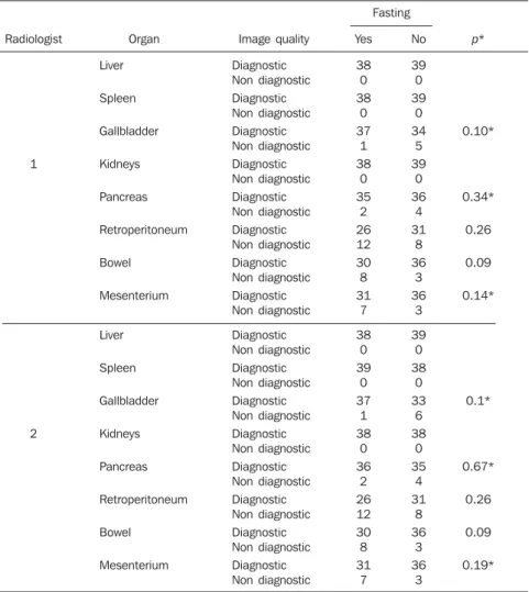
Fasting has shown to be advantageous only in the evaluation of the gallbladder by the sonographer 2 (p = 0.032), this ad-vantage being not observed by the sono-grapher 1.
Table 1 Relation between alimentary status and quality of sonographic images defined as “diagnostic” and “non-diagnostic”. Radiologist 1 2 Organ Liver Spleen Gallbladder Kidneys Pancreas Retroperitoneum Bowel Mesenterium Liver Spleen Gallbladder Kidneys Pancreas Retroperitoneum Bowel Mesenterium Image quality Diagnostic Non diagnostic Diagnostic Non diagnostic Diagnostic Non diagnostic Diagnostic Non diagnostic Diagnostic Non diagnostic Diagnostic Non diagnostic Diagnostic Non diagnostic Diagnostic Non diagnostic Diagnostic Non diagnostic Diagnostic Non diagnostic Diagnostic Non diagnostic Diagnostic Non diagnostic Diagnostic Non diagnostic Diagnostic Non diagnostic Diagnostic Non diagnostic Diagnostic Non diagnostic Yes 38 0 38 0 37 1 38 0 35 2 26 12 30 8 31 7 38 0 39 0 37 1 38 0 36 2 26 12 30 8 31 7 No 39 0 39 0 34 5 39 0 36 4 31 8 36 3 36 3 39 0 38 0 33 6 38 0 35 4 31 8 36 3 36 3 p* 0.10* 0.34* 0.26 0.09 0.14* 0.1* 0.67* 0.26 0.09 0.19* Fasting
351
Do children need fasting before abdominal ultrasonography?
Radiol Bras. 2009 Nov/Dez;42(6):349–352
phy in cases of abdominal emergency(15,16).
Such phenomenon contributes for an ex-tension of the waiting time for the previ-ously scheduled inpatients, considering that, usually, patients coming from the emergency department are examined at the intervals between previously scheduled studies.
The results of the present study demon-strate that previous fasting in children to be submitted to abdominal ultrasonography does not contribute to the acquisition of higher quality images. A higher score was observed only on sonographic images of the gallbladder, but solely by one of the sonographers. Nevertheless, no difference was observed as the images were classified into “diagnostic” or “non-diagnostic”.
Studies on this topic approaching spe-cifically children are not available in the lit-erature, but the results of the present study are similar to those observed with adults. Windler et al. have observed that the weight/height ratio was the most relevant determining factor in the evaluation of ab-dominal sonographic images quality in their study(7). Also, they have observed that
fasting contributed for higher quality im-ages only in the evaluation of the biliary tract. Additionally, images of the right kid-ney achieved higher scores in nonfasting patients, a finding that has not been ob-served in the present study. The inclusion of adult individuals in the mentioned study may explain this small discrepancy be-tween the results. Abdominal sonographic images of children are usually better be-cause of the small dimensions of the pedi-atric abdomen. Sinan et al. have not found any difference in scores among adult indi-viduals submitted to previous fasting and those previously fed(8). The number of
“di-agnostic” images in such study, however, was lower than in the present one, a find-ing that also may be attributed to the inclu-sion of adult individuals.
Fasting may be problematic in some cases. This practice should not be adopted if good quality images can be obtained in nonfasting individuals. At the unit of ultra-sonography of large hospitals, high num-ber of patients coming from the emergency department and inpatients with complica-tions requiring other further imaging stud-ies replace previously scheduled patients,
extending considerably the waiting time, much more than expected since the num-ber of unexpected examinations may be high. Hunger may be extremely distressing for children, and may lead to hypoglycemia and dehydration. Weep and irritability may even impair the study performance. Many patients may even refuse to wait, giving up submitting to the examination, which may lead to delayed diagnosis.
The present study had several limita-tions. The evaluation of sonographic im-ages through scores does not reflect the actual accuracy of the method. Ideally, it would be necessary to evaluate the perfor-mance of the method in the detection of specific diseases such as retroperitoneal lymphadenomegaly, cholelithiasis and thickening of intestinal loops. Also, clini-cally relevant situations, such as detection and staging of neoplasms and evaluation of inflammatory bowel diseases should be included. A situation where fasting could be useful is the evaluation of neonatal cholestatic icterus, since the evaluation of the gallbladder is required in this case. A previous meal may impair the study of this organ because of the vesical contractil-ity(12). Another limitation is the absence of
data regarding body mass index or body weight that presumably could influence the results. Additionally, the adopted scoring system is subjective and may affect the study reproducibility.
Although the present study has not been designed to evaluate the quality of sono-graphic scoring systems, the interobserver agreement obtained indicates the necessity of developing other systems for evaluating the quality of sonographic images. How-ever, the system adopted has been utilized in several studies with similar objec-tives(7,8,17,18). Studies approaching
interob-server agreement on any scoring system are not available in the literature. Such studies are required in addition to the development of better-defined systems for evaluating the quality of sonographic images.
CONCLUSION
The authors conclude that the practice of fasting was not essential for the acqui-sition of quality abdominal sonographic images in the evaluated children. Further
Table 2 Interobserver agreement related to the organ after images classification into “diagnostic” and “non-diagnostic”.
Organ
Liver
Spleen
Kidneys
Gallbladder
Pancreas
Retroperitoneum
Mesenterium
Bowel
Kappa (CI 95%)
1.0*
1.0*
1.0*
0.51 (0.32–0.71)
0.27 (0.05–0.5)
0.46 (0.23–0.68)
1.0 (0.77–1.22)
0.17 (–0.05–0.39)
CI, confidence interval; * Agreement ratio. No statistically significant difference was observed between the groups of “di-agnostic” and “non-di“di-agnostic” images (Table 1). Ages did not demonstrate any significant correlation with the scores ob-served in fasting and nonfasting patients.
The interobserver agreement in relation to the classification of images into “diag-nostic” and “non-diag“diag-nostic” is shown on Table 2.
DISCUSSION
The prescription of any type of previous preparation for an imaging study is aimed at making the procedure safer (like in the case where an antiallergenic preparation is prescribed before imaging studies requir-ing iodinated contrast injection), or im-proving the images quality, providing higher safety and diagnostic efficacy(14).
However, any prescription of either a drug or simply fasting must be based on studies that demonstrate an actual benefit from its practice.
ultrasonogra-352
Rabelo LAAA et al.
Radiol Bras. 2009 Nov/Dez;42(6):349–352
studies evaluating other variables such as age and body mass index, besides usual clinical situations are required. Also, it is important to note the need for developing better methods for evaluating the quality of abdominal sonographic images.
REFERENCES
1. Strouse PJ. Sonographic evaluation of the child with lower abdominal or pelvic pain. Radiol Clin North Am. 2006;44:911–23.
2. Levy JA, Noble VE. Bedside ultrasound in pediatric emergency medicine. Pediatrics. 2008; 121:e1404–12.
3. Haber HP. Cystic fibrosis in children and young adults: findings on routine abdominal sonography. AJR Am J Roentgenol. 2007;189:89–99. 4. Rocha SMS, Ferrer APS, Oliveira IRS, et al.
De-terminação do tamanho do fígado de crianças normais, entre 0 e 7 anos, por ultrassonografia. Radiol Bras. 2009;42:7–13.
5. Eppich WJ, Zonfrillo MR. Emergency department evaluation and management of blunt abdominal
trauma in children. Curr Opin Pediatr. 2007;19: 265–9.
6. Costa JD, Leão ARS, Santos JEM, et al. Quanti-ficação do fluxo portal em indivíduos sadios: comparação entre ressonância magnética e ultra-som Doppler. Radiol Bras. 2008;41:219–24.
7. Windler EE, Lempp FL. US of the upper abdo-men: factors influencing image quality. Radiol-ogy. 1985;157:513–5.
8. Sinan T, Leven H, Sheikh M. Is fasting a neces-sary preparation for abdominal ultrasound? BMC Med Imaging. 2003;3:1.
9. Devos AS, Meradji M, Blickman JG. The small bowel. In: Devos AS, Blickman JG, editors. Ra-diological imaging of the digestive tract in infants and children. Berlin: Springer; 2008. p. 167–91. 10. Sommer G, Filly RA, Laing FC. Use of sime-thicone as a patient preparation for abdominal sonography. Radiology. 1977;125:219–21.
11. Scortegagna Junior E, Leão ARS, Santos JEM, et al. Avaliação da concordância entre ressonância magnética e ultra-sonografia na classificação de fibrose periportal em esquistossomóticos, se-gundo a classificação de Niamey. Radiol Bras. 2007;40:303–8.
12. Teixeira MS, Coelho CAR, Teixeira AS. Avalia-ção da contratilidade da vesícula biliar com leite materno e leite de vaca em lactentes. Radiol Bras. 2004;37:163–6.
13. Siegel MJ. Fígado. In: Siegel MJ, editor. Ultra-sonografia pediátrica. 3a ed. Rio de Janeiro:
Gua-nabara Koogan; 2003. p. 189–244.
14. Trindade R, Sumi DV, Kravetz WL, et al. Avalia-ção do conhecimento de médicos não-radiologis-tas sobre reações adversas aos contrastes iodados. Radiol Bras. 2007;40:321–8.
15. Cavalcanti AF, Menezes MR. Radiologia de emer-gência: perspectivas. Radiol Bras. 2001;34:v–vi.
16. Vabo KA, Torres Neto G, Santos AASMD, et al. Achados ultra-sonográficos abdominais em pa-cientes com dengue. Radiol Bras. 2004;37:159– 62.
17. Elam EA, Hunter TB, Hunt KR, et al. The lack of sonographic image degradation after barium up-per gastrointestinal examination. AJR Am J Roentgenol. 1989;153:993–4.