ABSTRACT
Histomorphometric assessment of bone necrosis
liquid nitrogen: an experimental study on rat
femurs
Fábio Wildson Gurgel COSTA1 2, Rosana Maria Andrade PESSOA3, Eduardo Costa
STUDART-SOARES4
1- DDS, MSc, Assistant Professor, Division of Stomatology, Department of Clinical Dentistry, Federal University of Ceará, Sobral, CE, Brazil.
2- MD, MSc, PhD, Associate Professor, Division of Histology, Department of Physiology and Pharmacology, School of Medicine, Federal University of Ceará, Fortaleza, CE, Brazil.
3- DDS, Graduate student, Division of Oral Surgery, Department of Clinical Dentistry, Federal University of Ceará, Fortaleza, CE, Brazil.
4- DDS, MSc, PhD, Associate Professor, Division of Oral Surgery, Department of Clinical Dentistry, Federal University of Ceará, Fortaleza, CE, Brazil.
Corresponding address: Fábio Wildson Gurgel Costa - Coordenação do Curso de Odontologia da Universidade Federal do Ceará - Campus Sobral - Av. - Comte. Maurocélio Rocha Pontes, s/nº Derby - 62041-040 - Sobral - Ceará - Brazil - Phone: +55 (88) 3613 2603 - Fax: +55 (88) 3613 2603 - e-mail: [email protected]
Received: September 24, 2009 - Accepted: April 30, 2010
O
on the femoral diaphysis of rats. Material and Methods: The femoral diaphyses of 42 ! " # $ ! ! % & ' # ( & %#")* + ),-#)*% + / ! ! %01#%% + )%1#%1 + / # 3& & # 5 / protocol produced more marked bone necrosis than the 1-min protocol. Although our results cannot be entirely extrapolated to clinical practice, they contribute to the understanding of the behavior of bone tissue submitted to different cycles of liquid nitrogen freezing and #
Key words: 5# 9# ; #
INTRODUCTION
$ <= >& <&=! # 5 destruction4. The locally lethal effects of this method are the result of cell dehydration and the formation of intracellular ice crystal, causing direct cytotoxic injury and secondary vascular ischemia11,12,13,25.
The maxillomandibular complex is prone to a variety of lesions that, although benign, might be locally aggressive9,10,22,24. Doubts exist regarding the best therapeutic approach in these cases, since ! !
although resulting in cure in most cases, may cause severe esthetic-functional impairment21. In this respect, adjuvant therapies, such as cryosurgery, increasing morbidity5,21,23.
diaphysis of rats using a standardized experimental model and histomorphometric parameters.
MATERIAL AND METHODS
Animals
K/ 1 &
(360-460 g), randomly chosen from the Central Animal House of the Federal University of Ceará, K'! 5S! V'! # /% h light/dark cycle at 23-25o5! # the Animal Care and Use Committee of the same X 1Y))1Z conducted according to recommended guidelines on animal experimentation.
Cryosurgery protocols
K' 5([/G5®-3 cryostat
(model #B-700, Brymill, imported from CRY-AC®, Brazil) using liquid nitrogen as coolant and ` # G / ' " ! ' 0
w V / ' " ! ' 1 #
Surgical procedures
' injection of 2.5% tribromoethane (0.1 mg/100 g Z# |! #"/ made along the lateral aspect of the right thigh from # # surface 1 cm from the head of the femur. Retractors adjacent soft tissues during the freezing process. G ! their initial position, and the soft tissues and skin # # | administered postoperatively.
Histologic preparation
> ! ! % & # ! $ 10% neutral buffered formaldehyde for 48 h at room ! $ "}
) # G $! ! ! $ that the femur could be sectioned in the sagittal # positioning a millimeter rule 1 cm from the femoral # ~ X%//&Z from the lateral border to the site of cryoapplication #
Histomorphometric analysis
K ! ; #%0 s (National Institutes of Health, Bethesda, MD, USA; YY##YYZ# central stained section that corresponded to the &# V light microscope (Leica Microsystems, Nussloch, Germany) connected to a Nikon Alphaphot-2 VS2 digital camera (Nikon, Tokyo, Japan). In each
! $ ' # )) $ XZ of bone necrosis, (2) extent of bone necrosis, (3) number of empty osteocyte lacunae, and (4) number of empty vessel channels. For depth and ! into micrometers using a Neubauer chamber (0.1
mm/0.0025 mm2Z $#
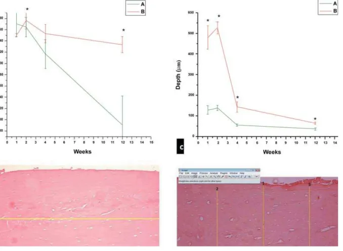
! $ ' the assessment of the extent of bone necrosis XK Z! ' performed for the evaluation of the depth of bone necrosis (Figure 1d). Empty osteocytes and vessel ! X<= <=Z empty and total vessel channels (Figure 2a).
Statistical analysis
G ! / 2-min protocol had been applied. Median, quartiles # ~ performed using the Origin 8.0 statistical program X ~! |! G! ~GZ# &/
and Dunn’s post-hoc
$#
G $ "} Xp<0.05).
RESULTS
One-minute protocol
The absolute peak extent of bone necrosis X),-#)*% Z & # 5 $ difference (p)#)"Z &! $ &# 3 & $ Xp<0.05) in mean depth of bone necrosis per histologic field for most ! &# ! & %#")* &# G & percentage of both empty osteocytes (Figure 2b) XK Z &! )#0 0.34, respectively.
Two-minute protocol
& )%1#%1 + & # G $ $ & Xp<0.05) (Figure 1a). Regarding the ! $
& Xp<0.05)
(Figure 1c). In addition, a mean depth of 436.424 + &# | osteocytes (Figure 2b) and vessel channels (Figure Z &! mean ratios of 0.64 and 0.71, respectively.
DISCUSSION
In an attempt to reduce or even to prevent esthetic-functional complications associated ! $ X5 Z X$ '
)o5Z
treatment1,17,21,23,26.
On the basis of morphofunctional phylogenetic
investigations18!
model using rat femurs for comparative studies in order to extrapolate the clinical use of cryotherapy # K ' ! since it permits better control of the quantity of tissue involved2.
The present study is the first employing histomorphometric parameters for the evaluation of the tissue response to cryosurgery. The Image J ! ! also been used for other purposes in dentistry as reported by Demirbas6 (2008).
Analysis of the extent of osteonecrosis in / $ $ # ! $ ! $ &# &#
/ ! $ difference in the mean extent of bone necrosis $ & / # ~! & ! $ $ & #
Comparison of the protocols of liquid nitrogen &! $
th &#
Another important parameter for the quantitative analysis of morphologic alterations resulting from cryotherapy is the percentage of necrotic # / ! $ & &# # 5 $ osteocytes for the 2-min protocol compared to the 1-min protocol, corresponding to peak osteocyte necrosis in the present study.
& ! corresponding to peak vascularization in the present study.
The depth of necrosis in animals is generally evaluated by thermographic imaging, a procedure $ the cryosurgical procedure14. Using this approach, Bradley and Fisher3 (1975) evaluated different ' / cavities of dry porcine mandibles. The authors observed that the use of nitrogen spray resulted in complete bone penetration, including the opposite cortex, after 5 min. In contrast, the use of closed probes resulted in a small area of cortical bone after ) # ! only immediate and in vitro !
in vivo longitudinal analysis using qualitative and
present study.
We also observed differences in the depth of $! & cryosurgery protocols. Using the 1-min protocol, $ $ $ &! observation of greater homogeneity in the band of necrotic cortical bone in the other groups. The & ! $ & $ & #
/ ! the 2-min protocol, a difference in mean necrosis & & # ! $ $ # $ $ &! $ the 1-min protocol.
! & $ & using the 2-min liquid nitrogen cryotherapy protocol / # $
15 X*,)Z
mandibles and observed marked bone necrosis in & # ! histologic study on rabbit femurs, Keijser14 (1999) demonstrated a clear demarcation of osteonecrosis & ! $ / study.
Clinically, bone fractures after cryotherapy have
mainly been reported for ameloblastoma involving ! odontogenic tumor1,5,7,16,17,21. Despite the absence ! suggest that the marked bone necrosis observed & contribute to the occurrence of this complication, a fact supporting the need for careful postoperative /#
CONCLUSIONS
$ the 2-min protocol produced more accentuated bone necrosis than the 1-min protocol. Although our results cannot be entirely extrapolated to clinical practice, they contribute to the understanding of the behavior of bone tissue submitted to different cycles of liquid nitrogen freezing and may serve # S important for the understanding of the mechanisms underlying the effects of cryosurgery.
REFERENCES
/ V |! K V! ~' ! K # ~ of the treatment and prognosis of the odontogenic keratocyst. Oral Surg Oral Med Oral Pathol Oral Radiol Endod. 2000;90:553-8. 2- Bradley PF. Modern trends in cryosurgery of bone in the maxillo-facial region. Int J Oral Surg. 1978;7:405-15.
3- Bradley PF, Fisher AD. The cryosurgery of bone: an experimental and clinical assessment. Br J Oral Surg. 1975;13:111-27. 4- Courage GR, Huebsch RF. Cold therapy revisited. J Am Dent Assoc. 1971;83:1070-3.
5- Curi MM, Dib LL, Pinto DS. Management of solid ameloblastoma # 9 ~ 9 Med Oral Pathol Oral Radiol Endod. 1997;84:339-44.
1/ G! S ~! > 3! G& V9! V # Mandibular bone changes in sickle cell anemia: fractal analysis. Oral Surg Oral Med Oral Pathol Oral Radiol Endod. 2008;106:e41-8. 7- Emmings FG, Neiders ME, Greene GW Jr, Koepf SW, Gage AA. K' # ; 9 ~# *11w%%"/ 55.
8- Fisher AD, Williams DF, Bradley PF. The effect of cryosurgery on the strength of bone. Br J Oral Surg. 1978;15:215-22.
9- Flórez-Moreno GA, Henao-Ruiz H, Santa-Sáenz DH, Castañeda-Peláez DA, Tobón-Arroyave SI. Cytomorphometric and # 9 ~ 9 9 3 9 Radiol Endod. 2008;105:625-32.
)/ >''/G 3! & G! 9& [! [' ! S, Sakashita H, et al. Keratocystic odontogenic tumour: a retrospective study of 183 cases. J Oral Sci. 2008;50:205-12. 11- Hafron J, Kaouk JH. Cryosurgical ablation of renal cell carcinoma. Cancer Control. 2007;14:211-7.
/ V! V ;5# crystallization during freezing. Cryobiology. 2004;48:8-21. 13- Hoffman NE, Bischof JC. The cryobiology of cryosurgical injury. Urology. 2002;60:40-9.
15- Kuylenstierna R, Anniko M, Lundquist PG, Nathanson A. Experimental cryosurgery on bone: a light and electron microscopical investigation. Criobiology. 1980;17:563-70. 16- Marciani RD, Trodahl JN, Suckiel MJ, Dubick MN. Cryotherapy in the treatment of ameloblastoma of the mandible: report of cases. J Oral Surg. 1977;35:289-95.
17- Morini P, Sanvitto LC, Dias WB. Criotherapy in ameloblastomas. Odontol Mod. 1982;9:28-38.
18- Najjar TA, Kahn D. Comparative study of healing and remodeling in various bones. J Oral Surg. 1977;35:375-9. 19- Natiella JR, Meenaghan MA, Rosa RA, Bessette RW, Gage AW. Cryosurgery of major and minor salivary gland: a light microscopic evaluation in the Rhesus monkey. J Oral Pathol. 1979;8:237-46. 20- Popken F, Land M, Erberich H, Bosse M, König DP, Eysel P. The vivo assessment of the probe and application of the method to bone in a sheep model. BMC Surg. 2003;3:3.
21- Salmassy DA, Pogrel MA. Liquid nitrogen cryosurgery and immediate bone grafting in the management of aggressive primary # ; 9 ~# **"w"0-,%/*)#
22- Sannomiya EK, Silva JV, Brito AA, Saez DM, Angelieri F, Dalben GS. Surgical planning for resection of an ameloblastoma and reconstruction of the mandible using a selective laser sintering 3D biomodel. Oral Surg Oral Med Oral Pathol Oral Radiol Endod. 2008;106:36-40.
23- Schmidt BL, Pogrel MA. Neurosensory changes after liquid nitrogen cryotherapy. J Oral Maxillofacial Surg. 2004;62:1183-7. %/ ~& >! V! ~ (! ~ B. Odontogenic myxoma of maxilla. Indian J Dent Res. 2008;19:62-5.
25- Theodorescu D. Cancer cryotherapy: evolution and biology. Rev Urol. 2004;6:S9-S19.
26- Webb DJ, Brockbank J. Treatment of the odontogenic keratocyst by combined enucleation and cryosurgery. Int J Oral Surg. 1984;13:506-10.