A new species of
Orobdella
(Hirudinida,
Arhynchobdellida, Gastrostomobdellidae) and
redescription of
O. kawakatsuorum
from Hokkaido,
Japan with the phylogenetic position of the new species
Takafumi Nakano1,†
1 Department of Zoology, Graduate School of Science, Kyoto University, Kyoto 606-8502, Japan
† urn:lsid:zoobank.org:author:FE6B9521-5C9E-43C5-B492-08B0D0590E1B
Corresponding author:Takafumi Nakano (nakano@zoo.zool.kyoto-u.ac.jp)
Academic editor:F. Govedich | Received 23 November 2011 | Accepted 31 January 2012 | Published 10 February 2012 urn:lsid:zoobank.org:pub:8F79801C-30DA-4F85-8CFF-4CEF42732286
Citation: Nakano T (2012) A new species of Orobdella (Hirudinida, Arhynchobdellida, Gastrostomobdellidae) and redescription of O. kawakatsuorum from Hokkaido, Japan with the phylogenetic position of the new species. ZooKeys 169: 9–30. doi: 10.3897/zookeys.169.2425
Abstract
A new quadrannulate Orobdella Oka, 1895 species, Orobdellakoikeisp. n., is described on the basis of six specimens collected from Hokkaido, Japan. In addition, an emended description of quadrannulate
Orobdellakawakatsuorum Richardson, 1975 is also provided. Orobdellakoikei difers from other quadran-nulate species of Orobdella in possessing the following combination of characters: color dorsally brown, IV uniannulate, male gonopore at XI b6, gastropore and female gonopore at XIII a1, 1/2 + 4 + 1/2 between gonopores, XXV triannulate, tubular but bulbous at junctions with gastropore and crop gastroporal duct, epididymides in XVII to XIX, and atrial cornua ovate. he phylogenetic position of the newly described species is estimated using mitochondrial COI, tRNACys, tRNAMet, 12S rDNA, tRNAVal and 16S rDNA markers. Orobdellakoikei is a sister taxon of O. kakawatsuorum according to the molecular phylogenetic analyses.
Keywords
Hirudinida, Hirudinea, Gastrostomobdellidae, Orobdellakawakatsuorum, new species, molecular phy-logeny, Japan
www.zookeys.org
Copyright Takafumi Nakano. This is an open access article distributed under the terms of the Creative Commons Attribution License 3.0 (CC-BY), which permits unrestricted use, distribution, and reproduction in any medium, provided the original author and source are credited.
introduction
he genus Orobdella Oka, 1895 consists of terrestrial gastroporous leeches in East Asia (Sawyer 1986). he species diversity of Orobdella has been revised recently, and now this genus includes eight species (Nakano 2010, 2011a, b). Among these species, only one quadrannulate species, Orobdellakawakatsuorum Richardson, 1975, has been known from Hokkaido, Japan (Richardson 1975). his species was described based on the two specimens collected from Sapporo, and its holotype has been deposited at the National Museum of Nature and Science, Tokyo (NSMT). Orobdellakawakatsuorum
is characterized especially by its possession of six annuli between gonopores and a sim-ple tubular gastroporal duct.
Quadrannulate Orobdella specimens were recently obtained from various places in Hokkaido. Most of these specimens were identiied as O. kawakatsuorum. However, several specimens difer from not only O. kawakatsuorum, but also the other quadran-nulate species, O. esulcata Nakano, 2010, O. tsushimensis Nakano, 2011, and O. whit-mani Oka, 1895, in several characterisics. herefore, they are described as a new species herein. In addition, an emended description of Orobdellakawakatsuorum is presented on the basis of its holotype and newly collected materials. he phylogenetic position of the new species is also estimated using mitochondrial COI, tRNACys, tRNAMet, 12S
rDNA, tRNAVal and 16S rDNA sequence data.
Materials and methods
For the taxonomic study, leeches were collected from Hokkaido, Japan (Fig. 1), under rocks along mountain or forest trails. Altitude and coordinates for localities were ob-tained using a Garmin eTrex GPS unit.
he preparation of the collected materials for the morphological and molecular analyses follows Nakano (2011b). Two measurements were taken: body length (BL) from the anterior margin of the oral sucker to the posterior margin of the caudal sucker, and maximum body width (BW). Examination, dissection, and drawing of the specimens were accomplished under stereoscopic microscopes with drawing tubes (Leica S6E, M125 and WILD HEERBRUGG TYP 308700).
he numbering convention is based on Moore (1927): body somites are denoted by Roman numerals and annuli in each somite are given alphanumeric designations.
For the molecular phylogenetic analyses, the sequence data of nine Orobdella spe-cies were newly obtained (Table 1). As outgroup, three Erpobdelliformes leeches, Gas-trostomobdella monticola Moore, 1929 (Gastrostomobdellidae), Erpobdella japonica
Pawłowski, 1962 (Erpobdellidae), and Mimobdella japonica Blanchard, 1897 (Salii-dae), were included.
Figure 1. Map showing the collection localities of Orobdellakoikei sp. n. and Orobdellakawakatsuorum
Richardson, 1975. Black triangles indicate the localities of O. koikei; black circles indicate those of O.
kawakatsuorum1 type locality of O. kawakatsuorum; and 2 type locality of O. koikei.
table 1. Samples used for the phylogenetic analyses. he information on voucher, collection locality, and GenBank accession numbers are indicated.
Species Voucher Locality COI 12S
Orobdellaesulcata KUZ Z29 Holotype
Kumamoto, Japan (32°48.60'N, 130°38.48'E)
AB679664 AB679665
Orobdellaesulcata KUZ Z170 Ikinoshima Isl., Japan (33°44.47'N, 129°42.25'E)
AB679666 AB679667
Orobdella dolichopharynx KUZ Z120 Holotype
Amamioshima Isl., Japan (28°17.18'N, 129°18.93'E)
AB679680 AB679681
Orobdella dolichopahrynx KUZ Z122 Kinsakubaru, Amamioshima Isl., Japan
AB679682 AB679683
Orobdella ijimai KUZ Z110 Topotype
Tochigi, Japan (36°46.98'N, 139°34.93'E)
AB679672 AB679673
Orobdella ijimai KUZ Z188 Nagano, Japan (36°12.44'N, 138°37.74'E)
AB679674 AB679675
Orobdellakawakatsuorum KUZ Z148 Toyotomi, Hokkaido, Japan (45°13.22'N, 141°41.07'E)
AB679692 AB679693
Orobdellakawakatsuorum KUZ Z150 Richirito Isl., Hokkaido, Japan (45°11.99'N, 141°14.26'E)
AB679694 AB679695
Orobdella kawakatsuorum KUZ Z152 Shari, Hokkaido, Japan (44°06.09'N, 145°06.09'E)
Species Voucher Locality COI 12S
Orobdellakawakatsuorum KUZ Z153 Ashoro, Hokkaido, Japan (43°23.70'N, 143°59.23'E)
AB679698 AB679699
Orobdellakawakatsuorum KUZ Z154 Kamikawa, Hokkaido, Japan (43°43.55'N, 142°57.53'E)
AB679700 AB679701
Orobdellakawakatsuorum KUZ Z159 Kyowa, Hokkaido, Japan (42°56.17'N, 140°35.57'E)
AB679702 AB679703
Orobdellakawakatsuorum KUZ Z167 Sapporo, Hokkaido, Japan (43°03.15'N, 141°18.71'E)
AB679704 AB679705
Orobdellakoikei KUZ Z145 Hiratori, Hokkaido, Japan (42°40.82'N, 142°25.44'E)
AB679684 AB679685
Orobdella koikei KUZ Z146 Shinhidaka, Hokkaido, Japan (42°42.86'N, 142°38.30'E)
AB679686 AB679687
Orobdella koikei KUZ Z156 Holotype
Kamikawa, Hokkaido, Japan (43°43.36'N, 142°56.85'E)
AB679688 AB679689
Orobdella koikei KUZ Z158 Mashike, Hokkaido, Japan (43°46.23'N, 141°30.63'E)
AB679690 AB679691
Orobdella octonaria KUZ Z177 Tokyo, Japan (35°42.94'N, 139°12.20'E)
AB679706 AB679707
Orobdella octonaria KUZ Z181 Topotype
Kanagawa, Japan (35°14.06'N, 139°04.21'E)
AB679708 AB679709
Orobdella shimadae KUZ Z128 Holotype
Okinawajima Isl., Japan (26°49.08'N, 128°16.90'E)
AB679676 AB679677
Orobdella shimadae KUZ Z138 Okinawajima Isl., Japan (26°40.20'N, 128°11.20'E)
AB679678 AB679679
Orobdella tsushimensis KUZ Z133 Tsushimajima Isl., Japan (34°34.66'N, 129°22.49'E)
AB679660 AB679661
Orobdella tsushimensis KUZ Z134 Holotype
Tsushimajima Isl., Japan (34°15.29'N, 129°17.28'E)
AB679662 AB679663
Orobdella whitmani KUZ Z45 Topotype
Gifu, Japan (35°25.65'N, 136°46.91'E)
AB679668 AB679669
Orobdella whitmani KUZ Z191 Shiga, Japan (35°39.63'N, 136°11.30'E)
AB679670 AB679671
Erpobdella japonica KUZ Z178 Nagano, Japan (36°12.43'N, 138°36.93'E) AB679654 AB679655 Gastrostomobdella monticola UNIMAS/ A03/ BH01/10
Kuching, Malaysia AB679656 AB679657
Mimobdella japonica KUZ Z179 Amamioshima Isl., Japan (28°26.53'N, 129°33.60'E)
AB679658 AB679659
PCR and DNA sequencing
sodium acetate (pH 5.2). Precipitated samples were dried and stored in TE bufer (10 mM Tris-HCl and 1 mM EDTA [pH 8.0]). Primer sets used in this study are listed in Table 2: for COI, LCO1490 and HCO2198 (Folmer et al. 1994), and LCO-in and HCO-out; for tRNACys, tRNAMet, 12S, tRNAVal and 16S (abbreviated 12S), 12SA-out
and 12SB-in, and 12SA-in and 12SB-out. All ampliication reactions were performed in a GeneAmp PCR System 2700 (Applied Biosystems) or a MyCycler (Bi-Rad Labo-ratories) using an Ex Taq Polymerase Kit (Takara Bio Inc.). Reaction mixtures were heated to 94°C for 5 min, followed by 35 cycles of 94°C (10 s), 42.5°C (20 s), and 72°C (1 min 13 s for COI, and 1 min for 12S) and a inal extension at 72°C for 6 min. he ampliied DNA fragments were puriied using polyethylene glycol (20% PEG 6000) precipitation.
table 2. PCR and cycle sequencing (CS) primers used in this study.
Gene Primer name Reaction Primer sequence (5’→ 3’) Source COI
1 LCO1490 PRC & CS GGTCAACAAATCATAAAGATATTGG Folmer et al. (1994) HCO2198 CS TAAACTTCAGGGTGACCAAAAAATCA Folmer et al. (1994)
2 LCO-in CS TCCAGAACGTATTCCATTATTTG his study
HCO-out PCR & CS TCTGGGTAGTCAGAATATCG his study tRNACys, tRNAMet, 12S rDNA, tRNAVal and 16S rDNA
1 12SA-out PCR & CS TTGATGAACAACATTAAATTGC his study
12SB-in CS TAAGCTGCACTTTGACCTGA his study
2 12SA-in CS AATTAAAACAAGGATTAGATACCC his study
12SB-out PCR & CS AACCCATAATGCAAAAGGTAC his study
All samples were sequenced in both directions. Sequencing reactions were per-formed using a BigDye Terminator v3.1 Cycle Sequencing Kit (Applied Biosystems). Each sequencing reaction mixture was incubated at 96°C for 2 min, followed by 40 cycles of 96°C (10 s), 50°C (5 s), and 60°C (45 s for COI, and 40 s for 12S). he prod-ucts were collected by ethanol precipitation and sequenced on an ABI 3130xl Genetic Analyzer (Applied Biosystems). Obtained sequences were edited using DNA BASER (Heracle Biosoft S.R.L.). hese sequence data were deposited in GenBank.
Phylogenetic analyses
substitu-tions. It was conirmed that COI and 12S did not show any signs of saturation (ti/tv rate ration of COI was 1.02, and that of 12S was 1.07). herefore, the concatenated sequences yielded a total of 1984 bp positions.
Phylogenetic tree were constructed using maximum likelihood (ML) and Bayesian inference (BI). Pairwise comparisons of Kimura-2 parameter (K2p) distance (Kimu-ra 1980) were also calculated using MEGA5. ML phylogenies were calculated using TREEFINDER v October 2008 (Jobb et al. 2004) with the tool package PHYLOGE-ARS v 2.0 (Tanabe 2008), and then non-parametric bootstrapping (Felsenstein 1985) was conducted with 500 replicates. he best-it models for each partition were selected using the Akaike Information Criterion (Akaike 1974) by using KAKUSAN4 (Tan-abe 2011). For the 1st position of COI, the Tamura-Nei model (TN93) with gamma distribution (+G) and proportion of invariant sites (+I) was selected. he transversion model (TVM)+I was selected for the 2nd position, the transition model (TIM)+G for the 3rd position of COI, and the general time reversal model (GTR)+G for 12S. BI and Bayesian posterior probabilities (BPPs) were estimated using the MPI version of MRBAYES v 3.1.2 (Altekar et al. 2004, Huelsenbeck et al. 2001, Ronquist and Huelsenbeck 2003). he best-it models for each partition were identiied using the Bayesian Information Criterion (Schwarz 1978) also by using KAKUSAN4: for COI 1st position, GTR+G+I; the Felsenstein 1981 model (F81)+I for COI 2nd position; the Hasegawa-Kishino-Yano model (HKY85)+G for COI 3rd position; and GTR+G for 12S. Two independent runs for four Markov chains were conducted for 1.5 million generations and the tree was sampled every 100 generations. Based on checking the pa-rameter estimates and convergence using TRACER v 1.5 (Rambaut and Drummond 2009), the irst 5,001 trees were discarded.
he nodes with bootstrap value (BS) higher than 70% were regarded as suiciently resolved (Hillis and Bull 1993). Nodes with BPP higher than 95% were considered statistically signiicant (Leaché and Reeder 2002).
Results
Taxonomy
Genus Orobdella Oka, 1895
Orobdellakoikei sp. n.
urn:lsid:zoobank.org:act:7DBE6F21-E4C3-4CBF-9469-2B13121F56D4 http://species-id.net/wiki/Orobdella_koikei
Figs 2–5
Diagnosis. In life, dorsal surface brown. Somites III and IV uniannulate, somites
Gas-troporal duct, tubular, but bulbous at junction with gastropore and at junction with crop. Male gonopore at XI b6, female gonopore at XIII a1 (slightly posterior to mid-dle of annulus), gonopores separated by 1/2 + 4 + 1/2. Paired epididymides in XVI/ XVII–XVII a2 to XIX a2/b5. Atrial cornua ovate.
Type materials. KUZ Z156, holotype, dissected, collected from under a rock
along a mountain trail at Sounkyo, Kamikawa, Hokkaido, Japan (43°43.36'N, 142° 56.85'E; Alt. 712 m), by Naoki Koike on 17 August, 2010.
Five paratypes collected from Hokkaido, Japan. Two specimens from the type lo-cality (43°43.36'N, 142°56.85'E; Alt. 712 m): KUZ Z157, dissected, by Naoki Koike on 17 August, 2010, and KUZ Z186, by TN on 19 September, 2011. KUZ Z145, dissected, from Hiratori (42°40.82'N, 142°25.44'E; Alt. 220 m), by Naoki Koike on 2 August, 2010. KUZ Z146, dissected, from Mt. Pisenaiyama, Shinhidaka (42°42.86'N, 142°38.30'E; Alt. 981 m), by Naoki Koike on 3 August, 2010. KUZ Z158, dissected, from Mt. Shokanbetsudake, Mashike (43°46.23'N, 141°30.63'E; Alt. 288 m), by Na-oki Koike on 18 August, 2010.
Etymology. he speciic name is a noun in the genitive case formed directly from
the name of Mr Naoki Koike, who collected many valuable specimens of Orobdella
leeches from Hokkaido.
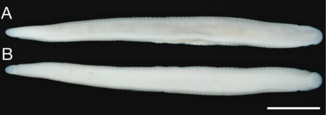
Description of holotype. Body irm, muscular, elongated, gaining regularly in
width in caudal direction, dorso-ventral depressed, sides nearly parallel from mid length to point just anterior to caudal sucker, BL 30.5 mm, BW 2.5 mm (Fig. 2). Caudal sucker ventral, oval, its diameter smaller than BW (Figs 2B, 3D). In life, dorsal surface brown, ventral surface grayish white. Color faded in preservative, without any dark lines (Fig. 2)
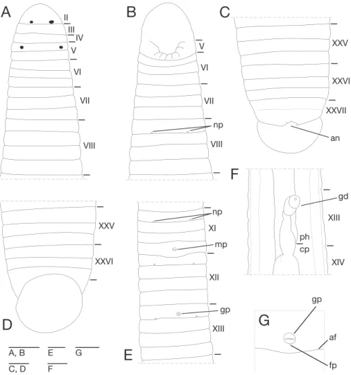
Somite I completely merged with prostomium (Fig. 3A). Somites II–IV unian-nulate (Fig. 3A). Somite V bianunian-nulate, (a1+a2) = a3 (Fig. 3A), V a3 forming posterior margin of oral sucker (Fig. 3B). Somites VI and VII triannulate (Fig. 3A–B). Somites VIII–XXIV quadrannulate, a1 = a2 = b5 = b6 (Fig. 3A–B, E). Somites XXV and XXVI triannulate (Fig. 3C–D), XXVI a3 being last complete annulus on venter (Fig. 3D). Somite XXVII uniannulate with one slight furrow on dorsal; anus behind it with no post-anal annulus (Fig. 3C).
Anterior ganglionic mass in VI a2 and a3. Ganglion VII in a2. Ganglion VIII in a2 and b5. Ganglion IX in a2. Ganglia X–XII in a2 and b5 of each somite (Fig. 4A). Ganglion XIII in b5 (Fig. 4A). Ganglia XIV and XV in a2 and b5 of each somite (Fig. 4A). Ganglia XVI–XXI in a2 of each somite (Fig. 4A). Ganglia XXII–XXIV in a1 and a2 of each somite. Ganglia XXV and XXVI in a1 of each somite. Posterior ganglionic mass in XXVII a2 and a3.
Figure 4.Orobdellakoikei sp. n., holotype, KUZ Z156 A Dorsal view of reproductive system includ-ing ventral nervous system B dorsal view of male atrium C lateral view of male atrium D ventral view of male atrium; and e dorsal view of female reproductive system including position of ganglion XIII. Scale bars, 1mm (A) and 0.25 mm (B–E). Abbreviations: ac, atrial cornu; at, atrium; cod, common oviduct; ed, ejaculatory duct; ep, epididymis; gp, gastropore; o, ovisac; od, oviduct; and ts, testisac.
posterior margin of a1 of each somite of VIII–XXIV (Fig. 3A, E). Papillae numerous, minute, hardly visible, one row on every annulus.
slightly posterior to middle of XIII a2 (Fig. 3E, G). Gastroporal duct, winding and bulbous at junction with gastropore, tubular but bulbous at junction with crop, join-ing with crop in XIV a1 (Fig. 3F). Intestine tubular, acaecate, in XIX b5/b6 to XXIII a2. Rectum, tubular, thin-walled.
Male gonopore at middle of XI b6 (Fig. 3E). Female gonopore located slightly posterior to middle of XIII a2, inconspicuous, located behind gastropore (Fig. 3G). Gonopores separated by 1/2 + 4 + 1/2 annuli (Fig. 3E). Testisacs multiple, one or two testisacs on each side in each annulus, in XIX a2/b5 to XXIV a1 (Fig. 4A). Paired epididymides in XVI a2/b5 to XIX a2/b5 (Fig. 4A). Ejaculatory bulbs absent. Ejacula-tory ducts in XI a2/b5 to XVI a2/b5, loosely coiled, each winding from each junction with epididymis, narrowing at junction with atrial cornu, then turning sharply inward toward atrial cornu without pre-atrial loop (Fig. 4A–D). Pair of atrial cornua in XI b5 and b6, muscular, ovate (Fig. 4B). Atrium short, muscular, globular in XI b6 (Fig. 4B–D). Penis sheath and penis absent. Ovisacs one pair, thin-walled, globular, in XIII a2 and b5 (Fig. 4A, E). Oviducts thin-walled, left oviduct crossing ventrally beneath nerve cord, both oviducts converging into common oviduct in XIII a2 (Fig. 4A, E). Common oviduct thin-walled, short, directly ascending to female gonopore (Fig. 4E).
Variation. In life, color generally same as holotype (Fig. 5). Somite III with slight furrow on dorsal (KUZ Z146). Somite IV with slight furrow on dorsal (KUZ Z158), or biannulate (KUZ Z146). Somite XXVI incomplete triannulate. Pharynx reaching to XIII b5/b6–XIV a1. Crop reaching to XIX b5–XX a1. Gastropore at middle of XIII. Gastroporal duct simple tubular (KUZ Z145 and Z146). Intestine reaching to XXIII a1–XXIV a2. Female gonopore at middle of XIII. Testisacs in XVIII a1–XIX a2/b5 to XXIII a2/b5. Epididymides in XVI/XVII–XVII a2 to XIX a2/b5. Right or left oviduct crossing ventrally beneath nerve cord.
Distribution. Known in mountainous regions of the central part of Hokkaido,
Japan (Fig. 1).
Remarks. he specimens examined in this study consist of small individuals.
However, testisacs and ovisacs of the holotype, of which BL is 30.5 mm, are devel-oped. In immature Orobdella specimens, testiscas are usually undeveloped, and hardly detected (Nakano pers. obs.). herefore, there is a possibility that the holotype of this species is a mature leech.
gonopores, annulation of XXV, morphology of the gastroporal duct and male atrium, and the length of epididymides. herefore, Orobdellakoikei can be treated as a distinct new species from Hokkaido.
Orobdellakawakatsuorum Richardson, 1975 http://species-id.net/wiki/Orobdella_kawakatsuorum
Figs 6–11
Orobdellakawakatsuorum Richardson, 1975: 42–51, igs 1, 2; Sawyer 1986: 680, 747.
Diagnosis. In life, dorsal surface grayish blue. Somites III and IV biannulate, somites VIII–XXV quadrannulate, somite XXVI triannulate, clitellum from X b5 to XIII a2. Pharynx reaching to XIV. Gastropore conspicuous in furrow of XIII a1/a2. Gastropo-ral duct, simple tubular. Male gonopore in furrow of XI b5/b6, female gonopore in furrow of XIII a1/a2, gonopores separated by 6 annuli. Paired epididymides in XVI a2/ b5–XVII b5 to XVI b5–XVII b6. Atrial cornua, coniform, undeveloped.
Material examined. NSMT-An 53, holotype, dissected by Richardson, LR,
col-lected from a home garden of Professor Masaharu Kawakatsu, Sapporo, Hokkaido, Japan, by Tetsuya Kawakatsu and Miyuki Kawakatsu on 1 June, 1974.
Emended description. Body irm, muscular, elongated, gaining regularly in width in caudal direction, dorso-ventral depressed, sides nearly parallel from mid length to point just anterior of caudal sucker (Figs 6, 7), maximu BL 111.64 (KUZ Z142), maximun BW 8.19 (KUZ Z154). Caudal sucker ventral, ova, its diameter smaller than BW (Figs 6, 7, 9D, 10D). In life, dorsal surface grayish blue, ventral surface bluish white (Fig. 8). Color faded in preservative, without any dark lines (Figs 6, 7).
Somite I completely merged with prostomium (Fig. 10A). Somite II uniannulate (Figs 9A, 10A). Somite III uniannulate in small specimens, biannulate in large speci-mens (Figs 9A, 10A). Somite IV generally biannulate (Figs 9A, 10A), but uniannulate in a few small specimens. Somite V biannulate, (a1 + a2) = a3, V a3 forming posterior margin of oral sucker (Figs 9A–B, 10A–B). Somites VI and VII triannulate, a1 = a2 = a3 (Figs 9A–B, 10A–B). Somites VIII–XXV quadrannulate, a1 = a2 = b5 = b6 (Figs 9A–E, 10A–E); X b5 being irst annulus of clitellum, XIII a2 being last annulus of clitellum. Somite XXVI triannulate, a1 = a2 = a3, a3 being last complete annulus on venter (Figs 9C–D, 10C–D); a3 with furrow on dorsal in large specimens (Fig. 10C). Figure 6.Orobdellakawakatsuorum Richardson, 1975, holotype, NSMT-An 53 A Dorsal and B ventral views. Scale bar, 5 mm.
Figure 8.Orobdellakawakatsuorum Richardson, 1975, collected from near the type locality, KUZ Z167, taken of live animal, dorsal view.
Figure 9.Orobdellakawakatsuorum Richardson, 1975, holotype, NSMT-An 53 A Dorsal view of somites I–IX B ventral view of somites I–IX C dorsal view of somites XXV–XXVII and caudal sucker D ventral view of somites XXV–XXVII and caudal sucker e ventral view of somites XI–XIII; and F ventral view of gastropore and female gonopore. Scale bars, 1 mm (A–E) and 0.25 mm (F). Abbreviations, see Fig. 3.
Somite XXVII uniannulate, or biannulate; anus behind it with no post-anal annulus (Fig. 9C, 10C).
in a1, XXV b6, XXV b6 and XXVI a1, or XXVI a2. Posterior ganglionic mass in XXVI a1–a3.
Intestine tubular, acaecate, in XIV a2–XIV/XV to XXIII b5–XIV b5/b6. Rectum, tu-bular, thin-walled.
Male gonopore generally in furrow of XI b5/6, or at anterior part of XI b6 (Figs 9E, 10E). Female gonopore in furrow of XIII a1/a2, located behind gastropore (Figs 9F, 10G). Gonopores separated by six annuli (Figs 9E, 10E). Testisacs multiple, two or three testisacs on each side in each annulus, in XVI b5–XVII b6 to XXIII a1–XXV b6 (Fig. 11A). Paired epididymides in XVI a2/b5–XVII b5 to XVI b5–XVII b6 (Fig. 11A). Ejaculatory bulbs absent. Ejaculatory ducts in XI b5 to XVI a2/b5–XVII b5, loosely coiled, each winding from each junction with epididymis, narrowing at junc-tion with atrial cornu, then turning inward toward atrial cornu without pre-atrial loop (Fig. 11A–D). Pair of atrial cornua in XI b5 and b6, undeveloped, coniform (Fig. 11B). Atrium short, muscular, globular in XI b5 and b6 (Fig. 11B–D). Ovisacs one pair, thin-walled, globular, in XIII a2 and b5 (Fig. 11A, E). Oviducts thin-walled, right or left oviduct crossing ventrally beneath nerve cord, both oviducts converging into common oviduct in XIII a2 (Fig. 11A, E). Common oviduct thin-walled, short, directly ascending to female gonopore (Fig. 11E).
Distribution. Known in mountainous regions of Hokkaido, Japan (Fig. 1).
Remarks. Richardson (1975) described that a gastropore of the holotype opened
at the middle of XIII a1, and the female gonopore in the furrow of XIII a1/a2. How-ever, both the gastropore and the female gonopore of O. kawakatsuorum are in the furrow of XIII a1/a2 on the basis of examination of the holotype and newly collected specimens. A gastropore of this species is coincident with a female gonopore. Richard-son also noted that a pair of nephririopores opened in XXV (XXIV in his paper). But it is rare for O. kawakatsuorum to possess 18 pairs of nephridiopores.
Phylogenetic relationships
he ML tree with ln L = -12757.40 (Fig. 12) was nearly identical to the obtained BI tree (not shown). Monophyly of the genus Orobdella was well supported (BS = 99%, BPP = 100%). Two Orobdella species from Hokkaido, Orobdella koikei and Orob-dellakawakatsuorum, formed a monophyletic group (BS = 100%, BPP = 100%). his clade was a sister taxon of the other Orobdella species. Monophyly of O. koikei and O.
kawakatsuorum was well supported (O. koikei: BS = 94%, BPP = 100%; O. kawakat-suorum: BS = 97%, BPP = 100%).
he COI sequence divergence between O. koikei and O. kawakatsuorum was tween 8.1–9.9% (mean = 9.0%). Intraspeciic variation of COI sequences ranged be-tween 4.8–8.1% (mean = 7.1%) in O. koikei, and 0.30–4.9% (mean = 3.7%) in O.
variance, p = 0.0096), but that of 12S did not have a signiicantly higher mean diver-gence (t-test with unequal variance, p = 0.22).
Monophyly of seven Orobdella species distributed in areas south of Hokkaido was recovered (BS = 95%, BPP = 100%). Monophyly of O. esulcata + O. shimadae + O.
dolichopharynx received strong support (BS = 95%, BPP = 100%), and that of O. jimai
+ O. octonaria was also recovered (BS = 92%, BPP = 100%).
Discussion
Orobdellakoikei difers from the four other quadrannulate species of the genus, O.
esulcata, O. kawakatsuorum, O. tsushimensis, and O. whitmani, in the following com-bination of characteristics (Table 3): 1) dorsal surface brown ; 2) IV uniannulate; 3) male gonopore at XI b6; 4) gastropore nad female gonopore at XIII a1; 5) gonopores separated by 1/2 + 4 + 1/2 annuli; 6) XXV triannulate; 7) gastroporal duct, tubular, but bulbous at junctions with gastropore and crop; 8) epididymides in XVII to XIX; and 9) atrial cornua ovate. Orobdellakoikei is easily distinguished from O. dolichophar-ynx Nakano, 2011, O. ijimai Oka, 1895, O. shimadae Nakano, 2011, and O. octonaria
Oka, 1895, in having mid-body somites that are quadrannulate; they are sexannulate in O. dolichopharynx, O. ijimai, and O. shimadae, and octannulate in O. octonaria.
(Nakano et al. in press). According to the phylogenetic analyses, several characteristics are considered to have evolved in parallel. Each of O. kawakatsuorum and O. esulcata
possess a tubular gastroporal duct (Nakano 2010). However, these two species are phylogenetically distant. he mid-body somite annulation of Orobdella leeches does not indicate phylogenetic relationships either. In the genus Orobdella, it is clear that the quadrannulate body somite is a plesiomorphic character. Sexannulate mid-body somite, which O. dolichopharynx, O. ijimai, and O. shimadae possess, evolved in parallel. Orobdellakawakatsuorum, and two species from Ryukyu Archipelago, Japan,
O. dolichopharynx and O. shimadae, possess rudimentary male atrial cornua (Nakano 2011b). However, undeveloped male atrial cornua do not indicate any phylogenetic relationships between O. kawakatsuorum and Ryukyu Orobdella species. hese char-acters are not useful for estimating phylogenetic relationships in the genus Orobdella, although they are suitable for the species level classiication.
Species delimitation in leeches based on genetic analyses, especially using COI DNA-barcode locus, has been discussed in many papers (see DeSalle et al. 2005 for review). he average sequence divergence of COI between O. koikei and O. kawakatsuo-rum was 9.0%, and that of 12S was 4.3%. Interspeciic genetic divergence of COI be-tween these two species showed a signiicantly higher value than that of the intraspeciic variation, although the intraspeciic genetic divergence of 12S sequences in O. koikei
(2.8–4.8%) was overlapped to a large extent with the interspeciic divergence of 12S (3.3–5.5%). hus, only the genetic distance of COI (mean = 9.0%) can be used as an indicator for deciding whether leeches are distinct species or not in the genus Orobdella, since O. koikei and O. kawakatsuorum are distributed syntopically at Sounkyo (Fig.1).
Gilyarov et al. (1969) reported quadrannulate Orobdella species from Primorsky Krai, Russia, as O. whitmani. Although they did not describe the detailed internal table 3. Comparisons of morphological characters between Orobdellakoikei sp. n. and four quadran-nulate congeneric species.
Character O. koikei sp. n. O. esulcata O. kawakatsuorum O. tsushimensis O. whitmani
Color brownish bluish bluish yellowish yellowish
Annulation of IV
uniannulate uniannulate biannulate unianulate uni- or biannulate Number
of annuli between gonopores
1/2 + 4 + 1/2 2/3 + 4 + 1/3 6 1/2 + 5 1/2 + 4 + 1/2
Annulation of XXV
triannulate quadrannulate quadrannulate quadrannulate quadrannulate
Gastroporal duct tubular, but bulbous at junctions with gastropore and crop tubular, but bulbous at junction with gastropore
simple tubular bottle-shaped bulbiform
anatomy of the specimen, the photograph of the ventral surface of their specimen (ig. 1 in their paper) clearly shows that the male gonopore opened in the furrow of XI/ XII and the female gonopore was at XIII a1. hus, the number of annuli between the gonopores was 4 + 1/2. his characteristic is not identical to those of the other known quadrannulate Orobdella species. here is a strong possibility that the quadrannulate
Orobdella species distributed in Primorsky Krai is an undescribed species. Primorsky Krai is located at the same latitude as Hokkaido, Japan. Clarifying the taxonomic sta-tus and phylogenetic position of Orobdella in Primorsky Krai will help to reveal the species diversity and the evolutionary history of the genus Orobdella.
Key to the known species of the genus Orobdella
1 Mid-body somites more than quadrannulate ...2
– Mid-body somites quadrannulate ...5
2 Mid-body somites sexannulate ...3
– Mid-body somites octannulate ...Orobdella octonaria Oka, 1895
3 Pharynx reaching to XVI ...4
– Pharynx reaching to XIV, gonopores separated by 1/2 + 7 + 1/2 annuli ... ...Orobdella ijimai Oka, 1895
4 Gonopores separated by 8 annuli ...Orobdella dolichopharynx Nakano, 2011
– Gonopores separated by 9 annuli ...Orobdella shimadae Nakano, 2011
5 Color yellowish ...6
– Color grayish blue or brown ...7
6 Gonopores separated by 1/2 + 5 annuli, gastroporal duct bottle-shaped ... ...Orobdella tsushimensis Nakano, 2011
– Gonopores separated by 1/2 + 4 + 1/2 annuli, gastroporal duct bulbiform .... ...Orobdella whimtani Oka, 1895
7 Color grayish blue ...8
– Color brown, gonopores separated by 1/2 + 4 + 1/2 annuli ... ...Orobdella koikei sp.n.
8 Gonopores separated by 2/3 + 4 + 1/3 annuli, gastroporal duct tubular, but bulbous at junction with gastropore ...Orobdella esulcata Nakano, 2010
– Gonopores separated by 6 annuli, gastroporal duct simple tubular ... ...Orobdella kawakatsuorum Richardson, 1975
Acknowledgements
he author is grateful to Naoki Koike and Naoyuki Nakahama for sending me valuable
manuscript. I am also grateful to Dr Zainnudin Ramlah (UNIMAS) for permitting me to use the Gastrostomobdella specimen for molecular analyses, to Koshiro Eto, Masa-hiro Nishi (Amami Mongoosebusters), Dr Kanto Nishikawa (KU) and Taku Shimada (Ant Room) for providing specimens, to Dr Elizabeth Nakajima (KU) for checking the English of this text, to two anonymous reviewers and Dr Fredric R. Govedich (Southern Utah University) for their constructive comments for this manuscript, and to Eri Kawaguchi (KU) for her technical support. I also express my sincere thanks to Ekgachai Jeratthitikul, Nobuto Kikukawa and Kazuki Kurita for their help in collect-ing specimens in the ield. his study was inancially support in part by a Grant-in Aid for Biodiversity and Evolutionary Research of Global COE (A06) from MEXT, Japan, to Kyoto University.
References
Akaike H (1974) A new look at the statistical model identiication. IEEE Transactions on Au-tomatic Control 19: 716–723. doi: 10.1109/TAC.1974.1100705
Altekar G, Dwarkadas S, Huelsenbeck JP, Ronquist F (2004) Parallel Metropolis coupled Mark-ov chain Monte Carlo for Bayesian phylogenetic inference. Bioinformatics 20: 407–415. doi: 10.1093/bioinformatics/btg427
Blanchard R (1897) Hirudinées du Musée de Leyde. Notes from the Leyden Museum 19: 73–113.
Castresana J (2000) Selection of conserved blocks from multiple alignments for their use in phylogenetic analysis. Molecular Biology and Evolution 17: 540–552.
DeSalle R, Egan MG, Siddall ME (2005) he unholy trinitiy: taxonomy, species delimitation and DNA barcoding. Philosophical Transactions of the Royal Society of London B Bio-logical Sciences 360: 1905–1916. doi: 10.1098/rstb.2005.1722
Felsenstein J (1985) Conidence limits on phylogenies: an approach using the bootstrap. Evolu-tion 39: 783–791. doi: 10.2307/2408678
Folmer O, Black M, Hoeh W, Lutz R, Vrijenhoek R (1994) DNA primers for ampliication of mitochondrial cytochrome c oxidase subunit I from diverse metazoan invertebrates. Mo-lecular Marine Biology and Biotechnology 3: 294–299.
Gilyarov MS, Lukin EI, Perel TS (1969) he irst terrestrial leech in fauna of the USSR, Orob-della whitmani Oka (Hirudinea, Herpobdellidae), a Tertiary relict of forests of the southern Maritime Region. Doklady Akademii Nauk SSSR 188: 235–237.
Hillis DM, Bull JJ (1993) An empirical test of bootstrapping as a method for assessing coni-dence in phylogenetic analysis. Systematic Biology 42: 182–192.
Hofacker IL, Fekete M, Stadler PF (2002) Secondary structure prediction for aligned RNA sequences. Journal of Molecular Biology 319: 1059–1066. doi: 10.1016/S0022-2836(02)00308-X
Jobb G, von Haeseler A, Strimmer K (2004) TREEFINDER: a powerful graphical analy-sis environment for molecular phylogenetics. BMC Evolutionary Biology 4: 18. doi: 10.1186/1471-2148-4-18
Katoh K, Toh H (2008) Improved accuracy of multiple ncRNA alignment by incorporating structural information into a MAFFT-based framework. BMC Bioinformatics 9: 212. doi: 10.1186/1471-2105-9-212
Kimura M (1980) A simple method for estimating evolutionary rates of base substitutions through comparative studies of nucleotide sequences. Journal of Molecular Evolution 16: 111–120. doi: 10.1007/BF01731581
Leaché AD, Reeder TW (2002) Molecular systematics of the eastern fence lizard (Sceloporus undulatus): a comparison of parsimony, likelihood, and Bayesian approaches. Systematic Biology 51: 44–68. doi: 10.1080/106351502753475871
McCaskill JS (1990) he equilibrium partition function and base pair binding probabilities for RNA secondary structure. Biopolymers 29: 1105–1119. doi: 10.1002/bip.360290621 Moore JP (1927) he segmentation (metamerism and annulation) of the Hirudinea. In: Hard-ing WA, Moore JP (Eds) he Fauna of British India, includHard-ing Ceylon and Burma Hir-udinea. Taylor and Francis, London, 1–12.
Moore JP (1929) Leeches from Borneo with descriptions of new species. Proceedings of the Academy of Natural Sciences of Philadelphia 81: 267–295.
Nakano T (2010) A new species of the genus Orobdella (Hirudinida: Arhynchobdellida: Gas-trostomobdellidae) from Kumamoto, Japan, and a redescription of O. whitmani with the designation of the lectotype. Zoological Science 27: 880–887. doi: 10.2108/zsj.27.880 Nakano T (2011a) A new species of Orobdella (Hirudinida: Arhynchobdellida:
Gastrostomob-dellidae) from Tsushima Island, Japan. Species Diversity 16: 39–47.
Nakano T (2011b) Redescription of Orobdella ijimai (Hirudinida: Arhynchobdellida: Gastros-tomobdellidae), and two new species of Orobdella from the Ryukyu Archipelago, Japan. Zootaxa 2998: 1–15.
Nakano T, Ramlah Z, Hikida T (in press) Phylogenetic position of gastrostomobdellid leeches (Hirudinida, Arhynchobdellida, Erpobdelliformes) and a new family for the genus Orob-della. Zoologica Scripta. doi: 10.1111/j.1463-6409.2011.00506.x
Okamoto T, Motokawa J, Toda M, Hikida T (2006) Parapatric distribution of the lizards Plestiodon (formerly Eumeces) latiscutatus and P. japonicus (Reptilia: Scinidae) around the Izu Peninsula, central Japan, and its biogeographic implications Zoological Science 23: 419–425. doi: 10.2108/zsj.23.419
Pawłowski LK (1962) O występowaniu pijawki Erpobdella octoculata (L.) w Japonii. Zeszyty Naukowe Uniwersytetu Łódzkiego Nauki Matematiczno-przyrodnicze Seria II 12: 127–136. Rambaut A, Drummond AJ (2009) Tracer. 1.5: http://tree.bio.ed.ac.uk/software/tracer/ Richardson LR (1975) A new species of terricolous leeches in Japan (Gastrostomobdellidae,
Orobdella). Bulletin of the National Science Museum Series A (Zoology) 1: 39–56. Ronquist F, Huelsenbeck JP (2003) MrBayes 3: Bayesian phylogenetic inference under mixed
Schwarz G (1978) Estimating the dimension of a model. he Annals of Statistics 6: 461–464. doi: 10.1214/aos/1176344136
Tabei Y, Kiryu H, Kin T, Asai K (2008) A fast structural multiple alignment method for long RNA sequences. BMC Bioinformatics 9: 33. doi: 10.1186/1471-2105-9-33
Tamura K, Peterson D, Peterson N, Stecher G, Nei M, Kumar S (2011) MEGA5: molecu-lar evolutionary genetics analysis using maximum likelihood, evolutionary distance, and maximum parsimony methods. Molecular Biology and Evolution 28: 2731–2739. doi: 10.1093/molbev/msr121
Tanabe AS (2008) Phylogears. 2.0: http://www.ifthdimension.jp/