INTRODUCTION
Genera AnteholostichaBerger, 2003 and Metaurostylopsis Song et al., 2001 belong to the family Urostylidae Bütschli, 1889. This family is characterized by having one or more marginal rows of left and right marginal cirri and the midven-tral cirri which arranged as a “zig-zag” file (Borror, 1979; Borror and Wicklow, 1983; Lynn, 2008).
Diverse and polyphyletic species in the Holosticha com-plex (Gao et al., 2010) were split up into four genera, Holosti-cha, Anteholosticha, Caudiholosticha and Biholostichaby Berger (2003). Recently Li et al. (2009) added another genus Nothoholostichato this complex, and therefore there are five genera in the Holosticha complex at present. The genus Ante-holostichacan be distinguished from other genera in Holo-sticha complex by its lack of caudal cirri and several other apomorphic characters, e.g., anterior end of left marginal row curved rightwards, proximalmost membranelles widened (Berger, 2003). Recently a Chinese population of A. pulchra
was reported by Li et al. (2007).
The genus Metaurostylopsiswas established by Song et al. (2001). The characteristics of this genus are as follows: clear-ly differentiated frontal, buccal, transverse cirri and frontoter-minal cirral row present; caudal cirri absent; more than one left and right marginal row (Song et al., 2001). Metaurosty-lopsis struederkypkeaewas newly established by Shao et al. (2008).
Here, we redescribe two urostylid ciliates, A. pulchraand M. struederkypkeae, collected from Korea. For precise iden-tification and description, we conducted live observation, protargol impregnation and small subunit ribosomal DNA (SSU rDNA) sequence analysis.
MATERIALS AND METHODS
Anteholosticha pulchra was isolated from Incheon Harbor (salinity, 30.6‰; temperature, 폭0.6�C; 37�26′N, 126�35′E), http://dx.doi.org/10.5635/ASED.2012.28.1.020
Redescription of Two Urostylid Ciliates
(Ciliophora: Urostylida),
Anteholosticha pulchra
and
Metaurostylopsis struederkypkeae
from Korea
Kyung-Min Park, Jae-Ho Jung, Gi-Sik Min*
Department of Biological Sciences, Inha University, Incheon 402-751, Korea
ABSTRACT
Two urostylid ciliates, Anteholosticha pulchra(Kahl, 1932) Berger, 2003 and Metaurostylopsis struederkyp-keaeShao et al., 2008, new to Korea, were collected from the Yellow Sea and the East Sea, Korea, respec-tively. They were identified based on live observation and protargol impregnation. Taxonomical characters of A. pulchraare as follows: 190-300×30-55µm size in vivo; contractile vacuole located on the left side of the posterior 1/4 of the cell; spherical-reddish granules at cirral bases and around dorsal bristles, somewhat sparsely distributed throughout the cell surface; four frontal and two frontoterminal cirri; four dorsal kineties; caudal cirri absent. Metaurostylopsis struederkypkeaeis characterized as follows: 80-110×40-50µm sizein vivo; caudal cirri absent; two types of cortical granules: type 1, yellow-green arranged along the ventral cirral rows and dorsal kineties; type 2, small and reddish, with an irregular arrangement; four frontal, four to eight frontoterminal, and two to six transverse cirri; five to seven left and three to five right marginal rows. Sequences of small subunit ribosomal DNA were determined from both species, and pairwise distances with their relatives were analyzed.
Keywords: Anteholosticha pulchra, Metaurostylopsis struederkypkeae, urostylid ciliate, marine, Korea
This is an Open Access article distributed under the terms of the Creative Commons Attribution Non-Commercial License (http://creativecommons.org/ licenses/by-nc/3.0/) which permits unrestricted non-commercial use, distribution, and reproduction in any medium, provided the original work is properly cited.
*To whom correspondence should be addressed Tel: 82-32-860-7692, Fax: 82-32-874-6737 E-mail: mingisik@inha.ac.kr
Incheon, Korea at January of 2011. Metaurostylopsis strue-derkypkeaewas collected from the lagoon of Youngrang (salinity, 14.8‰; temperature, 18.3�C; 38�13′N, 128�35′E), Sokcho, Korea at June of 2011. These two populations were maintained in petri dishes with rice grains to enrich bacteria growth as a food resource. The specimens were observed in vivounder a light microscope (Leica DM2500, Wetzlar, Ger-many) at a magnification of 50× to 1,000×. Protargol im-pregnation was performed to reveal the infraciliatures (Foiss-ner, 1991). Drawing was performed under a light microscope at a magnification of 1,000×. Terminology and classification were mainly followed according to Lynn (2008).
DNA extraction, amplification and sequencing of nearly complete SSU rDNA were performed according to Jung et al. (2011). Our sequences and retrieved sequences from Gen-Bank were aligned using BioEdit (Hall, 1999), and pairwise genetic distance based on Kimura two-parameter inference was obtained using MEGA 4.0 (Kumar et al., 2004).
RESULTS
Phylum Ciliophora Doflein, 1901 Class Spirotrichea Bütschli, 1889 Order Urostylida Jankowski, 1979 Family Urostylidae Bütschli, 1889 Genus AnteholostichaBerger, 2003
1*Anteholosticha pulchra(Kahl, 1932) Berger, 2003
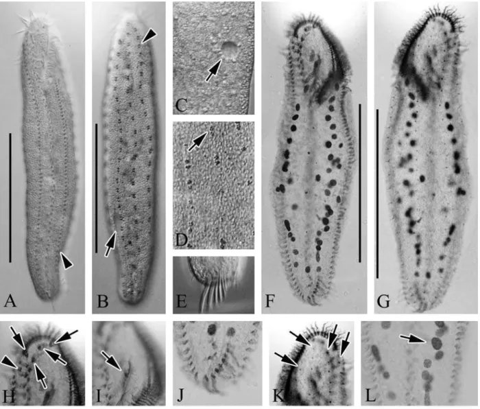
(Table 1, Figs. 1A-E, 2)
Keronopsis pulchraKahl, 1932: 573, fig. 104; Kahl, 1933: 109, fig. 16.
Holosticha(Keronopsis) pulchra: Berger, 2001: 36.
Anteholosticha pulchra: Berger, 2003: 377; Berger, 2006: 435, fig. 90; Li et al., 2007: 113, figs. 4-7.
Description.Live cell size usually 190-300×30-55µm, slen-der in shape (Figs. 1A, 2A, B), flexible but not contractile, an-terior and posan-terior ends rounded, contractile vacuole
approxi-Korean name: 1*홍 전열 충(신칭)
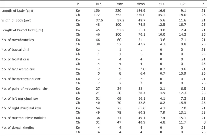
Table 1.Comparison of morphometric data from Anteholosticha pulchrawith Li et al. (2007)
P Min Max Mean SD CV n
Length of body (µm) Ko 150 220 184.9 16.9 9.1 21
Ch 172 352 250.0 45.1 18.0 25
Width of body (µm) Ko 37.5 57.5 48.7 5.6 11.6 21
Ch 48 100 74.8 12.5 16.7 25
Length of buccal field (µm) Ko 45 57.5 51.1 3.8 7.4 21
Ch 46 100 70.1 10.0 14.3 25
No. of membranelles Ko 46 60 51 3.6 7.1 21
Ch 38 57 47.7 4.2 8.8 25
No. of buccal cirri Ko 1 1 1 0 0 21
Ch 1 1 1 0 0 25
No. of frontal cirri Ko 4 4 4 0 0 21
Ch 4 4 4 0 0 25
No. of transverse cirri Ko 7 9 7.8 0.7 9.6 21
Ch 5 8 6.4 0.7 10.9 25
No. of frontoterminal cirri Ko 2 2 2 0 0 21
Ch 2 2 2 0 0 25
No. of pairs of midventral cirri Ko 27 34 32 2.1 6.5 21
Ch 21 38 28.4 4.9 17.3 25
No. of left marginal row Ko 51 68 56.1 4.1 7.3 21
Ch 40 70 52.8 8.2 15.5 25
No. of right marginal row Ko 54 73 61.6 4.3 7.0 21
Ch 49 75 60.3 7.9 13.1 25
No. of macronuclear nodules Ko 38 71 49.1 7.4 15.1 21
Ch 31 47 40.9 4.8 11.7 8
No. of dorsal kineties Ko 4 4 4 0 0 21
Ch 4 4 4 0 0 25
All data compared protargol-impregnated specimens of Korean population (Ko) and Chinese population (Ch). Frontal cirri including parabuccal cirrus (III/2).
mately 10µm in diameter, positioned on left side of posterior 1/4 of cell (Figs. 1A, 2B, C, arrow). Spherical-reddish corti-cal granules at cirral bases and around dorsal bristles, ca. 1 µm across, some sparsely distributed throughout cell surface (Figs. 1B, C, 2D, arrow). Four frontal cirri each located in anterior ventral area, ca. 18µm (Fig. 2H, arrows), and two frontoterminal located near anterior end of right marginal row (Fig. 2H, arrowhead) and seven to nine transverse cirri (Fig. 2E, J), respectively ca. 10 and 20µm in length, with one left and right marginal row located on left and right side (left marginal cirri ca. 56, right marginal cirri ca. 61). Buccal cirrus positioned near paroral membrane (Fig. 2I, arrow),
cau-dal cirri absent. Four dorsal kineties extended from anterior to posterior area (Fig. 2G, K, arrows) and approximately 49 macronuclear nodules scattered in body (Figs. 1E, 2L).
46-60 membranelles in adoral zone, ca. 25-35% of body length, and thick buccal lip. Long midventral pairs extending to transverse cirri, in a “zig-zag” pattern, 27-34 pairs of cirri. Food vacuoles colorless and sparsely located on cell. Distribution.Germany (Kiel, the Baltic Sea), China (Laiz-hou, the Yellow Sea), and Korea (this study).
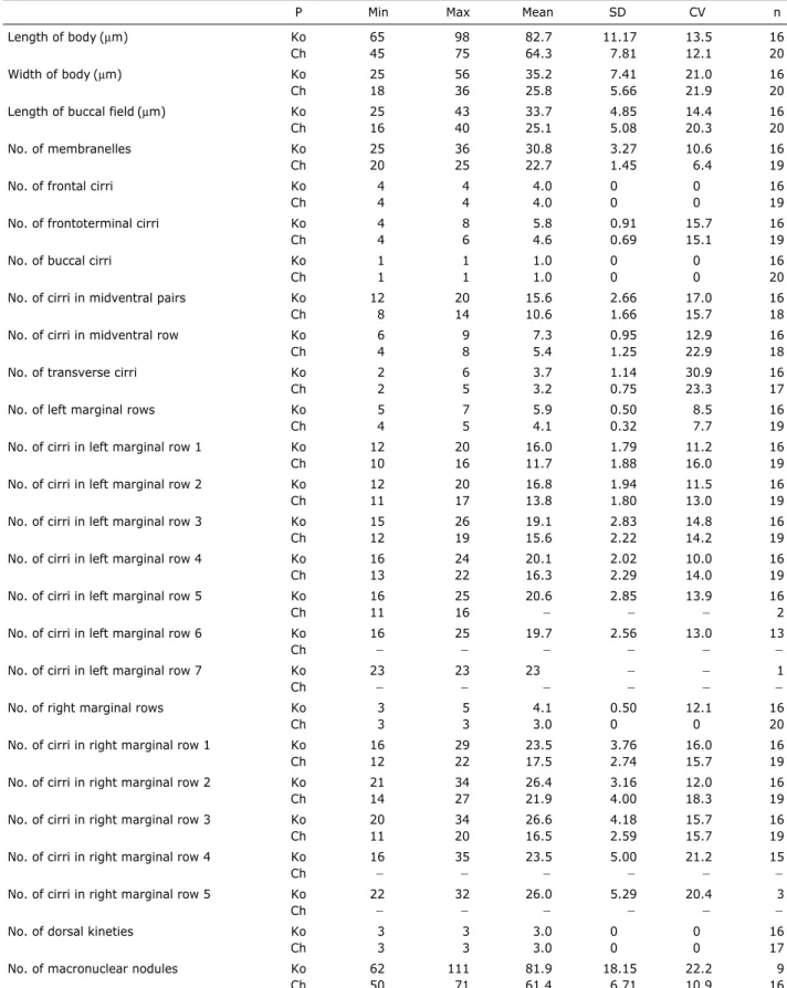
Table 2.Comparison of morphometric data from Metaurostylopsis struederkypkeaewith Shao et al. (2008)
P Min Max Mean SD CV n
Length of body (µm) Ko 65 98 82.7 11.17 13.5 16
Ch 45 75 64.3 7.81 12.1 20
Width of body (µm) Ko 25 56 35.2 7.41 21.0 16
Ch 18 36 25.8 5.66 21.9 20
Length of buccal field (µm) Ko 25 43 33.7 4.85 14.4 16
Ch 16 40 25.1 5.08 20.3 20
No. of membranelles Ko 25 36 30.8 3.27 10.6 16
Ch 20 25 22.7 1.45 6.4 19
No. of frontal cirri Ko 4 4 4.0 0 0 16
Ch 4 4 4.0 0 0 19
No. of frontoterminal cirri Ko 4 8 5.8 0.91 15.7 16
Ch 4 6 4.6 0.69 15.1 19
No. of buccal cirri Ko 1 1 1.0 0 0 16
Ch 1 1 1.0 0 0 20
No. of cirri in midventral pairs Ko 12 20 15.6 2.66 17.0 16
Ch 8 14 10.6 1.66 15.7 18
No. of cirri in midventral row Ko 6 9 7.3 0.95 12.9 16
Ch 4 8 5.4 1.25 22.9 18
No. of transverse cirri Ko 2 6 3.7 1.14 30.9 16
Ch 2 5 3.2 0.75 23.3 17
No. of left marginal rows Ko 5 7 5.9 0.50 8.5 16
Ch 4 5 4.1 0.32 7.7 19
No. of cirri in left marginal row 1 Ko 12 20 16.0 1.79 11.2 16
Ch 10 16 11.7 1.88 16.0 19
No. of cirri in left marginal row 2 Ko 12 20 16.8 1.94 11.5 16
Ch 11 17 13.8 1.80 13.0 19
No. of cirri in left marginal row 3 Ko 15 26 19.1 2.83 14.8 16
Ch 12 19 15.6 2.22 14.2 19
No. of cirri in left marginal row 4 Ko 16 24 20.1 2.02 10.0 16
Ch 13 22 16.3 2.29 14.0 19
No. of cirri in left marginal row 5 Ko 16 25 20.6 2.85 13.9 16
Ch 11 16 폭 폭 폭 2
No. of cirri in left marginal row 6 Ko 16 25 19.7 2.56 13.0 13
Ch 폭 폭 폭 폭 폭 폭
No. of cirri in left marginal row 7 Ko 23 23 23 폭 폭 1
Ch 폭 폭 폭 폭 폭 폭
No. of right marginal rows Ko 3 5 4.1 0.50 12.1 16
Ch 3 3 3.0 0 0 20
No. of cirri in right marginal row 1 Ko 16 29 23.5 3.76 16.0 16
Ch 12 22 17.5 2.74 15.7 19
No. of cirri in right marginal row 2 Ko 21 34 26.4 3.16 12.0 16
Ch 14 27 21.9 4.00 18.3 19
No. of cirri in right marginal row 3 Ko 20 34 26.6 4.18 15.7 16
Ch 11 20 16.5 2.59 15.7 19
No. of cirri in right marginal row 4 Ko 16 35 23.5 5.00 21.2 15
Ch 폭 폭 폭 폭 폭 폭
No. of cirri in right marginal row 5 Ko 22 32 26.0 5.29 20.4 3
Ch 폭 폭 폭 폭 폭 폭
No. of dorsal kineties Ko 3 3 3.0 0 0 16
Ch 3 3 3.0 0 0 17
No. of macronuclear nodules Ko 62 111 81.9 18.15 22.2 9
Ch 50 71 61.4 6.71 10.9 16
All data compared protargol-impregnated specimens of Korean population (Ko) and Chinese population (Ch).
ording to the relativeness of both morphology and phylogeny (Berger, 2006; Gao et al., 2010; Yi and Song, 2011). Among ca. 40 species known from this genus, following five species showed closest relationship with Anteholosticha pulchrain morphology: A. gracilis, A. xanthichroma, A. australis, A. sigmoideaand A. monilata.
Anteholosticha gracilisis most similar to A. pulchra be-cause both species have four frontal and two frontoterminal cirri as marine ciliates. However Anteholosticha pulchra dif-fers from A. gracilismainly in size (190-300×30-55µm vs. 100-150×30-40µm in vivo), position of contractile vacuole (posterior 1/4 vs. anterior 1/3), color of cortical granules (re-ddish vs. yellow-greenish), number of dorsal kineties (four vs. three), adoral membranelles (46-60 vs. 24-30) and pair of midventral cirri (27-34 vs. 17-24) (Hu and Suzuki, 2004). An-teholosticha pulchracan be distinguished from other species by the position of contractile vacuoles: it locates around 1/4 region from the posterior margin in A. pulchra; around 1/3 from the anterior margin in A. gracilis; ahead of mid-body in A. xanthichroma, A. australis, A. sigmoideaand A. monilata (Foissner and Didier, 1981; Foissner, 1982; Wirnsberger and Foissner, 1987; Blatterer and Foissner, 1988; Hu and Suzuki, 2004).
Nothoholosticha fasciolatransferred from the genus Ante-holosticha by Li et al. (2009) because this species has no frontoterminal cirri. However two species are similar in fol-lowing characteristics, e.g., one buccal cirrus and long belt-like body shape. However, A. pulchradiffers from N. fasci-ola in having frontoterminal cirri (vs. absent), four frontal cirri (vs. six), four dorsal kineties (vs. three), reddish cortical granules (vs. bright orange) and the contractile vacuole posi-tion (located around 1/5 region from the posterior margin in N. fasciola) (Li et al., 2009).
The Korean population corresponds well with Chinese population reported by Li et al. (2007), and showed high sim-ilarity each other in morphometric characteristics (Table 1). The SSU rDNA sequence of Korean A. pulchrais 1,650 bp in length (GenBank accession no: JN880476). Inter-specific genetic variation among five species (A. pulchra, A. gracilis, A. monilata, A. pseudomonilata, and N. fasciola) are ranged from 3.3% to 6.6% (Table 3).
1*Genus MetaurostylopsisSong, Petz and Warren, 2001
2*Metaurostylopsis struederkypkeaeShao et al., 2008
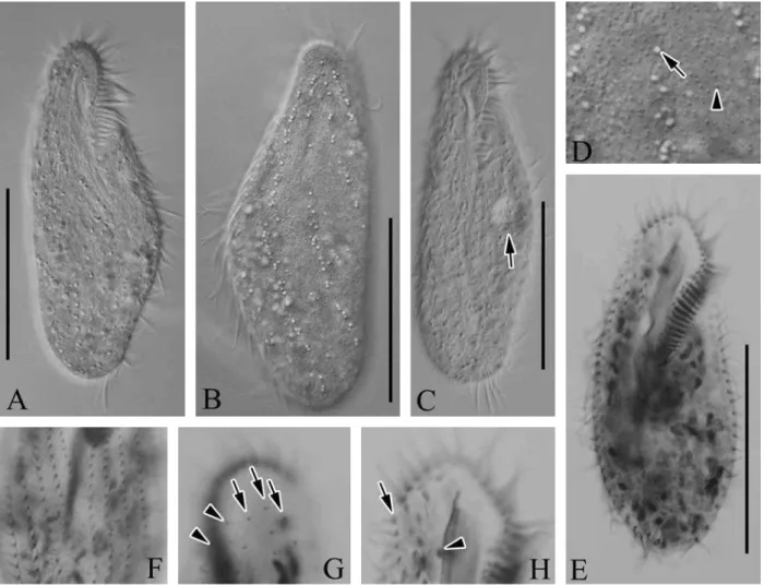
(Table 2, Figs. 1F-L, 3)
Metaurostylopsis struederkypkeae Shao et al., 2008: 289, figs. 1-20.
Description.Live cell size usually 80-110×40-50µm (Figs. 1F, 3A-C), slender in shape and slightly contractile, with an-terior and posan-terior ends rounded, and contractile vacuole approximately 10µm in diameter located on left side of equa-torial level of cell (Fig. 3C, arrow). Two types of cortical granules: one small and reddish, approximately 0.5µm across, spread irregularly on surface (Figs. 1G, H, 3D, arrowhead), and other one yellow-green, approximately 1.5µm across, situated at base of cirri, with some cortical granules clustered along cirral rows and dorsal kineties (Figs. 1G, H, 3D, arrow). Color of cells appeared reddish under low magnification due to numerous small cortical granules. Lengths of transverse and frontal cirri 15µm and 13µm, respectively. Other soma-tic cirri, midventral, left and right marginal cirri, ca. 10µm in length. Four frontal cirri and four to eight frontoterminal cirri located near distal portion of adoral membranelles (Fig. 3H, arrow). Two to six transverse cirri, and one buccal cirrus located in middle of next paroral membrane (Fig. 3H, arrow-head). Left and right marginal rows composed of five to seven and three to five, respectively (Fig. 3F). 12-20 midventral pairs, and six to nine unpaired midventral cirri between mid-ventral pairs and transverse cirri, three dorsal kineties (Fig. 3G, arrows), and separately two dikinetids (Fig. 3G, arrow-heads) on anterior dorsal side. Macronuclear nodules, ovoid and ellipsoid shape, ca. 82. Membranelles in adoral zone con-sisted of 25-36 membranelles, and portion of adoral zone is ca. 35-45% in length. Paroral and endoral membrane are equal level in length.
Distribution.China (Qingdao, the Yellow Sea) and Korea (this study).
Remarks.There are six species in this genus: Metaurostylop-sis marinaSong et al., 2001, M. rubra Song and Wilbert, 2002, M. songiLei et al., 2005, M. salinaLei et al., 2005, M. struederkypkeaeShao et al., 2008, and M. cheniChen et al., 2011.
Metaurostylopsis struederkypkeaeand M. rubrahave red-dish cell color. However M. struederkypkeaeis distinguished from M. rubraby the number of right marginal rows (3-5 vs. 6-7) and the type of cortical granules (two vs. one) and size (80-110×40-50µm vs. 150-300×50-90µm in vivo) (Song and Wilbert, 2002). Metaurostylopsis struederkypkeaeis distinguished from M. marinaby the body shape (slender vs. oval), the cell color (reddish vs. colorless) and the type of cortical granules (two vs. one) (Song et al., 2001).
The Korean population of M. struederkypkeaecorresponds with the original description by Shao et al. (2008) and mor-phometric comparisons highly overlap (Table 2); however, there are a few differences. The Korean population has more
number of left and right marginal rows, midventral pairs, and macronuclear nodules than those of the Chinese population (Shao et al., 2008).
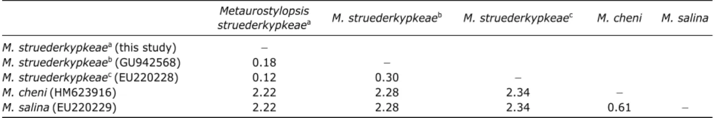
The SSU rDNA sequence of M. struederkypkeaeis 1,652 bp in length (GenBank accession no: JN880477). The
inter-specific pairwise distances between M. struederkypkeaeand its relatives (M. cheniand M. salina) are more than 2%, while intra-specific variation are less than 0.3% among two Korean and one Chinese M. struederkypkeaepopulations (Table 4). Fig. 3.Micrographs of Metaurostylopsis struederkypkeaefrom live (A-D) and protargol impregnated (E-H) specimen. A, B, Live ven-tral and dorsal views; C, Contractile vacuole (arrow); D, Yellow-green cortical granules (arrow), small reddish cortical granules (arrow-head); E, Ventral view of protargol-impregnated specimen; F, Ventral view, left and right marginal rows; G, Dorsal view, typically three dorsal kineties (arrows) and two dikinetids (arrowheads); H, Frontoterminal (arrow) and buccal (arrowhead) cirri. Scale bars: A-C, E==50µm.
Table 3.Kimura two-parameter pairwise distances (%) among the five species in Anteholosticha-Nothoholostichaassemblage based on small subunit ribosomal DNA sequences
A. pulchra A. gracilis A. monilata A. pseudomonilata N. fasciola
A. pulchra(this study) 폭
A. gracilis(FJ775713) 6.58 폭
A. monilata(GU942567) 5.04 4.10 폭
A. pseudomonilata(HM568416) 3.96 5.19 3.29 폭
ACKNOWLEDGMENTS
This study was supported by the Invasive Species Mana-gement Program in Marine Ecosystem, Korean Ministry of Land, Transport & Maritime Affairs of Korean Government, and also funded by the National Fisheries Research & Devel-opment Institute (NFRDI) of Korea and Polar Academic Pro-gram (PAP), KOPRI.
REFERENCES
Berger H, 2001. Catalogue of ciliate names 1. Hypotrichs. Verlag Helmut Berger, Salzburg, pp. 1-206.
Berger H, 2003. Redefinition of Holosticha Wrzesniowski, 1877 (Ciliophora, Hypotricha). European Journal of Protistology, 39:373-379.
Berger H, 2006. Monograph of the Urostyloidea (Ciliophora, Hypotricha). Springer, Dordrecht, pp. 1-1304.
Blatterer H, Foissner W, 1988. Beitrag zur terricolen Ciliaten-fauna (Protozoa: Ciliophora) Australiens. Stapfia, 17:1-84. Borror AC, 1979. Redefinition of the Urostylidae (Ciliophora,
Hypotrichida) on the basis of morphogenetic characters. The Journal of Eurkaryotic Microbiology, 26:544-550.
Borror AC, Wicklow BJ, 1983. The suborder Urostylina Janko-wski (Ciliophora, Hypotrichida): morphology, systematics and identification of species. Acta Protozoologica, 22:97-126.
Chen X, Huang J, Song W, 2011. Ontogeny and phylogeny of Metaurostylopsis chenisp. n. (Protozoa, Ciliophora), with estimating the systematic position of Metaurostylopsis. Zoo-logica Scripta, 40:99-111.
Foissner W, 1982. Ecology and taxonomy of the hypotrichida (Protozoa: Ciliophora) of some Austrian soil. Archiv Für Protistenkunde, 126:19-143.
Foissner W, 1991. Basic light and scanning electron microscopic methods for taxonomic studies of ciliated protozoa. Euro-pean Journal of Protistology, 27:313-330.
Foissner W, Didier P, 1981. Morphologie und infraciliatur eini-ger kinetofragminophorer und hypotricher ciliaten aus den Fliessgewässern von Besse-en-Chandesse (Frankreich). An-nales de la Station limnologique de Besse, 15:254-275.
Gao F, Yi Z, Gong J, Al-Rasheid KAS, Song W, 2010. Molecu-lar phylogeny and species separation of five morphologically similar Holosticha-complex ciliates (Protozoa, Ciliophora) using ARDRA riboprinting and multigene sequence data. Chinese Journal of Oceanology and Limnology, 28:542-548. Hall TA, 1999. BioEdit: a user-friendly biological sequence alignment editor and analysis program for Windows 95/98/ NT. Nucleic Acids Symposium Series, 41:95-98.
Hu X, Suzuki T, 2004. A new species of Holosticha (Ciliophora: Hypotrichida) from the coastal waters of Nagasaki, Japan: Holosticha nagasakiensis sp. nov. Journal of the Marine Biological Association of the United Kingdom, 84:9-13. Jung JH, Baek YS, Min GS, 2011. New record of two
Apokero-nopsisspecies (Ciliophora: Urostylida: Pseudokeronopsidae) from Korea. Korean Journal of Systematic Zoology, 27:115-122.
Kahl A, 1932. Urtiere oder Protozoa I: Wimpertiere oder Ciliata (Infusoria), 3. Spirotricha. Die Tierwelt Deutschlands, 25: 399-650.
Kahl A, 1933. Ciliata libera et ectocommensalia. Tierwelt der Nord- und Ostsee, 23:29-146.
Kumar S, Tamura K, Nei M, 2004. MEGA3: Integrated software for molecular evolutionary genetics analysis and sequence alignment. Briefings in Bioinformatics, 5:150-163. Li L, Khan SN, Ji D, Shin MK, Berger H, 2011. Morphology
and small subunit (SSU) rRNA gene sequence of the new brackish water ciliate Neobakuella flavan. g., n. sp. (Cilio-phora, Spirotricha, Bakuellidae) and SSU rRNA gene sequ-ences of six additional hypotrichs from Korea. The Journal of Eukaryotic Microbiology, 58:339-351.
Li L, Song W, Hu X, 2007. Two marine hypotrichs from north China, with description of Spiroamphisiella hembergerigen. nov., spec. nov. (Ciliophora, Hypotricha). Acta Protozoolo-gica, 46:107-120.
Li L, Zhang Q, Hu X, Warren A, Al-Rasheid KAS, Al-Khed-heiry AA, Song W, 2009. A redescription of the marine hy-potrichous ciliate, Nothoholosticha fasciola(Kahl, 1932) nov gen., nov comb. (Ciliophora: Urostylida) with brief notes on its cellular reorganization and SS rRNA gene sequence. European Journal of Protistology, 45:237-248.
Lynn DH, 2008. The ciliated protozoa: characterization, classi-fication, and guide to the literature. Springer, New York, pp. 1-605.
Table 4.Kimura two-parameter pairwise distance (%) based on small subunit ribosomal DNA sequences
Metaurostylopsis
M. struederkypkeaeb M. struederkypkeaec M. cheni M. salina
struederkypkeaea
M. struederkypkeaea(this study) 폭
M. struederkypkeaeb(GU942568) 0.18 폭
M. struederkypkeaec(EU220228) 0.12 0.30 폭
M. cheni(HM623916) 2.22 2.28 2.34 폭
M. salina(EU220229) 2.22 2.28 2.34 0.61 폭
Shao C, Song W, Al-Rasheid KAS, Yi Z, Chen X, Al-Farraj SA, Al-Quraishy SA, 2008. Morphology and infraciliature of two new marine urostylid ciliates: Metaurostylopsis strue-derkypkeaen. sp. and Thigmokeronopsis stoeckin. sp. (Cili-ophora, Hypotrichida) from China. The Journal of Eukaryot-ic MEukaryot-icrobiology, 55:289-296.
Song W, Petz W, Warren A, 2001. Morphology and morphoge-nesis of the poorly-known marine urostylid ciliate, Metau-rostylopsis marina(Kahl, 1932) nov. gen., nov. comb. (tozoa, Ciliophora, Hypotrichida). European Journal of Pro-tistology, 37:63-76.
Song W, Wilbert N, 2002. Faunistic studies on marine ciliates from the Antarctic benthic area, including descriptions of one epizoic form, 6 new species and, 2 new genera
(Proto-zoa: Ciliophora). Acta Protozoologica, 41:23-61.
Wirnsberger E, Foissner W, 1987. Morphologie von Holosticha xanthichromasp. n. und die variabilität der infraciliatur in der gattung Holosticha(Ciliophora, Hypotrichida). Acta Protozoologica, 26:1-8.
Yi Z, Song W, 2011. Evolution of the order Urostylida (Proto-zoa, Ciliophora): new hypotheses based on multi-gene infor-mation and identification of localized incongruence. PLoS ONE, 6:e17471.