INTRODUCTION
Congenital cataracts are the major cause of preventable and/or treatable childhood blindness(1,2), accounting for 8%-39% of cases of childhood blindness(3). The worldwide prevalence of congenital cataracts has been estimated to be between 1 and 15 per 10,000 children(1).
Surgical treatment for congenital cataract has developed over the last few decades, allowing cataract removal in children younger than 1 year. Congenital cataract deprives the retina of light during periods of greater neural plasticity, and surgery is the primary
treat-ment to prevent amblyopia; however, it only partially solves the problem because the aphakic eye has a refractive error that must be corrected(4,5).
Biometric calculation of a growing eye is challenging, and intrao-cular lenses (IOLs) implanted for emmetropia will produce a myopic shift with axial growth(6). Portable devices for biometric calculation may not be available for some of congenital/developmental cata-ract surgeons. Therefore, the selection of the IOL power at the time of implantation is based on tables of biometric measurements by age in normal eyes, which have biometric characteristics that may
Submitted for publication: 17 November, 2014 Accepted for publication: November 2, 2015
1 Department of Ophthalmology, Otorhinolaryngology and Head and Neck Surgery, Botucatu Medical School, Universidade Estadual Paulista (UNESP), Botucatu, SP, Brazil.
Funding: This study was supported by Research program for the Brazilian National Health System (PPSUS-SP), National Council for Scientic and Technological Development- Brazil (CNPq), and S̃o Paulo Research Foundation-Brazil (FAPESP).
Disclosure of potential conflicts of interest: None of the authors have any potential conflict of interest to disclose.
Corresponding author: Antonio Carlos Lottelli Rodrigues. Faculdade de Medicina de Botucatu - UNESP. Depto. OFT/ORL/CCP. Rubĩo Júnior, s/n, Botucatu, SP - 18618-970 - Brazil E-mail: [email protected]
Approved by the following research ethics committee: Botucatu Medical School Ethics Committee under number 3358.
ABSTRACT
Purpose: To evaluate the ocular axial length (AL) and keratometry (K) in Brazilian children with congenital/developmental cataract, assess the differences and evolution of AL and K according to age, and establish functional models of AL and K as function of age.
Methods: Children with congenital/developmental cataract aged 1.5 months old
to 8 years old and no other ocular diseases were included. All eyes with unilateral cataract, the left eyes from children with bilateral cataracts, and healthy eyes from children with unilateral cataract were analyzed. After the administration of anesthesia, K was measured with a portable automatic keratometer, and AL was measured with a contact biometer. Cataract surgery was performed immediately after the measurements were taken. The data were statistically analyzed, and a linear regression with an age logarithm was used to model the relationship.
Results: Forty-four eyes with cataract were included in this analysis, comprising
15 eyes with unilateral cataract and 29 left eyes from children with bilateral cataracts. The mean age was 27.3 months with a mean AL of 20.63 ± 2.11 mm and a mean K of 44.94 ± 2.44 D. The K value was significantly steeper and the AL value was significantly shorter in younger children (P<0.001). No significant differences were found neither between eyes with unilateral and bilateral cataracts nor between eyes with unilateral cataract and their corresponding healthy eyes (P>0.05). Conclusion: The values of K and AL significantly change with age, especially during the first 6 months of life. A linear functional relationship between K and AL with the logarithm of age and between K and AL was established.
Keywords: Cataract/congenital;Cornea/pathology; Axial length, eye; Lenses,
in-traocular; Corneal topography/methods
RESUMO
Objetivo: Avaliar o comprimento axial (AL) e a ceratometria (K) de olhos de crianças brasileiras com catarata congênita/desenvolvimento, analisar diferenças e evoluções de acordo com a idade e estabelecer modelos funcionais de comprimento axial e ceratometria em função da idade e entre eles.
Métodos: Crianças com catarata congênita/desenvolvimento com idade de 1,5 meses a 8 anos de idade e sem outras doenças oculares foram incluídas. Todos os olhos com catarata unilateral, o olho esquerdo de crianças com catarata bilateral e o olho sadio de crianças com catarata unilateral foram analisados. Após a administração de anestesia, a ceratometria foi obtida com um ceratômetro automático portátil e o comprimento axial medido com um biômetro de contato. Em seguida, a cirurgia de catarata foi realizada. Os dados foram analisados estatisticamente, a regressão linear com o logaritmo da idade foi utilizado para modelar os relacionamentos.
Resultados: Todos os olhos com catarata unilateral (n=15) e um olho selecionados aleatoriamente a partir dos casos bilaterais (n=29) foram incluídos na análise (total= 44 olhos). A idade média foi de 27,3 meses, as médias do comprimento axial e da ceratometria foram respectivamente 20,63 ± 2,11 mm e 44,94 ± 2,44 dioptrias. A ce-ratometria foi significativamente mais curvo e comprimento axial significantemente mais curto em crianças mais jovens (P<0,001). Não foram encontradas diferenças sig nificativas na comparação entre os olhos com cataratas unilaterais e bilaterais e com-parando os olhos com catarata unilateral a correspondentes olhos saudáveis (P>0,05).
Conclusão: Os valores de ceratometria e comprimento axial mudam significativamente com a idade, principalmente nos primeiros seis meses de vida. Foi estabelecida uma relação funcional linear entre comprimento axial e ceratometria com o logaritmo da idade e entre ceratometria e comprimento axial.
Descritores: Catarata/congênito; Córnea/patologia; Comprimento axial do olho; Lentes intraoculares; Topografia da córnea/métodos
Congenital and developmental cataract: axial length and keratometry study in
Brazilian children
Catarata congênita e do desenvolvimento: estudo do comprimento axial e da ceratometria em
crianças brasileiras
differ from the characteristics of eyes with cataracts, or on tables of biometric data in cataract eyes collected from populations in specific ethnicity (5,7).
The objective of this study was to evaluate the ocular axial length (AL) and keratometry (K) in Brazilian children with congenital/deve-lopmental cataract to assess the differences and evolution according to age as well as to establish functional models of AL and K in function of age and between K and AL.
METHODS
This retrospective observational study was approved by the Botucatu Medical School Ethics Committee; we analyzed the medi-cal records of patients undergoing cataract surgery for congenital/ developmental cataract at the Clinical Hospital of Botucatu Medical School, São Paulo, Brazil.
Prematurely born children as well as those with glaucoma, retinal disease, microphthalmia, traumatic cataract, pathological myopia, lens subluxation, or other ocular diseases were not included.
Ocular biometric data were obtained under general anesthesia by an experienced ophthalmologist, and cataract surgery was per-formed immediately after these measurements were made. The keratometric measurements were obtained using a regularly calibra-ted handheld autorefract keratometer (Retinomax K-Plus 2; Righton, Tokyo, Japan). AL measurement was performed with a contact ultrasonic biometer (model 1000A; Sonomed, New York, NY, USA). The ultrasound velocity used was 1532 m/s for the anterior chamber, 1641 m/s for the lens, and 1532 m/s for the vitreous.
All eyes with unilateral cataract and the left eyes in children with bilateral cataracts were selected and included in the analysis. In chil-dren with unilateral cataract, the affected eyes were compared with the healthy eye. The biometric data were transferred to Microsoft Excel 2007 (Redmond, WA, USA). The statistical analyses for the paired samples were performed using Student’s t-test.
The assessment of the relationship between the child’s age in months and the ocular biometric measurements was calculated with Pearson’s correlations. The linear regression analysis using the logarithm of a patient’s age as an independent variable was adjusted to obtain the estimates of the mean ocular parameters (K and AL). We considered P values <0.05 as statistically significant.
RESULTS
A total of 44 children were included in the study, and 28 (63%) were male. The median and mean age at the time of cataract sur-gery were 12.5 months and 27.3 months, respectively (range: 1.5-92 months). Twenty-nine children (66%) had bilateral cataracts, and 15 children (34%) had a unilateral cataract.
The eyes with unilateral cataract were not significantly different in terms of the mean AL and K with the left eyes in the bilateral cases (P>0.05) (Table 1).
The 44 analyzed eyes had a mean AL of 20.63 mm (range 17.27 mm- 24.96 mm) and a mean K of 44.94 D (range 40.00 D-53.00 D). The calculations of the mean, standard deviation, confidence interval, median, and variable range for AL and K according to the age groups are shown in table 2. The mean of AL increased with age, whereas the mean of K decreased. These relationships were statistically significant (P<0.001). If the AL observations were divided into those from chil-dren under and over 12 months old, the relative variability expressed by the coefficient of variation showed homogeneity of the response between the two age groups.
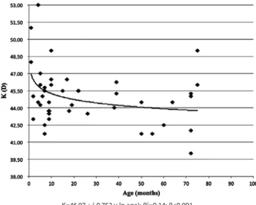
The linear regression analyses performed using the natural logarithm (ln) of the patient’s age (in months) as an independent variable are shown below by the following equations: AL=16.66 + (1.476 × ln age); R2=0.74, P<0.001 and K=46.97 + (-0.752 × ln age); R2=0.14, P<0.001.
A graphical representation of AL and K according to the patients’ age and the fitted curves derived from the model above are shown in figures 1 and 2. AL and K change markedly with an increase in age.
Table 1. Axial length (AL) and mean keratometry (K) values according to the laterality of the cataracts
Groups n Age (months) AL (mm) K (D)
Unilateral 15 Mean ± SD 27.7 ± 27.1 20.6 ± 2.0 44.8 ± 2.7
Median 18 20.8 44.5
Range 2-75 17.9-24.7 41.7-53.0
Bilateral 29 Mean ± SD 27.1 ± 27.7 20.6 ± 2.2 45.0 ± 2.4
Median 10 19.9 45.0
Range 1-92 17.3-25.0 40.0-51.0
P value 0.960 0.990 0.629
AL= axial length; K= keratometry; D= diopter; SD= standard deviation; Student’s t-test.
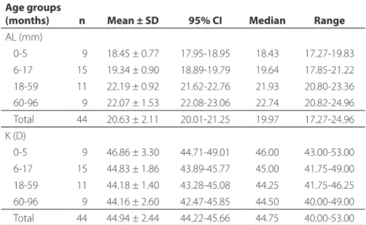
Table 2. Mean, standard deviation, conidence interval, median, and range of the axial length (AL) and keratometry (K) values according to the age group of 44 eyes with unilateral cataract and one randomly chosen eye from the bilateral cataract cases
Age groups
(months) n Mean ± SD 95% CI Median Range
AL (mm)
0-5 09 18.45 ± 0.77 17.95-18.95 18.43 17.27-19.83
6-17 15 19.34 ± 0.90 18.89-19.79 19.64 17.85-21.22
18-59 11 22.19 ± 0.92 21.62-22.76 21.93 20.80-23.36
60-96 09 22.07 ± 1.53 22.08-23.06 22.74 20.82-24.96
Total 44 20.63 ± 2.11 20.01-21.25 19.97 17.27-24.96
K (D)
0-5 09 46.86 ± 3.30 44.71-49.01 46.00 43.00-53.00
6-17 15 44.83 ± 1.86 43.89-45.77 45.00 41.75-49.00
18-59 11 44.18 ± 1.40 43.28-45.08 44.25 41.75-46.25
60-96 09 44.16 ± 2.60 42.47-45.85 44.50 40.00-49.00
Total 44 44.94 ± 2.44 44.22-45.66 44.75 40.00-53.00
AL= axial length; K= keratometry; D= diopter; SD= standard deviation; CI= confidence interval; Student’s t-test.
AL=16.66 + (1.476 × ln age); R2=0.74; P<0.001.
Figure 1. Distribution of axial length (AL) values of 44 eyes of children with unilateral
The highest rates of ocular axial growth and corneal flattening are observed in the first 6 months of life.
The dispersion values of AL according to K and the inverse rela-tionship between them are shown in figure 3. The equation provided for the graph is K=52.91 -0.385 × AL (mm), with R2=0.11 (P<0.05).
Table 3 shows a comparison of the biometric measurements between eyes with a unilateral cataract and the corresponding healthy eye, with no significant differences found in the AL and K values (P>0.05).
Tests of the two regressions for the AL and K logarithm models showed that there were no statistically significant differences bet-ween eyes with cataract from different genders, eyes with unilateral cataract and the ones with bilateral cataracts, and normal eyes and eyes with cataract in children with unilateral cataract (P>0.05).
Table 4 estimates AL and K according to age (in months) based on the following equations: AL=16.66 + (1.476 × ln age) and K=46.97 + (-0.752 × ln age), from figures 1 and 2 and the IOL for emmetropia and
for minimizing myopic shift in adulthood, using suggestions from some authors(8), according to age, based on the estimated values of AL and K.
DISCUSSION
The development of cataract surgery in children has created the need to study the biometric and refractive changes that occur with ocular growth(9,10). In the first months of extra-uterine life, the human eye experiences axial growth accompanied by flattening of the corneal curvature(11,12). When this process occurs uneventfully, the decrease of the refractive power of the lens focuses the image on the retina because of the increased AL. During this rapid biometric change, emmetropia is maintained(13).
Eyes presenting cataract have different biometric measurements from normal eyes, and various factors, such as the child’s age at surgery, aphakia, pseudophakia, cataract laterality, and visual depri-vation have been reported to influence axial growth(12).
K=46.97 + (-0.752 × ln age); R2=0.14; P<0.001.
Figure 2. Distribution of the mean keratometry (K) values of 44 eyes of children with
unilateral cataracts and randomly selected eyes in the bilateral cases.
K=52.91-0.385 × AL; R2=0.11; P<0.05.
Figure 3. Distribution of the mean keratometry (K) values for axial length (AL) values.
Table 3. Comparison of the mean, median, and standard deviation of axial length (AL) and the mean keratometry (K) values of both eyes of 15 children with unilateral cataract
n AL (mm) K (D)
Cataract eye 15 Mean ± SD 20.6 ± 2.0 44.8 ± 2.7
Median 20.8 44.5
Range 17.9-24.7 41.7-53.0
Healthy eye 15 Mean ± SD 21.3 ± 1.7 44.4 ± 1.5
Median 20.9 44.5
Range 18.8-24.8 42.0-48.5
P value 0.061 0.649
AL= axial length; K= keratometry; mm= millimeter; D= diopter; SD= standard deviation. Student’s t-test.
Table 4. Axial length (AL) and keratometry (K) values estimated by age according to the linear regression analysis performed using the natural logarithm (ln) of the patient’s age as an independent variable: AL=16.66 + (1.476 × ln age) and K=46.97 + (-0.752 × ln age), as well as the intraocular lens (IOL) power calculated for emmetropia based on the values of K and AL using the Hofer Q formula with pACD=5.26 (A-constant=118.5). The indicated desired refraction to minimize late myopia shift8 and the ideal IOL power to obtain the desired refraction
are also demonstrated
Age (months)
AL
(mm) K (D)
IOL to emmetropia
(D)
Desired refraction
(D)
Ideal IOL power for desired refraction (D)
3 18.28 46.14 +41.36 +9.00 +26.90
6 19.30 45.62 +35.73 +8.00 +23.11
9 19.90 45.32 +32.95 +7.00 +22.06
12 20.33 45.10 +31.14 +6.00 +21.93
18 20.93 44.80 +28.81 +6.00 +19.53
24 21.35 44.58 +27.31 +5.00 +19.67
30 21.68 44.41 +26.18 +5.00 +18.51
36 21.95 44.28 +25.29 +5.00 +17.58
42 22.18 44.16 +24.56 +5.00 +16.82
48 22.37 44.06 +23.97 +4.00 +17.87
54 22.55 43.97 +23.42 +4.00 +17.30
60 22.70 43.89 +22.97 +3.00 +18.45
66 22.84 43.82 +22.56 +3.00 +18.03
72 22.97 43.75 +22.19 +2.00 +19.21
Researchers have reported the existence of a passive ocular-stret ching component that is genetically determined and an active component that is observed when the image is not formed on the retina(13). Supporting this hypothesis, several studies have observed increased AL in eyes with visual deprivation(14). This increase occurs in human eyes as well as in the eyes of primates and other animals(6,13).
Several studies have demonstrated flattening of the K values in ol-der children(11,15). A previous study reported mean K values of 47.50 D in newborns and 43.69 D in children aged 2-4 years and concluded that K values reach adulthood values at the age of approximately 3 years (16). In the present study, the eyes with congenital and develop-mental cataract showed K values that were significantly more curved in children younger than 6 months old. Although it presented a poor correlation (R2=0.14), the value of K was inversely related to the ages of the children up to 18 months. After this age, corneal flattening tends to stabilize.
It has been demonstrated that normal eyes have a smaller myopic shift in comparison to aphakic eyes(11). The reason for this difference is that phakic eyes exhibit a decline in the refractive power of the crystalline lens from +34.4 D to +18.8 D with growth, which does not occur with an implanted IOL(11). Therefore, children with pseudophakia might present a large myopic change in adulthood if the implanted IOL aims for emmetropia at the time of surgery(6).
Two papers have described K and AL measurements in North American children with congenital cataract(5,17). Further, a study in Italy showed the AL and K values of a Caucasian pediatric population with congenital/developmental cataract(7). Databanks containing biometric information from eyes with pediatric cataract allow esti-mation of the ocular values of AL and K based on age. These data facilitate the selection of IOL power in children with cataract when it is not possible to measure AL and K.
In accordance with the results from another study(18), Brazilian children showed corneal flattening and increased AL with age. In the study by Ingaki(18), there was no significant difference between K and AL in a comparison of eyes with unilateral and bilateral cata-racts. The same results were obtained from comparing healthy eyes and affected eyes in children with unilateral cataract. Additionally, in studies of AL and K in North American children, lower AL and greater K were found in eyes with unilateral cataract compared with the AL and K values in healthy eyes, although the difference in the K value was not statistically significant(5,17). As visually impaired eyes tend to have greater axial growth(12), the absence of differences between the AL value of healthy eyes and that of eyes in children with unilateral cataract might have resulted from later diagnosis and treatment in our sample, which could have resulted in worsened visual prognosis for these eyes(19).
Regarding gender, previous studies have reported steeper corneas and shorter AL in girls than in boys; however, these results were not observed in our sample(5,17).
To avoid the development of high myopia with axial growth, some authors recommend that after the calculation of the IOL for emmetropia, 20% of the IOL power should be subtracted in young children (<8 months of age), and 10% should be subtracted in chil-dren aged between 2 and 3 years(16). Others propose tables with residual hyperopia from +12.00 to +0.50 based on the age of the chil-dren, from 3 months to 14 years(8). After the surgical treatment, optical correction or contact lenses are prescribed for residual hyperopia, which decreases with age. Table 4 shows suggestions, based on our data, for IOL implantation according to age for emmetropia and for minimizing late myopia using the suggestions from some authors(8) when AL and K cannot be measured.
When K measurement is not possible because of the unavailabi-lity of manual keratometers, refractive powers of 28, 27, 26, 24, and 22 of the implanted IOL are suggested for ALs of 17, 18, 19, 20, and 21 mm, respectively(20). We suggest that when only AL can be mea-sured, the K value should be calculated using the equation: K=52.91-0.385 × AL (mm) (Figure 3) or based on age.
The selection of the power of the IOL for implantation in a gro-wing eye represents a major challenge. The use of a published table alone to decide IOL power is not recommended. The tables are only intended as a starting point toward appropriate IOL power selection, which is a multifactorial decision customized for each child based on many variables, particularly age, laterality, amblyopia status, likely compliance with glasses, and a family history of myopia(8).
The main limitation of this study is the K values. The measure-ments were performed with a manual keratometer which, although reproducible in awake patients, may not be as reliable in our sample where most of the measurements were performed on patients under anesthesia without fixation. These conditions probably influenced these values and their relationships with age and AL, generating a poor correlation (R2=0.11). These findings have already been descri-bed by other authors, who also found weaker relationships between K and age, with R2 varying from 0.31(17) to 0.20(7), as well as between K and AL (R2=0.32)(17), despite the larger sample sizes. Due to the difficulty of obtaining these values, the measurement method used in the present study still seems to be the best manner to obtain K values; however, a device that allows more accurate measurements may establish prediction models with more precise estimates of K in relation to age and AL.
CONCLUSION
The values of K and AL change significantly with age, especially in the first 6 months of life. A linear functional relationship between K and AL with the logarithm of age and between K and AL was established for this Brazilian pediatric sample with congenital/deve-lopmental cataract.
ACKNOWLEDGMENT
Research program for the Brazilian National Health System (PPSUS- SP), National Council for Scientific and Technological Development-Bra zil (CNPq), and São Paulo Research Foundation-Development-Brazil (FAPESP).
REFERENCES
1. Foster A, Gilbert C, Rahi J. Epidemiology of cataract in childhood: a global perspective. J Cataract Refract Surg. 1997;23(1):601-4.
2. Trivedi RH, Wilson ME. Childhood blindness and pediatric cataract. J Cataract Refract Surg. 2005:52-4.
3. Trivedi RH, Wilson ME. Epidemiology of pediatric cataract and associated blindness. In: Wilson ME, Trivedi RH, Pandey SK, editors. Pediatric Cataract Surgery: techniques, complications and management. Baltimore, Maryland: Lippincott Williams & Wilkins; 2005. p.18-22.
4. Wright KW. Visual development, amblyopia, and sensory adaptations. In: Wright KW, editor. Pediatric Ophthalmology and Strabismus. New York: Springer-Verlag; 2003. p.157-71.
5. Trivedi RH, Wilson ME. Biometry data from Caucasian and African- American catarac-tous pediatric eyes. Invest Ophthalmol Vis Sci. 2007;48(10):4671-8.
6. Dahan E, Drusedau MU. Choice of lens and dioptric power in pediatric pseudophakia. J Cataract Refract Surg. 1997;23 Suppl 1:618-23.
7. Capozzi P, Morini C, Piga S, Cuttini M, Vadalà P. Corneal curvature and axial length values in children with congenital/infantile cataract in the first 42 months of life. Invest Ophthalmol Vis Sci. 2008;49(11):4774-8.
8. Trivedi RH, Wilson ME. Pediatric cataract: preoperative issues and considerations. In: Wilson ME, Saunders RA, Trivedi RH, editors. Pediatric Ophthalmology: Current Thought and A Practical guide. Heidelberg, Germany: Springer; 2009. p. 311-24.
9. Flitcroft DI, Knight-Nanan D, Bowell R, Lanigan B, O’Keefe M. Intraocular lenses in children: changes in axial length, corneal curvature, and refraction. Br J Ophthalmol. 1999;83(3):265-9.
10. Rabin J, Van Sluyters RC, Malach R. Emmetropization: a vision-dependent phenomenon. Invest Ophthalmol Vis Sci. 1981;20(4):561-4.
11. Gordon RA, Donzis PB. Refractive development of the human eye. Arch Ophthalmol. 1985;103(6):785-9.
12. Vasavada AR, Raj SM, Nihalani B. Rate of axial growth after congenital cataract surgery. Am J Ophthalmol. 2004;138(6):915-24.
13. Brown NP, Koretz JF, Bron AJ. The development and maintenance of emmetropia. Eye (Lond). 1999;13(Pt 1):83-92.
eye compared to the unoperated fellow eye in children with bilateral cataracts. J AAPOS. 2014;18(2):173-7.
15. Asbell PA, Chiang B, Somers ME, Morgan KS. Keratometry in children. CLAO J. 1990;16(2):99-102.
16. Ehlers N, Sorensen T, Bramsen T, Poulsen EH. Central corneal thickness in newborns and children. Acta Ophthalmol (Copenh). 1976;54(3):285-90.
17. Trivedi RH, Wilson ME. Keratometry in pediatric eyes with cataract. Arch Ophthalmol. 2008;126(1):38-42.
18. Inagaki Y. The rapid change of corneal curvature in the neonatal period and infancy. Arch Ophthalmol. 1986;104(7):1026-7.
19. Rodrigues AC, Prado RB, Miguel L. Implementation of red reflex exam in children in the area of Botucatu Medical School--São Paulo, Brazil. Arq Bras Oftalmol. 2012;75(5): 337-40.