Clinical and electroencephalographic
characteristics of benign occipital
epilepsy of childhood in two
tertiary Brazilian hospitals
Soniza Vieira Alves-Leon1, Renata Gomes Nunes2,
Maria Emilia Cosenza Andraus3, José Carlos Biagini Junior4,
Marta Hemb5, Maria Alice Genofre6
ABSTRACT
This study intended to investigate the clinical and electroencephalographic benign occipital epilepsy of childhood (BOEC) characteristics in a population sample of patients from two tertiary Brazilian hospitals. We analyzed retrospectively 4912 electroencephalograms (EEGs) records, and the included patients were submitted to a new clinical and EEG evaluation. Were included 12 (0.92%) patients; 4 (33.3%) with criteria for early BOEC; 6 (50%) for late form and 2 (16.7%) with superimposed early and late onset forms. After new investigation, 2 (16.7%) had normal EEG; 4 (33.3%) had paroxysms over the occipital region; 3 (25%) over the temporal posterior regions and 3 (25%) over the posterior regions. Sharp waves were the predominant change, occurring in 8 (66.6%); spike and slow wave complexes in 1 (8.3%) and sharp and slow wave complexes in 1 (8.3%). Vomiting, headache and visual hallucinations were the most common ictal manifestations, presented in 100% of patients with superimposed forms. Vomiting were absent in the late form and headache was present in all forms of BOEC.
Key words: occipital epilepsy, epilepsy of childhood, benign epilepsy of childhood, benign occipital epilepsy.
Características clínicas e eletrencefalográficas da epilepsia occipital benigna da infância em dois hospitais terciários brasileiros
RESUMO
Este estudo teve como objetivo investigar as características clínicas e eletrencefalográficas da epilepsia occipital benigna da infância (EOBI) em uma amostra populacional de pacientes de dois hospitais terciários brasileiros. Foram analisados retrospectivamente 4912 registros de eletrencefalograma (EEG). Os pacientes incluídos foram submetidos a nova avaliação clínica e eletrencefalográfica. Foram incluídos 12 (0,92%) pacientes; 4 (33,3%), com critérios para EOBI de início precoce; 6 (50%) para a forma tardia e 2 (16,7%), com superimposição de formas de início precoce e tardio. Após nova investigação, 2 (16,7%) apresentaram EEG normal; 4 (33,3%) paroxismos sobre a região occipital; 3 (25%) sobre a região temporal posterior e 3 (25%) sobre as regiões posteriores. Ondas agudas foram a alteração predominante, ocorrendo em 8 (66,6%); complexos espícula e onda lenta em 1 (8,3%) e complexos onda aguda e onda lenta em 1 (8,3%). Vômitos, cefaléia
Correspondence
Soniza Vieira Alves-Leon Programa de Epilepsias Hospital Universitário Clementino Fraga Filho UFRJ Unidade de Vídeo-EEG Enfermarias 10C2 e 10C4 Rua Prof. Rodolpho Paulo Rocco 255 21941-617 Rio de Janeiro RJ - Brasil E-mail: [email protected]
Received 29 June 2010 Received in final form 12 April 2011 Accepted 19 April 2011
1Epilepsy Program, Service of Neurology, Hospital Universitário Clementino Fraga Filho, Universidade Federal do Rio de
Janeiro (UFRJ), Rio de Janeiro RJ, Brazil. Associate Professor of Neurology and Permanent Professor of Strictu Sensu Post Graduation Program in Neurology, Universidade Federal do Estado do Rio de Janeiro (UNIRIO), Rio de Janeiro RJ, Brazil; 2Neurologist and Clinical Neurofisiologist, Doctor in Neurology UFRJ; 3Electroencephalography Section, Department
of Neurology, 24th and 25th Infirmaries, Santa Casa da Misericórdia do Rio de Janeiro, Rio de Janeiro RJ, Brazil. Collaborative
Professor of Strictu Sensu Post Graduation Program in Neurology, UNIRIO; 4Neurologist and Clinical Neurofisiologist; 5Pediatric
e alucinações visuais foram as manifestações ictais mais comuns, estando presentes em 100% dos pacientes com formas superimpostas de EOBI. Vômitos não foram relatados na forma tardia e cefaléia esteve presente em todas as formas de EOBI.
Palavras-chave: epilepsia occipital, epilepsia da infância, epilepsia benigna da infância, epilepsia occipital benigna.
In 1950, Gastaut1 described the benign occipital epi-lepsy of childhood (BOEC), with visual seizures and oc-cipital epileptiform paroxysms reactive to ocular opening and, in 1982, proposed it as a new epileptic syndrome2. As a consequence of this fact, there were considerable work done on this subject, some reairming Gastaut’s idea and others contesting its benign course or even the existence of the syndrome, as well as its speciicity to the reactivity of occipital paroxysms and the neuropsycho-logical proile3. In 1989, Panayiotopoulos4,5 proposed that the syndrome should be divided in the early and the late onset variants. Again, new reports against and in favor this theory were published. In 1989, he International League Against Epilepsy (ILAE)6 recognized BOEC as a syndrome, classifying it in the group of partial epilepsy age and localization-related and, in 20017, admitted that the early variant is diferent from the late one8.
Clinical features
According to the clinical features and the prognosis, the BOEC can be classiied in two types: an early one or Panayiotopoulos syndrome and a late one or idiopathic childhood epilepsy of Gastaut7.
In the early BOEC, the onset is between 1 and 12 years-old, and the main ictal indings are vomiting and/ or deviation of the eyes, which can progress to hemi or generalized seizures. here is also a high occurrence of partial status epilepticus that sometimes can be the only clinical event4,5,9-22. Characteristically, even after the most severe seizures and status, the child is normal after a few hours of sleep22. Sometimes, this Panayiotopoulos syndrome has eluded recognition because emetic and other ictal autonomic manifestations are dismissed as non-epileptic events of other diseases23,24. Encephalitis is a common example of a misdiagnosis for Panayioto-poulos syndrome23. he pathophysiology of Panayioto-poulos syndrome is unknown, but it is likely that they are due to difuse maturation-related epileptogenicity activa-tion susceptible for children emetic centers and the hy-pothalamus25. Converging evidence from multiple and independent studies has documented Panyiotopoulos syndrome as a model of childhood autonomic epilepsy, wich is common and benign23.
In the late BOEC, the onset is between 3 and 16 years-old, and the main ictal indings are visual seizures,
as elementary visual hallucinations, complex visual hal-lucinations and visual illusions, blindness or partial vi-sual loss, and non vivi-sual seizures, as deviation of the eyes and oculoclonic seizures, forced eyelid closure and eyelid blinking, sensory hallucinations of ocular movements and pain1,2,26. Migraine with visual aura is a common mis-diagnosis for this syndrome23.
here are few studies of inheritance involvement, al-though Kuzniecky and Rosenblatt found 3 children of a family with early BOEC27. Nagendran et al.28 found dif-ferent types of benign epilepsies of childhood in the same family, but yet there is still lack of enough studies of the genetic basis of late BOEC.
Sometimes, the same child may present other idio-pathic epilepsy of childhood associated with BOEC8,29. hese inding suggest a maturation process involvement, and in the case of benign epilepsy with centro-temporal spikes and BOEC, for example, a close genetic relation-ship, or even a common marker with variable pheno-types (idiopathic partial epilepsies with rolandic and oc-cipital spikes appearing in the same children)23,25.
Electroencephalographic indings
he electroencephalographic indings are similar in both variants, and consist in the majority of high voltage spike-wave complexes in a normal background activity, bilateral and synchronous, over the posterior regions, predominantly in the occipital lobes2,4,5,9,12. Some authors describe the high prevalence of changes on the foci lo-calization as the child is growing-up20.
oc-cipital region. Ictal EEG abnormalities did not change during the diferent seizure manifestations. he end of the seizure was abrupt21.
In the late BOEC, some patients may have only random occipital spikes; others may have occipital spikes only in sleep records and some have a consistently normal interictal EEG23. he ictal EEG indings were de-scribed in the late BOEC during the visual seizure as fast paroxystic epileptiform activity localized in the occipital regions, with occasional spreading2,19.
Sometimes, because of the frequent EEG occipital spikes in the both variants, the diferential diagnosis be-tween them may become difficult, based on the EEG indings23.
Neuroimaging studies
here are few systematic studies in the literature re-lated to the age and neurologic normality demonstrated by neuroimaging studies in order to discard small or-ganic lesions.
Our purpose was to evaluate the clinical and electro-encephalographic characteristics of benign occipital epi-lepsy of childhood in a sample population of children and adolescents from two tertiary Brazilian hospitals.
METHOD
We performed a bidirectional study. First, we made a retrospective study, analyzing 4029 EEGs of 600 epileptic patients and clinical notes of the requesting physician, from 1995 to 2004, at the Instituto Fernandes Figueira (IFF), a pediatric institute. hese EEGs were performed using an analogic EEG, Berger’s type. his cohort was associated with another retrospective study of 818 EEG records from 2001 to 2005 of 503 patients at the Hos-pital Universitário Clementino Fraga Filho, Universidade Federal do Rio de Janeiro (HUCFF/UFRJ). hese EEGs were performed using a digital Neurotec® EEG equip-ment, using especially longitudinal and referential mon-tages and 24 channels. We analyzed EEG records of a total of 1103 patients with epilepsy. Later, we recaptured the patients who met criteria for BOEC and they were submitted to a new clinical, neuroimaging (computed tomography (CT) scan and/or magnetic resonance im-aging (MRI) of the brain) and electroencephalographic evaluation, this time at the HUCFF/UFRJ (using the dig-ital 24 channels Neurotec® equipment to perform the EEGs). he EEGs had a minimum duration of 40 min-utes, and included records of spontaneous sleep, inter-mittent photic stimulation and hyperventilation.
The inclusion criteria were clinical and electroen-cephalographic, as: [1] age of onset of seizures from 1 to 16 years-old; [2] normal development, neurological and mental state; [3] normal brain imaging studies; [4]
normal background EEG, except for postictal records; [5] EEG with posterior discharges; [6] ictal emetic symp-toms; [7] visual hallucinations; [8] onset as partial status epilepticus; [9] eyes deviation.
We excluded patients with: [1] abnormal neurolog-ical development; [2] abnormal brain imaging studies; [3] abnormal background EEG; [4] children with febrile seizures, but without epilepsy development, considering that febrile seizures can be viewed as a syndrome of re-active seizures, and not as a true epileptic syndrome, ac-cording Engel7.
In the new evaluation, we analyzed age, gender dis-tribution, age onset of the seizure, clinical indings, EEG indings in awake and asleep records and as well as re-sponse to antiepileptic drugs. Two neurologists, titular members of the Brazilian Clinical Neurophysiology So-ciety analyzed, together and simultaneously, all EEG re-cords and clinical features.
his study protocol has been approved by the Sci-entifc Investigation Committee (Comitê de Investigação Cientíica - CIC) and the Research Ethic Committee (Co-mitê de Ética em Pesquisa - CEP) of the HUCFF/UFRJ. All participants signed an Informed Consent Term.
RESULTS
Analyzing the data of the IFF, we found 81 records from patients with history and EEG indings of occip-ital epilepsy, but only 14 children met the clinical and EEG criteria for BOEC. Eight of these 14 patients who met the BOEC inclusion criteria in the review of the his-tory, were not traced. Six underwent new clinical, EEG and imaging evaluation, and 1 of these was excluded, due to brain tumor found in the new CT scan investigation. From 2001 to 2005, out of 503 patients in the HUCFF/UFRJ outcome Epilepsy Unit, 8 met the inclu-sion criteria, and 1 of them was excluded after the MRI study, due to neurocysticercosis.
So, the total number of patients who met the inclu-sion criteria for this study was 12. Ten of them were male. Six of these children turned out to fall in the late BOEC category, and 2 turned out to fall with super-imposition of early and late onset, all of them having mainly elementary visual hallucinations (Tables 1 and 2). Four children met the criteria for early BOEC with ictal emetic symptoms, in 1 of them accompanied by devia-tion of the eye and other with elementary visual halluci-nations and emetic symptoms (Table 2).
Vomiting, headache and visual hallucinations were presented in 100% of patients with superimposed forms. Vomiting were absent in the late form and headache was present in all forms of BOEC (Table 2).
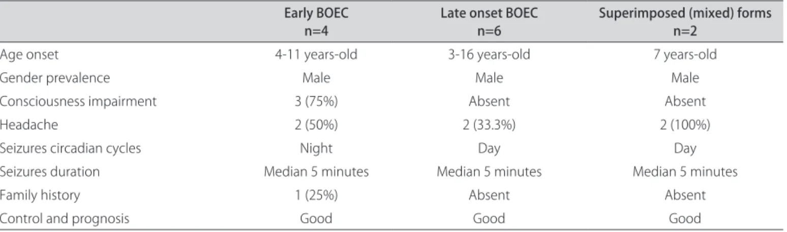
occip-Table 1. Results (n=12) of early versus late onset and mixed forms of BOEC patients analysis.
Early BOEC
n=4 Late onset BOEC n=6 Superimposed (mixed) forms n=2
Age onset 4-11 years-old 3-16 years-old 7 years-old
Gender prevalence Male Male Male
Consciousness impairment 3 (75%) Absent Absent
Headache 2 (50%) 2 (33.3%) 2 (100%)
Seizures circadian cycles Night Day Day
Seizures duration Median 5 minutes Median 5 minutes Median 5 minutes
Family history 1 (25%) Absent Absent
Control and prognosis Good Good Good
BOEC: benign occipital epilepsy of childhood.
Table 2. Results of ictal phenomenology described by patients with early, late and mixed forms of BOEC (n=12).
Early BOEC
n=4 Late onset BOECn=6 Superimposed (mixed forms) of BOECn=2
Vomiting 4 (100%) Absent 2 (100%)
Headache 2 (50%) 2 (50%) 2 (100%)
Tonic deviation of the eyes 1 (25%) Absent Absent
Secondarily generalized seizures Absent 1 (17%) Absent
Focal status Absent Absent Absent
Visual hallucinations Absent 6 (100%) 2 (100%)
Amaurosis Absent Absent Absent
Oculo-clonic seizures Absent Absent Absent
Opening and closing sustained eyes Absent Absent Absent
BOEC: benign occipital epilepsy of childhood.
Fig 1. Patient 1, a 5 years-old boy with crisis of visual elemen-tary hallucinations. The EEG shows discharges of occipital bilateral spike and wave complexes, with right predominance (ellipses).
Fig 2. Patient 10, 4 years-old, with crisis of vomiting and head-ache. The EEG shows interictal epileptiform discharges of bilat-eral occipital sharp and slow wave complexes, with right predom-inance (ellipses).
ital region in 4 EEGs (33.3%), with predominance in one side (right or left equally). he temporal posterior re-gions were involved in 3 (25%) cases, bilateral in 1, on the right in 1 and on the left in 1. he posterior regions (including posterior temporal, parietal and occipital
he voltage was high in all EEGs.
he paroxysm reactivity was demonstrated in EEGs of 7 patients, where the discharges disappeared with eye opening, and appeared with eye closure.
In our series, the paroxysms improved during sleep. Hyperventilation and intermittent photic stimulation did not show additional abnormalities.
All the patients have normal neuroimaging studies, which was an inclusion criterion.
DISCUSSION
Despite the heterogeneity of the BOEC data in the lit-erature reviewed, it is reasonable to conclude that BOEC exists as an electroclinical entity, being divided in two types: early BOEC or Panayiotopoulos syndrome and late BOEC or Gastaut type. Both types occur at similar age, 1 to 12 years in the early BOEC and 3 to 16 years in the late one. he two types are mainly diferentiated by their ictal clinical manifestation, that in the early one is vom-iting and tonic deviation of the eyes, during the sleep
and in the late BOEC symptoms are visual seizures (il-lusions, hallucinations, amaurosis) and non-visual sei-zures (eye deviation and oculoclonic seisei-zures). Postictal cephalalgia is more frequent in late BOEC. BOEC has a benign course, especially the early one, which frequently appears as a single seizure. According to the literature, the frequency of BOEC is high, representing 20-25% of the benign partial epilepsies of childhood. In our series few cases of BOEC were found23. We found that the early variant is being super diagnosed in those cases in which it occurs as a single seizure and with normal EEG. It is seeing with the increased number of publications after Panayiotopoulos’ articles.
Although some people consider the late BOEC worse than the early one, Caraballo et al.31 found, during a pro-spective study with adolescents, that its course is also benign. As the syndrome occurs in children with age ranging from 1 to 16 years and the prognosis is good, it seems to result from an evolution of the same mat-urational process which also involves the rolandic and Table 3. Results of follow up of the patients (n=12) with BOEC.
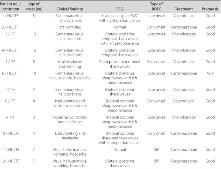
Patient no. /
Institution onset (yo)Age of Clinical indings EEG Type of BOEC Treatment Prognosis
1 / HUCFF 5 Elementary visual
hallucinations with right predominanceBilateral occipital SWC Late onset Valproic acid Good
2 / HUCFF 11 Ictal vomiting Normal Early onset Carbamazepine Good
3 / IFF 12 Elementary visual
hallucinations temporal sharp waves Bilateral posterior
with left predominance
Late onset Phenobarbital Good
4 / HUCFF 16 Elementary visual
hallucinations temporal sharp wavesBilateral posterior Late onset Phenobarbital Good
5 / IFF 5 Ictal headache
and vomiting Right posterior temporal sharp waves Early onset Valproic acid Good
6 / HUCFF 10 Elementary visual
hallucinations, headache sharp waves with left Bilateral posterior predominance
Late onset Carbamazepine NCT
7 / IFF 7 Elementary visual
hallucinations Bilateral posterior sharp waves Late onset Valproic acid Good
8 / IFF 8 Ictal vomiting and
tonic eye deviation sharp waves with left Bilateral occipital
predominance
Early onset Valproic acid Good
9 I IFF 3 Visual hallucinations
and headache sharp waves with left Bilateral occipital
predominance
Late onset Phenobarbital Good
10 / HUCFF 4 Ictal vomiting and
headache sharp-and slow waves Bilateral occipital
with right predominance
Early onset Carbamazepine Good
11 / HUCFF 7 Visual hallucinations,
vomiting, headache Normal SIF Carbamazepine Good
12 / HUCFF 7 Visual hallucinations,
vomiting, headache Bilateral posterior sharp waves SIF Carbamazepine Good
absence epilepsy, which they sometimes could be as-sociated with. he neurological examination should be normal in BOEC patients, like the neuroimaging studies. In our study all the patients had normal CT scan and three of them had also normal brain MRI studies. In re-lation to the interictal paroxysms, there is some general agreement about the localization and morphology of the electroencephalographic abnormalities with the re-viewed articles. We found the paroxysms localization as the most concordant parameter.
he interictal electroencephalographic indings are similar in the two types of BOEC showing occipital or posterior temporal epileptiform paroxysms that disap-pear with eye opening although this reactivity is not pathognomonic. In our series, the interictal epilepti-form discharges were similar to the rolandic epilepsy dis-charges, with a predominance of sharp and high voltage waves, which appeared in 77% of the altered EEG.
here is a lot to learn about this syndrome. Possible genetic links with rolandic phenotype may provide fur-ther information about the Panayiotopoulos suggestion of an age related continuum of benign childhood sei-zure susceptibility syndromes23. Taylor et al.29, exploring the clinical features, classiication and clinical genetics of these two BOEC syndromes, using twin and multiplex family studies to determine whether they are indeed dis-tinct, found that BOEC may be an electro-clinical spec-trum with Panayiotopoulos and Gastaut syndromes, with many cases showing mixed features, the same described by Genizi et al.8. Family studies showed both focal and generalized features, reinforcing that these are not dis-crete categories of idiopathic epilepsies and are likely to share genetic determinants29. According to Panayioto-poulos et al.23, benign childhood focal seizures and re-lated epileptic syndromes would need proper multi-dis-ciplinary re-assessment in an evidence-based manner.
his study can contribute to the literature with de-scription of BOEC in a cohort of patients from two ter-tiary hospitals, adding new cases to other published records and outlining the demographic and electroen-cephalographic indings of our population. Although few cases have been found and reviewed, their characteris-tics could be described in details. he benign course of BOEC may have contributed to the patients have been rarely referred to tertiary hospitals.
REFERENCES
1. Gastaut H. Electrographic detection of a sub-cortical mechanism in some partial epilepsy; clinical significance of the “areo-thalamic sector”. Rev Neurol (Paris) 1950;83:396-401.
2. Gastaut H. A new type of epilepsy: benign partial epilepsy of childhood with occipital spike-waves. Clin Electroencephalogr 1982;13:13-22. 3. Germano E, Gagliano A, Magazu A, et al. Benign childhood epilepsy with
occipital paroxysms: neuropsychological indings. Epilepsy Res 2005;64: 137-150.
4. Panayiotopoulos CP. Benign childhood epilepsy with occipital paroxysms: a 15-year prospective study. Ann Neurol 1989;26:51-56.
5. Panayiotopoulos CP. Benign nocturnal childhood occipital epilepsy: a new syndrome with nocturnal seizures, tonic deviation of the eyes, and vomiting. J Child Neurol 1989;4:43-49.
6. Proposal for revised classiication of epilepsies and epileptic syndromes. Commission on Classiication and Terminology of the International League Against Epilepsy. Epilepsia 1989;30:389-399.
7. Engel J Jr. A proposed diagnostic scheme for people with epileptic seizures and with epilepsy: report of the ILAE Task Force on Classiication and Ter-minology. Epilepsia 2001;42:796-803.
8. Genizi J, Zelnik N, Ravid S, Shahar E. Childhood epilepsy with occipital paroxysms: diiculties in distinct segregation into either the early-onset or late-onset epilepsy subtypes. J Child Neurol 2007;22:588-592.
9. Ohtsu M, Oguni H, Hayashi K, Funatsuka M, Imai K, Osawa M. EEG in children with early-onset benign occipital seizure susceptibility syndrome: Panayio-topoulos syndrome. Epilepsia 2003;44:435-442.
10. Martinovic Z. Panayiotopoulos syndrome or early-onset benign childhood occipital epilepsy. Epilepsia 2002;43:1270-1271.
11. Panayiotopoulos CP. Panayiotopoulos syndrome. Lancet 2001;358:68-69. 12. Camield P, Camield C. Epileptic syndromes in childhood: clinical features,
outcomes, and treatment. Epilepsia 2002;43(Suppl 3):S27-S32.
13. Deonna T, Ziegler AL, Despland PA, van Melle G. Partial epilepsy in neu-rologically normal children: clinical syndromes and prognosis. Epilepsia 1986;27:241-247.
14. Panayiotopoulos CP. Vomiting as an ictal manifestation of epileptic sei-zures and syndromes. J Neurol Neurosurg Psychiatry 1988;51:1448-1451. 15. Kivity S, Lerman P. Stormy onset with prolonged loss of consciousness in
benign childhood epilepsy with occipital paroxysms. J Neurol Neurosurg Psychiatry 1992;55:45-48.
16. Ferrie CD, Beaumanoir A, Guerrini R, et al. Early-onset benign occipital seizure susceptibility syndrome. Epilepsia 1997;38:285-293.
17. Panayiotopoulos CP. Benign childhood epileptic syndromes with occipital spikes: new classiication proposed by the International League Against Epilepsy. J Child Neurol 2000;15:548-552.
18. Van den Hout BM, Van der Meij W, Wieneke GH, Van Hufelen AC, Van Nieu-wenhuizen O. Seizure semiology of occipital lobe epilepsy in children. Epilepsia 1997;38:1188-1191.
19. Verrotti A, Domizio S, Guerra M, Sabatino G, Morgese G, Chiarelli F. Child-hood epilepsy with occipital paroxysms and benign nocturnal childChild-hood occipital epilepsy. J Child Neurol 2000;15:218-221.
20. Caraballo R, Cersosimo R, Medina C, Fejerman N. Panayiotopoulos-type benign childhood occipital epilepsy: a prospective study. Neurology 2000;55:1096-1100.
21. Vigevano F, Lispi ML, Ricci S. Early onset benign occipital susceptibility syndrome: video-EEG documentation of an illustrative case. Clin Neuro-physiol 2000;111(Suppl 2):S81-S86.
22. Panayiotopoulos CP. The benign occipital epilepsies of childhood: how many syndromes? Epilepsia 1999;40:1320-1323.
23. Panayiotopoulos CP, Michael M, Sanders S, Valeta T, Koutroumanidis M. Benign childhood focal epilepsies: assesment of established and newly recognized syndromes. Brain 2008;131:2264-2286.
24. Morin L, Smail A, Mercier J-C, Titomanlio L. Clinical reasoning: a child with pulsatile headache and vomiting. Neurology 2009;72:69-71.
25. Panayiotopoulos CP. Autonomic seizures and autonomic status epilep-ticus peculiar to childhood: diagnosis and management. Epilepsy Behav 2004;5:286-295.
26. Di Bonaventura C, Giallonardo AT, Fattouch J, Manfredi M. Symptoms in focal sensory seizures. Clinical and electroencephalographic features. Seizure 2005;14:1-9.
27. Kuzniecky R, Rosenblatt B. Benign occipital epilepsy: a family study. Epilepsia 1987;28:346-350.
28. Nagendran K, Prior PF, Rossiter MA. Benign occipital epilepsy of childhood: a family study. J R Soc Med 1990;83:804-805.
29. Taylor I, Berkovic SF, Kivity S, Schefer IE. Benign occipital epilepsies of child-hood: clinical features and genetics. Brain 2008;131:2287-2294. 30. Maher J, Ronen GM, Ogunyemi AO, Goulden KJ. Occipital paroxysmal
discharges suppressed by eye opening: variability in clinical and seizure manifestations in childhood. Epilepsia 1995;36:52-57.