CHRONIC INFLAMMATORY DEMYELINATING
POLYRADICULONEUROPATHY IN CHRONIC
GRAFT-VERSUS-HOST DISEASE FOLLOWING
ALLOGENEIC HEMATOPOIETIC STEM
CELL TRANSPLANTATION
Case report
Paulo José Lorenzoni, Rosana Herminia Scola, Ana Lucila Moreira Carsten,
Ana Paula Trentin, Hélio A.G. Teive, Ricardo Pasquini, Lineu C. Werneck
ABSTRACT - The chronic inflammatory demyelinating polyradiculoneuropathy (CIDP) is an unusual but im-portant complication of hematopoietic stem cell transplantation (HSCT) rarely reported to date. We de-scribe a 17-year-old woman with a diagnosis of acute myeloid leukemia due to Fanconi’s anemia who was submitted to allogeneic HSCT and developed CIDP as part of graft-versus-host disease. Investigation showed high cerebrospinal fluid protein; electrophysiological studies revealed sensory-motor demyelinating poly-radiculoneuropathy; muscle and nerve biopsy were compatible with CIDP.
KEY WORDS: graft-versus-host disease, hematopoietic stem cell transplant, bone marrow transplantation, neuropathy, polyneuropathy.
Polirradiculoneuropatia desmielinizante inflamatória crônica na doença do enxerto contra o hospedeiro após transplante de células hematopoiéticas alogênicas: relato de caso
RESUMO - A polirradiculoneuropatia desmielinizante inflamatória crônica (CIDP) é uma incomum, porém, importante complicação do transplante de células hematopoiéticas (HSCT) raramente relatada até a data. Nós descrevemos uma mulher de 17 anos com diagnóstico de leucemia mielóide aguda por anemia de Fan-coni que foi submetida à HSCT e desenvolveu CIDP como parte da doença do enxerto contra o hospedeiro. A investigação mostrou elevação na proteína no líquor; estudo eletrofisiológico revelando polirradiculo-neuropatia desmielinizante sensitivo-motora; e biópsia de músculo e nervo compatível com CIDP. PALAVRAS-CHAVE: doença do enxerto contra o hospedeiro, transplante de células hematopoiéticas, trans-plante de medula óssea, neuropatia, polineuropatia.
Neurology and Bone Marrow Transplantation Services, Internal Medicine Department, Hospital de Clínicas, Federal University of Paraná (UFPR), Curitiba PR, Brazil.
Received 24 November 2006, received in final form 1 March 2007. Accepted 23 April 2007.
Dra. Rosana Herminia Scola - Serviço de Doenças Neuromusculares / Hospital de Clínicas da UFPR - Rua General Carneiro 181 / 3º andar - 80060-900 Curitiba PR - Brasil. E-mail: scola@hc.ufpr.br
Hematopoietic stem cell transplantation (HSCT) has found a place in the treatment of a variety of haematological disorders, including lymphomas, leukemias and multiple myeloma1. Graft-versus-host
disease (GVHD) remains a major cause of morbidity and mortality in HSCT recipients. GVHD may occur in acute or chronic forms, with symptoms arising before or after the 100th day after transplantation2,3.
Chron-ic GVHD symptoms affect predominantly the skin, mucosae and liver, and are due to activation of do-nor immunological cells against host tissues2-4
.
Neu-romuscular complications have rarely been reported after HSCT, including neuropathies (axonal neurop-athy, brachial plexopathy and polyradiculoneuropa-thy), myopathies and dysfunction of the motor end-plate1,5-8
. Peripheral neuropathy as a complication of tissue transplantation has not received as much at-tention as other neurological complications6,8
(CIDP) with increased cerebrospinal fluid (CSF) pro-tein in a setting of otherwise stable chronic GVHD.
CASE
We present a 17-year-old woman with a pancytopenia that had been followed up for 7 years and, after gingival bleeding, received a diagnosis of acute myeloid leukemia (AML) due to Fanconi’s anemia, in 2002. She underwent an allogeneic HLA-matched bone marrow transplant and developed symptoms of acute GVHD, and kidney toxici-ty caused by cyclosporin. Chronic, progressive, GVHD de-veloped and was managed with prednisone. Eight months post-transplant she was diagnosis as having meningitis caused by Haemophillus influenzae and also complained of bilateral auditory impairment.
At 10 months after transplant she complained of sen-sory disturbance (stocking-glove pattern) and distal weak-ness, with progression to the upper limbs. In 30 days the patient developed a flaccid tetraparesis. On general phys-ical examination, she had oral and palatal mucosae white ulcers and multiple hypochromic skin lesions. Neurological examination revealed bilateral hearing loss, diffuse muscle
atrophy and hypotonia, generalized pain on muscle palpa-tion, muscle strength grade 3 (MRC scale) in proximal and distal limbs, absent deep tendon reflexes and bilateral flex-or plantar response. Pain, temperature, vibration, joint po-sition sense, pinprick and light touch were impaired distal-ly in the arms and legs. Examination of coordination and equilibrium was not possible. Muscle pain limited gait ex-amination. Laboratory tests showed normal blood counts, normal serum potassium and creatine kinase, aspartate aminotransferase 76 U/L (normal<35 U/L), alanine amino-transferase 108 U/L (normal<35 U/L) and gamma-glutamyl transpeptidase 173 U/L (normal<30 U/L). Laboratory eval-uation for HIV and cytomegalovirus (CMV) infections, vi-ral hepatitis, paraproteinemias and autoimmune disorders was unremarkable except for positive antinuclear antibod-ies 1:160 (normal<140) with diffuse pattern. CSF analysis showed 12 leukocytes/mm3 with lymphocytic predomi-nance, glucose 62 mg/dL, protein 250 mg/dL.
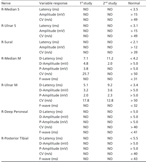
The initial nerve conduction studies (NCS) revealed a marked slow motor nerve conduction velocity with low compound muscle action potential amplitude at the up-per limb. The sensory nerve action potentials were not
de-Table 1. Sensory and motor nerve conduction studies.
Nerve Variable response 1st study 2nd study Normal
R-Median S Latency (ms) Amplitude (mV) CV (m/s) NO NO NO NO NO NO < 3.5 > 15 > 49
R-Ulnar S Latency (ms)
Amplitude (mV) CV (m/s) NO NO NO NO NO NO < 3.1 > 15 > 49
R-Sural Latency (ms)
Amplitude (mV) CV (m/s) NO NO NO NO NO NO < 2.1 > 12 > 39
R-Median M D-Latency (ms) D-Amplitude (mV) P-Amplitude (mV) CV (m/s) F-wave (ms) 7.1 4.8 3.8 25.7 NO 11.2 2.0 NO NO NO < 4.2 > 5.0 > 5.0 > 50 < 31
R-Ulnar M D-Latency (ms)
D-Amplitude (mV) P-Amplitude (mV) CV (m/s) F-wave (ms) 5.1 3.2 2.0 17.8 NO 9.2 3.6 2.3 12.8 NO < 3.4 > 5.0 > 5.0 > 50 < 32
R-Deep Peroneal D-Latency (ms) D-Amplitude (mV) P-Amplitude (mV) CV (m/s) F-wave (ms) NO NO NO NO NO NO NO NO NO NO < 5.0 > 5.0 > 5.0 > 40 < 41
R-Posterior Tibial D-Latency (ms) D-Amplitude (mV) P-Amplitude (mV) CV (m/s) F-wave (ms) NO NO NO NO NO NO NO NO NO NO < 5.5 > 5.0 > 5.0 > 40 < 43
tected in the right median, ulnar and sural nerves (Table 1). Needle electromyography (NE) showed diminished recruit-ment pattern in first dorsal interosseus, extensor digitorum communis, biceps brachialis, tibialis anterior and quadriceps femoris muscles. This electrophysiological pattern is indica-tive of a sensory-motor demyelinating polyradiculoneurop-athy, consistent with CIDP (Table 1).
The patient received intravenous immunoglobulin (400 mg/kg for 5 days) with partial recovery, following to eight sessions of plasmapheresis with recovery. Clinical improve-ment occurred, and at discharge the patient was able to leave the hospital walking without help.
She was admitted again 12 months after HSCT with reduced strength associated with calf pain. CFS analysis showed 3.3 leukocytes/mm3, glucose 47 mg/dL, protein 430 mg/dL. Sorological reactions for CMV, IgM toxoplasmosis, VDRL test and PCR for herpes virus (HSV) were negative. The second electrophysiological study showed a worsened motor NCS, including conduction block, and NE findings, confirming the diagnosis of CIDP (Table 1).
She had a sural nerve and gastrocnemic muscle biopsies frozen in liquid nitrogen, cut in a cryostat and stained his-tologically and histochemically according to standard pro-cedures9. The nerve biopsy had mild inflammatory perivas-cular lymphomononuclear infiltration in the endoneurium and epineurium, a reduction in the number of large myelin-ized fibers in some sectors of the fascicles, asymmetrical ax-onal degeneration within fascicles, occasiax-onal presence of myelin ovoid, and compact and disarranged myelin sheath in most of the material. The muscle biopsy had inflamma-tory perivascular lymphomononuclear infiltrate with inva-sion of the media. The inflammatory reaction spread from infiltrated vessels to adjacent muscle fibers and some had necrosis with phagocytosis.
With the diagnosis of CIDP (relapsing-remitting form) associated with GVHD after HSCT, the oral prednisone dose was increased to 1 mg/kg/day and mofetil mycophenolate was added. There was a substantial improvement in
mus-cle strength, besides the improvement in other manifesta-tions of GVHD.
All studies were done following informed consent.
DISCUSSION
CIDP is an unusual but important complication of HSCT, rarely reported to date. To the best of our knowledge a consistent case of CIDP as a manifesta-tion of GVHD was first reported in 1991 and was fol-lowed by other eight cases in the following decade, but CIDP after HSCT by AML had not been described (Table 2)5-8,10,11. The case reported has neurological
examination as well as laboratorial analysis, electro-physiological studies and histopathological exami-nation compatible with ‘CIDP’ (relapsing-remitting form)12-14
.
CIDP is a clinical syndrome based on a physiologi-cal and pathologiphysiologi-cal concept as followed: (1) cliniphysiologi-cal features of chronic progressive or relapsing and re-mitting, symmetrical, sensory and motor polyradicu-loneuropathy causing weakness of proximal and dis-tal muscles; (2) CSF protein concentration is almost always increased; (3) electrophysiological evidence of demyelination is required for the diagnosis, but axonal degeneration can occurs in evolution; (4) his-tological examination reveals demyelination with variable inflammatory infiltrates12-14. In addition to
this core clinical picture, pure motor, pure sensory, multifocal sensory and motor and multifocal motor forms have been described as subcategories or sepa-rate entities12,13
. Also, the CIDP after HSCT can occurs as a chronic progressive or relapsing and remitting CIDP6,7.
Table 2. Reports of patients with CIDP after HSCT.
Author Age/Sex Underlying
disorder
Latency* CSF protein
Treatment Outcome
Adams et al.5
5/F MOP 4 years NP P+CS Recovery
Amato et al.6 31/M
44/M 29/M 43/M
CML CML AA NHL
6 months 8 months 2 weeks 1 month
NP
↑
NP
↑
PSL+AZP+CS+IvIg PP+PSL+CS PSL+CS+IvIg PP+PSL
Recovery Recovery Recovery Recovery
Griggs et al.10 42/M NHL 3 years ↑ PP Recovery
Nagashima et al.7
32/M NHL 5 years NL MPS Partial recovery
Openshaw et al.11 36/M 21/M
CML HL
7 days 16 days
↑ ↑
P+CP+PP+IvIg P+CP+PP+IvIg
Death Death
Peter et al.1 62/M MM 1 month NP NP Recovery
Present case 17/F AML 10 months ↑ IvIg+PP+P+MM Recovery
Increased spinal fluid protein occurs in at least 90% of patients with CIDP13
. Therefore, increased protein levels can be used as a supportive but not mandatory criterion for the diagnosis13. Increased
CSF protein without pleocytosis is usually present in patients with peripheral neuropathy associated with chronic GHVD6,15. Of the published CSF analyses,
there was no evidence of blood-brain barrier disrup-tion with increased CFS protein from chronic myeloid leukemia patients studied during HSCT and chronic GVHD, but there is report of increased CSF protein secondary to CIDP after HSCT (Table 2)6,7,10,11,16
. The diagnosis of CIDP associated with chronic GVHD may require histological confirmation, which can be obtained in nerve biopsy specimens. The char-acteristic lesions of CIDP consist of patchy regions of demyelination and edema with variable inflammato-ry infiltrates14
. The inflammatory infiltrate are found in both the endoneurium and the epineurium but, in contrast to vasculitic neuropathy, are more abun-dant in the endoneurium12
. The histological analysis of the nerve can showed perivascular inflammatory cells in 54.5% of the patients with CIDP17. The
inflam-matory reaction in the endoneurial infiltrates is made of mononuclear cells, mainly lymphocytes and mac-rophages cells12,14. In long stand disease is reported
chronic inflammation in the perineurium and numer-ous onion bulbs in the endoneurium12
.
In this case, the nerve biopsy showed mild endo-neurial inflammatory infiltrates, axonal alterations with asymmetric nerve fiber loss and reduction in the number of large myelinated axons compatible with CIDP, but onion bulbs not had been found probably due to short evolution time. The nerve ischemia sec-ondary to inflammatory processes could induce acute axonal asymmetrical degeneration within the fascicle, that can occurs in CIDP, but had not been described to date in CIDP by chronic GVHD after HSCT15,18.
There are a few studies about muscle biopsy pat-tern in CIDP with focus on the specific muscular ab-normalities in this disorder. Nevertheless, we believe that specific muscle findings can be similar to nerve findings vary according to the time when the study is performed and the severity of the disease. Therefore, abnormalities as inflammatory perivascular infiltrate adjacent at muscle fibers can occur, as in our case.
The electrophysiological manifestation in chronic GVHD after HSCT may present as neuropathy (demy-elinating and/or axonal neuropathy, sensory and/or motor neuropathy, multiplex mononeuropathy and polyneuropathy), myopathy and dysfunction of the
motor end-plate5,19-21. The electrophysiological
evi-dence of primary demyelination is required for the diagnosis of CIDP, according to strict diagnostic cri-teria, but as the disease advances, axonal degenera-tion becomes superimposed13,14
. The CIDP features associated with chronic GHVD can be found, but was not described previously associated with AML after HSCT6.
Neuropathies associated with GHVD had been re-ported2,4,6,7,10,21
. Temporary imbalances in the mecha-nisms of immune regulation, known to occur after immune reconstitution, have been suspected in the pathogenesis of post-transplantation neuropathies1,5.
Both cell-mediated and antibody-mediated immune responses to glycolipid or myelin protein antigens have been implicated in the pathogenesis of CIDP12,14.
It is interesting to note that when there is an im-mune-mediated alteration involved in these cases, patients normally shows an improvement with the resolution of the GVHD itself, as occured in our pa-tient2,6. Until further studies are done, we can only
speculate that the pathogenesis of the GVHD-asso-ciated CIDP can be related to the development of nerve demyelination and inflammation secondary to immune-mediated lesion.
The management of CIDP with corticosteroids, intravenous immunoglobulin and plasma exchange each provide short term benefit and immunosuppres-sive drugs possible may make long-term benefits12-14
. Unfortunately, experience has been too limited to suggest specific regimens or the optimal sequence of immunosuppressant therapies in patients with CIDP associated with GVHD after HSCT.
REFERENCES
1. Peters G, Larner AJ. Chronic inflammatory demyelinating polyneurop-athy after autologous peripheral blood stem cell transplantation. J Pe-ripher Nerv Syst 2005;10:384-385.
2. Gabriel CM, Goldman JM, Lucas S, Hughes RA. Vasculitic neuropathy in association with chronic graft-versus-host disease. J Neurol Sci 1999; 168:68-70.
3. Krouwer HGJ, Wijdicks EFM. Neurologic complications of bone mar-row transplantation. Neurol Clin 2003;21:319-352.
4. Fleming DR. Graft-vs-host disease: what is the evidence? Evidence-based. Oncology 2002;3:2-6.
5. Adams C, August C, Maguire H, Sladky JT. Neuromuscular complica-tions of bone marrow transplantation. Pediatr Neurol 1995;12:58-61. 6. Amato AA, Barohn RJ, Sahenk Z, Tutschka PJ, Mendell JR.
Polyneurop-athy complicating bone marrow and solid organ transplantation. Neu-rology 1993;43:1513-1518.
7. Nagashima T, Sato F, Chuma T, et al. Chronic demyelinating polyneu-ropathy in graft-versus-host disease following allogeneic bone marrow transplantation. Neuropathology 2002;22:1-8.
8. Sostak P, Padovan CS, Yousry TA, et al. Prospective evaluation of neu-rological complications after allogeneic bone marrow transplantation. Neurology 2003;60:842-848.
10. Griggs JJ, Commichau CS, Rapoport AP, Griggs RC. Chronic inflam-matory demyelinating polyneuropathy in non-Hodgkin’s lymphoma. Am J Hematol 1997;54:332-334.
11. Openshaw H, Hinton DR, Slatkin NE, Bierman PJ, Hoffman FM, Sny-der DS. Exacerbation of inflammatory demyelinating polyneuropathy after bone marrow transplantation. Bone Marrow Transplant 1991;7: 411-414.
12. Hughes RAC, Allen D, Makowska A, Gregson NA. Pathogenesis of chronic inflammatory demyelinating polyradiculoneuropathy. J Periph-er NPeriph-erv Syst 2006;11:30-46.
13. Hughes RAC, Bouche P, Cornblath DR, et al. European Federation of Neurological Societies/Peripheral Nerve Society Guideline on manage-ment of chronic inflammatory demyelinating polyradiculoneuropathy: report of a joint task force of the European Federation of Neurological Societies and the Peripheral Nerve Society. J Peripher Nerv Syst 2005; 10:220-228.
14. Said G. Chronic inflammatory demyelinative polyneuropathy. Neuro-muscul Disord 2006;26:293-303.
15. Collins MP, Periquet MI. Non-systemic vasculitic neuropathy. Curr Opin Neurol 2004;17:587-598.
16. Almeida SM, Livramento JA, Pasquini R, et al. Blood-brain barrier evaluation in bone marrow transplantation. Arq Neuropsiquiatr 1997; 55:812-818.
17. Hirata MTA. Chronic idiopathic inflammatory demyelinating polyra-diculoneuropathy: clinical, diagnostic and therapeutical evaluation. Arq Neuropsiquiatr 1995;53:346.
18. Said G, Lacroix C. Primary and secondary vasculitic neuropathy. J Neu-rol 2005;252:633-641.
19. Bolger GB, Sullivan KM, Spence AM, et al. Myasthenia gravis after al-logeneic bone marrow transplantation: relationship to chronic graft-versus-host disease. Neurology 1986;36:1087-1091.
20. Couriel DR, Beguelin GZ, Giralt S, et al. Chronic graft-versus-host dis-ease manifesting as polymyositis: an uncommon presentation. Bone Marrow Transplant 2002;30:543-546.