ABSTRACT
Sao Paulo Med J. 2007;125(2):73-6.
ORIGINAL AR
TICLE
Ricardo Pires de Souza
Nestor de Barros
Ademar José de Oliveira Paes Junior
Olger de Souza Tornin
Abrão Rapoport
Giovanni Guido Cerri
Value of computed tomography
for evaluating the subglottis in
laryngeal and hypopharyngeal
squamous cell carcinoma
Departments of Head and Neck Surgery, Otorhinolaryngology and
Imaging, Hospital Heliópolis (Hosphel), São Paulo, Brazil
INTRODUCTION Squamous cell carcinoma (SCC) consists of epithelial tumors of mostly mucosal origin. SCC is the most common form of malignant tumor in the pharynx and hypopharynx, accounting for approximately 90%of the malignant tumors in these areas.1 It is potentially curable by treat-ment with surgery and radiotherapy, and these constitute the main therapeutic methods.2
The possible surgical treatments for laryn-geal and hypopharynlaryn-geal carcinoma include total laryngectomy, with lost of the voice, and partial laryngectomy, which preserves phona-tory functions.3
Indication of procedures that conserve the voice, such as radiotherapy and partial la-ryngectomy, depends on preoperative staging of the lesion such that its extent is not under-estimated4 and the principle of oncological radicality is consequently not disregarded.
Diagnostic and clinical staging of laryngeal and hypopharyngeal carcinoma are usually performed by clinical examination, indirect laryngoscopy, direct laryngoscopy and biopsy with histopathological examination.5
Laryngoscopic examination allows a close approach to the mucosal areas of the larynx and hypopharynx, as well as evaluation of the lesion extent in these areas.6 However, subglottic in-volvement may be an exception, since this is dif-fi cult or impossible to evaluate by laryngoscopic examination, especially in cases of large, bulky tumors or when the patient’s respiratory state does not allow for such examination.7
Subglottic involvement is a determining factor for contraindicating conservative partial surgery.8 Indication of radiotherapy in such cases is controversial.9-11
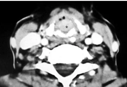
The subglottis is easily assessed by axial computed tomography (CT) at the level of the cricoid ring.12 The subglottic mucosa is closely connected to the surface of the internal cricoid cartilage, and any thickening or irregularity at this level is usually not identifi ed13-15(Figure 1).
OBJECTIVE The present study aimed to evaluate the occurrence of negative and false-positive results and the accuracy of staging by CT for detecting involvement of the subglottic laryngeal compartment, in cases of laryngeal and hypopharyngeal squamous cell carcinoma.
MATERIAL AND METHODS Sixty patients with laryngeal and hypopha-ryngeal squamous cell carcinoma were evaluated and treated at the Head and Neck Surgery Ser-vice of Hospital Heliópolis (Hosphel), São Paulo, Brazil, from 1991 to 1996.
Fifty-four patients (90.0%) were male and six were female (10.0%), with ages ranging from 37 to 76 years.
Fifty-four patients (91.6%) had a history of smoking and 48 (80.0%) had a history of alcohol consumption.
The most common initial complaint was hoarseness (n = 35/60 or 58.3%), followed by odynophagia (n = 12/60 or 20.0%), neck nodule (n = 7/60 or 11.6%), sore throat (n = 4/60 or 6.6%) and foreign body sensation in the throat (n = 2/60 or 3.3%).
The time elapsed between symptom appearance and the clinical diagnosis of cancer ranged from one to 48 months. For 31 patients (51.6%), this time was less than or equal to six months, and for 53 patients (88.3%) it was less than or equal to 12 months.
All patients underwent indirect laryngos-copy and 52 underwent direct laryngoslaryngos-copy. Forty-three patients underwent total laryngectomy and seventeen had some type of conservative surgery.
Among 17 tumors (28.3%) originat-ing from the hypopharynx, 16 (26.6%) were in the pyriform sinus and one (1.6%) in the posterior wall. There were 43 laryn-geal tumors (71.7%), and these were
dis-CONTEXT AND OBJECTIVE: Subglottic involvement in squamous cell carcinoma is a determining factor for contraindicating conservative partial surgery. The subglottis is easily identifi ed by axial computed tomography sections. The present study aimed to evaluate the occurrence of false-negative and false-positive results, and the overall accuracy of staging by computed tomography, in order to detect the involvement of the subglottic laryngeal compartment, in cases of laryngeal and hypopha-ryngeal squamous cell carcinoma.
DESIGN AND SETTING:Retrospective, non-ran-domized study of patients treated at Hospital Heliópolis, São Paulo, Brazil.
METHODS: Computed tomography scans were performed on third-generation equipment with 5-mm slice thickness. Afterwards, all patients underwent surgical and anatomopathological examinations as the gold standard procedures.
RESULTS: Among 60 patients, 14 were diag-nosed with subglottic extension by surgical and histopathological examination. There were three false-negative and no false-positive results from computed tomography scans. The sensitivity and negative predictive value were 100.0%. Accuracy was 95.0%, specifi city was 93.5% and positive predictive value was 82.4%.
CONCLUSIONS: Computed tomography could serve as a powerful auxiliary method for staging laryngeal and hypopharyngeal cancer. However, precautions should be taken in analyzing com-puted tomography scan data, because vegetating lesions may also be projected into the subglottic compartment, without real involvement of the sub-glottis, which may cause a false-positive result.
74
Sao Paulo Med J. 2007;125(2):73-6. tributed in the following order: vocal cords: n = 14 (23.3%); epiglottis: n = 10 (16.6%); ventricle: n = 9 (15.0%); ventricular band: n = 6 (10.0%); and aryepiglottic fold: n = 4 (6.6%).
The local staging or T parameter for the tumors, according to the TNM scale of the Union Internationale Contre le Cancer (UICC) classification (1992), was distributed thus: Tx: n = 3 (5.0%); T1a: n = 3 (5.0%); T1b: n = 1 (1.6%); T2: n = 15 (25.0%); T3: n = 28 (46.6%); and T4: n = 10 (16.6%).
None of the patients had previously un-dergone any kind of treatment.
All the CT scans were performed on third-generation equipment, with 5-mm slice thickness and identical increments, guided by digital profi le radiography. Intravenous contrast medium (iohexol 300 mg/ml) was administered to 58 patients (96.7%).
Involvement of the subglottic compart-ment was characterized when any irregularity or thickening of the soft tissue along the inner margin of the cricoid cartilage was identifi ed on the CT scan.
The results from the CT scan examina-tions were compared with the fi ndings from the direct and indirect laryngoscopy examina-tions, and with the surgical fi ndings and/or histopathological examinations. The surgical and histopathological fi ndings represented the gold standard.
RESULTS Intraoperative macroscopic evaluation and/or histopathological examination on the 60 surgical specimens showed subglottic involvement in 14 cases (Figure 2) out of this series (23.3%).
Among these 60 cases in the series, indirect laryngoscopy was unable to evalu-ate the subglottic compartment in 26 cases (43.3%).
Out of the 52 cases (86.6%) in which direct laryngoscopy was performed, this method was unable to evaluate the subglottis in 11 cases (21.2%).
Evaluation of the subglottic compartment by CT yielded the results presented in Table 1 and the sensitivity measurement calculations (specifi city, positive predictive value, negative predictive value, accuracy) in Table 2.
The three false-positive cases (5.0%) found by CT in the subglottic compartment evaluation resulted from bulky polyp lesions that were projected into the inner subglot-tis compartment, without real involvement (Figures 3 and 4).
Figure 1. Axial computed tomography scan at the cricoid cartilage (*) level. Normal appearance of subglottis. True-negative example. The scan shows left lymph node enlargement (star).
Figure 2. Axial computed tomography scan. Left vocal cord tumor (*). True-posi-tive case.
Figure 3. Axial computed tomography scan. Vegetative lesion (*) extending inferiorly and simulating glottis involve-ment. False-positive example.
75
Sao Paulo Med J. 2007;125(2):73-6.
Mafee et al. (1983),23 García-Alonso et al. (1992)24 and Kazkayasi et al. (1995)25 presented absence of false-negative fi ndings. Like these authors, we did not fi nd any false-negative results in our series of 60 patients. All 14 cases of subglottic involvement were correctly detected by CT scans, thus giving a negative predictive value of 100%. In our series, three false-positive results were found, caused by bulky lesions originating in the vocal cords that extended inferiorly and projected into the inner margin of the subglottis, without having any real involvement with this compartment. Two of these cases underwent conservative surgery after intraoperative evaluation. The third case underwent simple total laryngectomy. These false-positive results gave rise to a positive predictive value of 82.4%.
Conjointly with the absence of false-negative results, the three false-positive cases in this series resulted in accuracy of 95.0% in the subglottic evaluation of laryngeal and hypopharyngeal SCC by CT scans.
CONCLUSIONS According to these results, we conclude that computed tomography could serve as a powerful auxiliary method for staging laryn-geal and hypopharynlaryn-geal cancer. However, precautions should be taken in analyzing CT scan data, because vegetating lesions may also be projected into the inner subglottis compartment, without its real involvement, which may cause a false-positive result. DISCUSSION
Laryngeal and hypopharyngeal SCC repre-sent more than 90.0% of the malignant tumors in these regions. SCC usually progresses slowly and more frequently affects men, particularly in their sixth and seventh decades of life. It has been shown to present its highest incidence among smokers and individuals with high alcohol consumption. In our sample, nine times more men were affected than women, eleven times more smokers than non-smokers and four times more consumers of alcohol than non-consumers. The peak incidence of the disease (n = 23/60 or 38.3%) occurred within the age range from 51 to 60 years old.
SCC typically begins in the inner laryngeal and hypopharyngeal surfaces and presents in three different types: infiltrative, bulky or mixed. A local dissemination pattern for SCC can be found on mucosa surfaces or when there is deep invasion of structures in these organs, with consequent submucosal extension.
It is a potentially curable tumor. The survival rates depend on early diagnosis and adequate treatment for each situation.
The subglottic larynx or subglottis marks the transition from stratified squamous epithelium to respiratory epithelium. It con-sists of a small round segment of the airway that is situated immediately under the vocal cords and above the trachea. Although pri-mary carcinoma in the subglottis is quite rare, secondary invasion by carcinoma that began in other adjacent areas is frequent.
The diagnosis of SCC usually starts to emerge with the appearance of its symptoms: typically hoarseness caused by vocal cord involvement, or pain on deglutition (odynophagia), in cases of hypopharyngeal tu-mors. Sometimes, the primary tumor remains clinically silent and the initial manifestation is the appearance of a cervical nodule that represents metastasis to lymph nodes. The time elapsed between symptom appear-ance and making the defi nitive diagnosis is fundamental in determining the extent of tumor spreading.16,17
In addition to offering a means of ob-taining material for histopathological con-fi rmation of the tumor by biopsy, direct or indirect laryngoscopy should be capable of determining the diagnosis and precisely defi n-ing the local extent of the tumor. Nonetheless, laryngoscopy does not allow evaluation of the extent and depth of tumor invasion into the submucosa. It has to be considered that cricoid cartilage involvement, or invasion of deep tissues in the neck will necessitate total laryngectomy.18-20
It is diffi cult or impossible for laryngo-scopic examination to provide information about the subglottis and the deep structures of the larynx and hypopharynx. However, there is a great need to identify tumor extent in a very precise manner. Consequently, over the course of time, imaging examinations were added to the large number of auxiliary methods for tumor staging. CT scans were thus brought in as an auxiliary method for laryngeal and hypopharyngeal tumor staging, in primary tumor evaluations and for attempting to identify and characterize metastatic cervical lymph nodes.
Not all cases of laryngeal and hypopha-ryngeal SCC have an indication for CT scans. In cases with early identifi cation of small and superfi cial lesions that are clearly demonstrated by clinical examination and in which palpation is enough for defi ning the procedures to adopt, imaging examinations are not necessary.21
In the present study, the subglottis could be evaluated by axial CT scans obtained at cricoid ring level, in each of the 60 cases in this series. In accordance with this criterion, 17 cases in our sample were identifi ed as posi-tive, although three of them were shown to be false-positive by histopathological analysis. All 14 cases in which there was subglottic involvement were correctly detected.
Although there are some case reports on false-negative CT scans in the literature, such as Grandjean et al. (1993),22 other studies by
Table 1. Results from evaluation of the subglottic compartment by computed tomography
Cases n %
True-negative 43 71.7 True-positive 14 23.3 False-negative 0 0 False-positive 3 5.0
Confi dence interval (95.0%) = 0.728-1.012.
Table 2. Sensitivity, specifi city, positive predictive value, negative predictive value and accuracy of the computed tomography fi ndings on subglottic involvement
%
Sensitivity 100.0
Specifi city 93.5
Positive predictive value 82.4 Negative predictive value 100.0
76
Sao Paulo Med J. 2007;125(2):73-6.
1. Horowitz BL, Woodson GE, Bryan RN. CT of laryngeal tumors. Radiol Clin North Am. 1984;22(1):265-79.
2. Sexton CC, Anderson CG. Computed tomography in carcinoma of the larynx. Australas Radiol. 1984;28(4):330-4. 3. Curtin HD. Imaging of the larynx: current concepts. Radiology.
1989;173(1):1-11.
4. Prando D, Figueiredo MA, Albertotti CJ, Clemente Filho AS, Secaf F. Tomografia computadorizada da laringe. [Computed tomography of the larynx]. Rev Imagem. 1981;3(2):27-41.
5. Dubin J, Rieux D, Caron C, Desnos J. Utilisation de la to-modensitométrie dans le bilan d’extension de certains cancers laryngés. [Use of tomodensitometry in the evaluation of the extension of certain laryngeal cancers]. Ann Otolaryngol Chir Cervicofac. 1983;100(2):125-8.
6. Remacle M, Mazy G, Marbaix E, Synaeve P, Pringot J, Van den Eeckhaut J. L’apport de la tomodensitométrie en carcinologie cervi-co-faciale. [The contribution of tomodensitometry to cervicofacial oncology]. Acta Otorhinolaryngol Belg. 1983;36(3):453-61. 7. Scott M, Forsted DH, Rominger CJ, Brennan M. Computed
tomographic evaluation of laryngeal neoplasms. Radiology. 1981;140(1):141-4.
8. Weinstein GS, Laccourreye O, Brasnu D, Yousem DM. The role of computed tomography and magnetic resonance imaging in planning for conservation laryngeal surgery. Neuroimaging Clin N Am. 1996;6(2):497-504.
9. Lee WR, Mancuso AA, Saleh EM, Mendenhall WM, Parsons JT, Mil-lion RR. Can pretreatment computed tomography fi ndings predict local control in T3 squamous cell carcinoma of the glottic larynx treated with radiotherapy alone? Int J Radiat Oncol Biol Phys. 1993;25(4):683-7. 10. Mukherji SK, Mancuso AA, Mendenhall W, Kotzur IM, Kubilis
P. Can pretreatment CT predict local control of T2 glottic carcinomas treated with radiation therapy alone? AJNR Am J Neuroradiol. 1995;16(4):655-62.
11. Pameijer FA, Mancuso AA, Mendenhall WM, Parsons JT, Kubilis PS. Can pretreatment computed tomography predict local control in T3 squamous cell carcinoma of the glottic larynx treated with defi nitive radiotherapy? Int J Radiat Oncol Biol Phys. 1997;37(5):1011-21.
12. Mancuso AA, Calcaterra TC, Hanafee WN. Com-puted tomography of the larynx. Radiol Clin North Am. 1978;16(2):195-208.
13. Mancuso AA, Hanafee WN. A comparative evaluation of computed tomography and laryngography. Radiology. 1979;133(1):131-8.
14. Parsons C. Computed tomography of laryngeal tumors. J Belge Radiol. 1983;66(5):343-9.
15. Reid MH. Laryngeal carcinoma: high-resolution com-puted tomography and thick anatomic sections. Radiology. 1984;151(3):689-96.
16. Giron J, Joffre P, Serrés-Cousine O, Senac JP. CT and MR evaluation of laryngeal carcinomas. J Otolaryngol. 1993;22(4):284-93. 17. Thabet HM, Sessions DG, Gado MH, Gnepp DA, Harvey
JE, Talaat M. Comparison of clinical evaluation and computed tomographic diagnostic accuracy for tumors of the larynx and hypopharynx. Laryngoscope. 1996;106(5 Pt 1):589-94. 18. Lemoine C, Serrano E, Arrue P, Woisard V, Rolland V, Pessey JJ.
Apports comparés de l’endoscopie, de la tomodensitométrie et de l’I.R.M. dans le bilan pré-opératoire des cancers de l’endolarynx: à propos de 18 patients operes. [Comparative contribution of endoscopy, x-ray computed tomography and MRI in the preoperative evaluation of cancers of the endolarynx: apropos of 18 surgically-treated patients]. Rev Laryngol Otol Rhinol (Bord). 1992;113(3):173-8.
19. Zbären P. Becker M, Läng H. Pretherapeutic staging of hypopha-ryngeal carcinoma. Clinical fi ndings, computed tomography, and magnetic resonance imaging compared with histopathologic evalu-ation. Arch Otolaryngol Head Neck Surg. 1997:123(9):908-13.
20. Zbären P, Becker M, Läng H. Staging of laryngeal cancer: en-doscopy, computed tomography and magnetic resonance versus histopathology. Eur Arch Otorhinolaryngol. 1997;254(Suppl 1):S117-22.
21. Williams DW. Imaging of laryngeal cancer. Otolaryngol Clin North Am. 1997;30(1):35-58.
22. Grandjean E, Debry C, Briche D, el Jerrarri A, Gentine A, Conraux C. Corrélations anatomo-scanographiques des cancers du pharyngo-larynx. A propos d’une étude prospective portant sur 35 patients. [Correlations between anatomy and x-ray com-puted tomography in cancers of the pharyngo-larynx. Apropos of a prospective study of 35 patients]. Ann Otolaryngol Chir Cervicofac. 1993;110(3):135-41.
23. Mafee MF, Schild JA, Valvassori GE, Capek V. Computed tomography of the larynx: correlation with anatomic and pathologic studies in cases of laryngeal carcinoma. Radiology. 1983;147(1):123-8.
24. García Alonso Y, Alvarez Villa A, Baldo Sierra C, Montes No-riega B. La TC en el estudio del carcinoma de laringe. [CT in the study of laryngeal carcinoma]. Acta Otorrinolaringol Esp. 1992;43(3):153-8.
25. Kazkayasi M, Onder T, Ozkaptan Y, Can C, Pabuscu Y. Com-parison of preoperative computed tomographic fi ndings with postoperative histopathological fi ndings in laryngeal cancers. Eur Arch Otorhinolaryngol. 1995;252(6):325-31.
Sources of funding: Not declared
Confl ict of interest: Not declared
Date of fi rst submission:December 10, 2003
Last received:February 25, 2007
Accepted: February 28, 2007
REFERENCES
AUTHOR INFORMATION
Ricardo Pires de Souza, MD.Radiologist, Complexo Hospi-talar Heliópolis, São Paulo, Brazil.
Nestor de Barros, MD. Radiologist, Hospital das Clínicas da Faculdade de Medicina da Universidade de São Paulo, São Paulo, Brazil.
Ademar José de Oliveira Paes Junior, MD.Radiologist, Com-plexo Hospitalar Heliópolis, São Paulo, Brazil.
Olger de Souza Tornin, MD. Radiologist, Hospital das Clínicas da Faculdade de Medicina da Universidade de São Paulo, São Paulo, Brazil.
Abrão Rapoport, MD.Head and Neck Surgeon, Complexo Hospitalar Heliópolis, São Paulo, Brazil.
Giovanni Guido Cerri, MD. Radiologist, Hospital das Clínicas da Faculdade de Medicina da Universidade de São Paulo, São Paulo, Brazil.
Address for correspondence: Ricardo Pires de Souza
Rua Cônego Xavier, 276 — 10o andar
São Paulo (SP) — Brasil — CEP 04231-030 Tel. (+55) 2274-8224
E-mail: [email protected] E-mail: [email protected]
Copyright © 2007, Associação Paulista de Medicina
RESUMO
Valor da tomografi a computadorizada na avaliação da subglote no carcinoma espinocelular de laringe e hipofaringe
CONTEXTO E OBJETIVO: O envolvimento subglótico, em casos de carcinoma espinocelular, é fator determi-nante na contra-indicação da cirurgia parcial conservadora. A subglote pode ser facilmente identifi cada no cortes axiais da tomografi a computadorizada. O presente estudo tem por objetivos avaliar a ocor-rência de resultados falsos negativos, falsos positivos e a efi cácia global do estadiamento por tomografi a computadorizada na detecção do envolvimento do compartimento subglótico da laringe, em casos de carcinoma espinocelular de laringe e hipofaringe.
TIPO DE ESTUDO E LOCAL: Retrospectivo não randomizado, realizado no Hospital Heliópolis, São Paulo, Brasil.
MÉTODOS: Os exames de tomografi a computadorizada foram realizados em equipamentos de terceira geração, com cortes com espessura de 5 mm. Todos os pacientes foram posteriormente submetidos a cirurgia com comprovação anatomopatológica, sendo este o padrão ouro.
RESULTADOS: Dentre os 60 pacientes, o envolvimento subglótico ocorreu em 14, conforme observado no achado cirúrgico e ou anatomopatológico. A avaliação pela tomografi a computadorizada resultou em três casos falsos positivos. Não ocorreram resultados falsos negativos. A tomografi a computadorizada apresentou 100,0% de sensibilidade, 100,0% de valor preditivo negativo e 95,0% de efi cácia global. A especifi cidade foi de 93,5% e o valor preditivo positivo de 82,4%.
CONCLUSÕES:Consideramos que a TC pode ser um poderoso instrumento auxiliar no estadiamento do câncer de laringe e de hipofaringe, devendo-se ter precaução com lesões vegetantes que se projetam para a subglote sem seu verdadeiro envolvimento.