Rev Ass Med Brasil 2000; 46(2): 177-81 1 7 7
Original Article
Effect of combined treatment with calcitonin on bone
densitometry of patients with treated hypothyroidism
F. J. DA C. STAMATO, E. C. J. AMARANTE, R. P. FURLANETTO
Division of Endocrinology, Department of Medicine, Federal University of São Paulo (EPM-UNIFESP), São Paulo, SP, Brazil
SUMMARY – INTRODUCTION. Thyroid hormones (TH) may affect bone metabolism and turnover, inducing a loss of bone mass among hyperthyroid and in hypothyroid patients under hormone re-placement treatment. Thyroid dysfunction leads to changes in the dynamics of parathyroid hor-mone (PTH) and calcitonin (CT) secretion.
OBJECTIVE. The objective of the study was to determine the usefulness of CT as adjuvant the-rapy in the prevention of bone loss during the treatment of hypothyroidism.
MATERIAL AND METHODS. We studied 16 female patients with recently diagnosed primary hypo-thyroidism, divided into two groups: group G1 (n=8) submitted to treatment with thyroxine (L-T4), and Group 2 (n=8) that, in addition to being treated with L-T4, received a nasal CT spray. All
INTRODUCTION
Thyroid hormones (TH) affect bone metabolism and turnover, increasing the number of bone remo-deling cycles, activating and increasing the num-ber of osteoclasts and altering the relation between bone reabsorption and bone formation2,5,24,25. This effect induces a loss in bone mass in hyperthyroid individuals2,6,8,18,20,24 and also among hypothyroid patients submitted to hormonal replacement therapy2,5,7,18,29.
Parathyroid hormone (PTH) is released by the parathyroid glands depending on serum calcium and its major action is to stimulate bone reab-sorption through the osteoclasts1. In hypothy-roidism, basal PTH and its response to hypo-calcemia are higher than in normal individuals, and this exaggerated response does not normalize even after 6 months of replacement treatment with TH8-12,33-35.
Calcitonin (CT) is a hormone mainly produced by the parafollicular cells of the thyroid (C cells) and is a potent inhibitor of bone reabsorption; its secretion is stimulated by calcium. In hypothy-roidism there is a lower CT reserve and its res-ponse to a hypercalcemic stimulus is significantly
patients were submitted to determination of TSH, free T4, bone mineral densitometry (BMD) and total bone calcium (TBC) at the time of diagnosis, after 6 to 9 months of treatment, and after 12 months of treatment.
RESULTS. No statistical significant differences were detected in either group between the total BMD values obtained for the femur and lumbar spine before and after treatment. However, group G1 presented a statistical significant TBC loss after 12 months of treatment compared to initial values. In contrast, no TBC loss was observed in the group treated with LT-4 in combination with CT, a fact that may suggest that CT was responsible for the lower bone reabsorption during treatment of hypothyroidism.
KEYWORDS: Calcitonin. Osteoporosis. Hypothyroidism
reduced13, probably due to the destruction of C cells by the process of chronic thyroiditis. Hypo-thyroid patients, when starting hormonal repla-cement therapy, may present bone remodeling of high turnover owing to the action of TH. Mundy, in 197625, showed that CT inhibits TH-induced bone reabsorption in vitro. Since CT mainly acts by suppressing the osteoclastic activity of bone, it may be assumed that patients with osteoporosis involving a high turnover may benefit more rapi-dly from CT treatment than patients with less severe disorders of bone remodeling1,28.
The ain of the present study was to assess the possible loss of bone mass occurring during trea-tment of hypothyroidism and to clarify the pro-bable role of calcitonin in the prevention of osteo-penia and its possible use as an adjuvant in the treatment of hypothyroidism.
MATERIAL AND METHODS
thyro-tropin (TSH) and free thyroxine (T4L) concentra-tion and divided into two groups. The patients in group G1 (n=10) received L-thyroxine at the dose of 1.6 to 2.0 µg/kg/day, while the patients in group G2 (n=10) received L-thyroxine at the same dose in combination with calcitonin nasal spray at the dose of 100 IU three times a week. The patients were submitted to determination of TSH and T4L, to bone densitometry of the lumbar spine (BMDc), of the femur (BMDf) and of the entire body (BMDt) and to determination of total bone calcium (TBC) at the beginning of the study, after 6 to 9 months and after 12 months of treatment. Four patients (two from each group) were excluded from the evaluations because of lack of compliance with treatment. All other patients continued to be euthyroid during follow-up.
Laboratory determinations: serum T4L and TSH were determined by an ultrasensitive immu-nofluorimetric method, using commercial Delfia kits (Pharmacia, Turku, Finland).
Bone densitometry: Bone densitometry was de-termined by DEXA using a LUNAR DPX-L densi-tometer. The results are reported as g/cm2 and the z score standard deviation was used.
Statistical analysis: The Friedman test was applied to the changes obtained in BMDc, BMDf and BMDt data along time in each group. The initial and final BMDc, BMDf, BMDt, TBC and body weight values were compared by the Wilco-xon test in each group, and the Mann-Whitney test was used for comparison between groups. The level of significance was set at =.05 in all analyses.
RESULTS
No patients reported any complaints about complications and/or side effects caused by calci-tonin. No statistical significant variation in body weight occurred in the patients of either group during the study. The initial median weight of G1
patients was 63,87 Kg (range: 52,0 - 73,0) and the median at the end of the study was also 63,87 Kg (range: 51,0 to 75,0). The initial median weight of G2 patients was 66,75 Kg (range: 49, 0 to 77,0) and the median at the end of the study was 64,5 Kg (range: 49,0 to 74,0).
No significant differences between groups were observed with respect to the initial values of BMDc, BMDf, BMDt and TBC.
No significant differences in total, spinal or femoral BMD (reported as g/cm2 and z score) were observed during the study period within groups. Also, no significant differences were observed between initial and final values (Tables 1 and 2). Group G1 presented a significant loss (P<0.01) of total bone calcium during the study period (Table 3), with significantly lower values (p<0.02) at 12 months (median 815 - range 680 to 980 g) compared to initial values (median 867.5 - range 713-980 g). In group G2 there was no statistical difference between the initial (median 876.5 ; range 626-1057 g) and end values (median 837.5 ; range 607-1011 g) of TBC.
DISCUSSION
Osteoporosis is a highly common skeletal disor-der of multifactorial etiology which mainly affects women23 and with important effects in terms of patient morbidity and mortality. Because of the multifactorial nature of the disorder, whenever possible the association of osteopenic factors should be avoided and, depending on the cause, treatment should be combined with pro-osteogenic or antireabsorptive drugs.
Changes in thyroid function affect the osteo-mineral metabolism7,15,17,28,36, leading to changes in the dynamics of PTH and CT secretion8,11,13, with a direct action of TH on bone tissue25 as well as potentiation of the action of PTH on bone reab-sorption32. In hypothyroid subjects, the dynamics
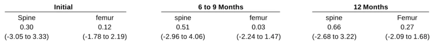
Tabela 2 – BMD (Zscore) of the lumbar spine (L2-L4) and of the femural neck in group G2I (median and range). Initial 6 to 9 Months 12 Months
Spine femur spine femur spine Femur
0.30 0.12 0.51 0.03 0.66 0.27
(-3.05 to 3.33) (-1.78 to 2.19) (-2.96 to 4.06) (-2.24 to 1.47) (-2.68 to 3.22) (-2.09 to 1.68)
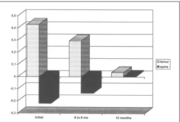
Tabela 1 – BMD (Zscore) of the lumbar spine (L2-L4) and of the femural neck in group G1 (median and range). Initial 6 to 9 Months 12 Months
Spine femur spine femur spine Femur
-0.22 0.43 -0.13 0.29 0.00 0.03
Rev Ass Med Brasil 2000; 46(2): 177-81 1 7 9
Graf. 2 – BMD (Z score) of the lumbar spine (L2-L4) and of the femural neck in group G2 (hypothyroid patients that received L-thyroxine in combination with calcitonin)
Graf. 1 – BMD (Z score) of the lumbar spine (L2-L4) and of the femural neck in group G1 (hypothyroid patients that received only L-thyroxine)
of PTH secretion continues to be altered even after 6 months of euthyroid status11, favoring the expo-sure of bone tissue to relatively high quantities of PTH associated with normal TH levels, with consequent greater bone reabsorption.
Calcitonin is a potent inhibitor of bone reab-sorption and its partial or total deficiency may represent the loss of an important bone-protecting factor among patients, with bone tissue becoming more vulnerable to the action of hormones that stimulate its reabsorption. In hypothyroidism there is a lower calcitonin reserve, with a signi-ficantly decreased response to the hypercalcemic stimulus13, which justifies the therapeutic use of CT in hypothyroid patients under hormonal repla-cement treatment.
CT is an effective therapeutic agent in various diseases characterized by accelerated bone reab-sorption23. Its therapeutic use in the treatment of osteoporosis is questionable, although this pa-thology is frequently associated with an increase in bone reabsorption. Results obtained in patients with accelerated bone reabsorption treated with CT have shown normalization of the loss of bone mass similar to that obtained with the use of other bone reabsorption inhibitors (estrogens and bis-phosphonates)31. Other reports have demonstrated an increase in bone mass in the vertebrae and long bones 16,22,26, a reduced fracture rate19,30 and relief of the syndromes accompanied by bone pain21.
Even though the evaluation of our patients did
not reveal a significant alteration of BMDt and of lumbar spine and femur BMD, the TBC mea-surement showed that hypothyroid female pa-tients presented a significant bone calcium loss after 1 year of replacement treatment with TH (group G1).
The combined use of CT in the treatment of hypothyroidism seems to be useful for the pre-vention of greater bone loss, especially in patients with other risk factors for osteopenia. This was demonstrated by the follow-up of our G2 patients treated with CT in combination with thyroid hormone, who did not show a significant TBC loss after 1 year of treatment.
Evaluation of TBC is considered to be the most sensitive determination for the assessment of bone mass. It is expressed in grams and may present alterations with a change in patient body
Tabela 3 – Total bone calcium (g) determined in group G1 patients
Patient no Initial at 6 to 9 months at 12 months
1 830 807 807
2 883 825 815
3 852 806 815
4 713 713 680
5 969 909 895
6 885 884 891
7 806 798 799
8 980 971 969
Tabela 4 – Total bone calcium (g) determined in group G2 patients
Patient no Initial at 6 to 9 months at 12 months
1 882 856 845
2 871 829 828
3 1011 950 957
4 1057 1007 1011
5 626 618 607
weight. The patients in the two groups studied here did not present significant differences in weight during the study.
On the basis of these data, we may infer that a loss of bone calcium occurs in hypothyroid pa-tients submitted to hormonal replacement, a fact that can be minimized when CT is administered in combination. Since CT is an antireabsorptive hormone, it is not expected to produce a signi-ficant increase in bone mass, but rather to main-tain bone mass by the prevention or reduction of later bone losses23. It has been demonstrated that the use of CT can stabilize or modestly increase the indices of cortical and trabecular bone mass and total bone calcium when administered to patients over a period of 1 to 2 years23,26,27. Burck-hardt & Burnand3 demonstrated that in all contro-lled studies in which CT was used there was a decrease in the rate of vertebral fractures, al-though the difference was not statistically signi-ficant.
As a potent antiosteoclastic drug, CT seems to be relatively harmless when compared to the potential complications caused by the other drugs used in the treatment or prevention of osteoporosis37. Among the side effects reported with the use of CT, the most important ones are nausea, gastric discomfort and skin rashes1, which, however, were subs-tantially reduced when the nasal spray was intro-duced4,37. None of the patients followed up by us complained about side effects of calcitonin.
Although our study followed the patients for one year, a more prolonged prospective study with a larger number of patients would be necessary to definitely confirm whether calcitonin is a hormo-ne that could, or should, be added to the treatment of hypothyroidism, especially among patients with proven osteopenia.
ACKNOWLEDGEMENTS
The authors thank Dr. Marcus Magliano from Sandoz S.A. for his colaboration in Calcitonin (Miacalcic ®) supplying.
RESUMO
Influência da terapêutica associada com cal-citonina sobre a densitometria óssea de pa-cientes com hipotiroidismo tratado.
INTRODUÇÃO. Os hormônios tiroidianos (HT)
podem influenciar o metabolismo e o “turnover” ósseo, induzindo perda de massa óssea em hiper-tiróideos e em hipótiroideos na vigência de repo-sição hormonal. As disfunções tiroidianas levam a alterações na dinâmica de secreção de
parator-mônio (PTH) e de calcitonina (CT).
OBJETIVO. Esclarecer a utilidade da CT como
terapêutica coadjuvante na prevenção de perda óssea durante o tratamento do hipotiroidismo.
MATERIALE MÉTODOS. Dezeseis pacientes do sexo
feminino com hipotiroidismo primário recém-diagnosticados, divididos em dois grupos: grupo G1 (n=8) tratado com tiroxina (L-T4) e grupo G2 (n=8) que recebeu, além de L-T4, CT “spray” nasal. Todos os pacientes foram avaliados com TSH, T4 livre, densitometria mineral óssea (BMD) e cálcio ósseo total (TBC) ao diagnóstico após 6 a 9 meses de terapêutica e com 12 meses de tratamento.
RESULTADOS. Em ambos os grupos não foram
encontradas mudanças estatisticamente signifi-cantes entre as medidas da BMD total antes e após o tratamento, assim como no fêmur e na coluna lombar. Entretanto, o grupo G1 apresentou perda significante do TBC após 12 meses de tratamento em relação aos valores iniciais. Já no grupo que usou terapêutica associada com CT, não houve perda de cálcio ósseo total, o que pode sugerir que a CT foi responsável por uma menor reabsorção óssea durante o tratamento do hipotiroidismo. [Rev Ass Med Bras 2000; 46(2): 177-81]
NOTICE
The authors hereby confirm that neither the manuscript nor any part of it has been published or is being considered for publication elsewhere. By signing this letter each of us acknow-ledges that he or she participated sufficiently in the work to take public responsibility for its content.
REFERÊNCIAS BIBLIOGRÁFICAS
1. Avioli, L.V. - Calcitonin therapy in osteoporotic syndromes.
Rheum Dis Clin North Am 20:777-785,1994.
2. Baran, D.T. & Braverman, L.E. Editorial: Thyroid hormones and bone mass. J Clin Endocrinol Metab 72:1182-3,1991. 3. Burckhardt, P. & Burnand, B. - The effect of treatment with
calcitonin on vertebral fracture rate in osteoporosis. Osteo-poros Int 3(1):24-30,1993.
4. Carstens, J.H. Jr.; Feinblatt, J.D. - Future horizons for calcitonin: a US perspective. Calcif Tissue Int 49:S2,1991. 5. Coindre, J.M.; David, J.P.; Rivière, L.; Goussot, J.F.; Roger, P.;
Mascarel, A.; Meunier, J.P. - Bone loss in hypothyroidism with hormone replacement.: A histomorphometric study. Arch Intern Med 146:48-53,1986.
6. Diamond, T.; Nery, L.; Hales, I. - A therapeutic dilemma: supressive doses of thyroxine significantly reduce bone mine-ral measurements in both premenopausal and postmenopau-sal women with thyroid carcinoma. J Clin Endocrinol Metab
72:1184-8,1991.
7. Franklyn, J.A.; Betteridge, J.; Daykin, J.; Holder, R.; Oates, G.D.; Parle, J.V.; et al. - Long-term thyroxine treatment and bone mineral density. Lancet 340:9-13,1992.
hipoti-Rev Ass Med Brasil 2000; 46(2): 177-81 1 8 1
roidismo. In: Congresso Brasileiro de Endocrinologia e Meta-bologia, 18, Rio de Janeiro,1988, Resumo dos trabalhos. Rio de Janeiro,1988,p.97 (Resumo, 225).
9. Furlanetto, R.P.; Castro, M.L.; Mesquita, C.H.; Kasamatsu, T.S.; Vieira, J.G.H. - Resposta do paratormônio à hipocal-cemia em pacientes com disfunções tiroidianas. In: Encontro Brasileiro de Tiróide, 3, Rio de Janeiro,1989,p.18 (Resumo, T18).
10. Furlanetto, R.P.; Castro, M.L.; Kasamatsu, T.S.; Brandão, C.M.A.; Vieira, J.G.H. - Parathyroid glands hyperresponsi-veness to EDT induced hypocalcemia in treated hypothyroid patients. In: Congresso Panamericano de Endocrinologia 12, Recife,1990. Endocrino 90. Recife,1990,p.113 (Resumo, 202). 11. Furlanetto, R.P.; Castro, M.L.; Kasamatsu, T.S.; Brandão,
C.M.A.; Vieira, J.G.H. - Persistent hyperactivity of the para-thyroid glands in treated hypopara-thyroid patients. Acta Endo-crinol (Copenh) 123:609-12,1990.
12. Furlanetto, R.P.; Castro, M.L.; Kasamatsu, T.S.; Brandão, C.M.A.; Vieira, J.G.H. - Parathyroid glands hyperresponsive-ness to EDTA-induced hypocalcemia in treated hypothyroid patients. In: International Thyroid Conference,10, Haia,1991, Abstract book. Haia,1991,p.169 (Abstract 278).
13. Furlanetto, R.P.; Brandão, C.M.A.; Kasamatsu, T.S.; Castro, M.L.; Vieira, J.G.H. - Calcitonin secretion in hypo and hyper-thyroidism. In: Latin-American Thyroid Congress,5, São Paulo,1991, Abstract book. São Paulo,1991,p.21 (Abstract 38). 14. Furlanetto, R.P.; Castro, M.L.; Mesquita, C.H.; Kasamatsu, T.S.; Vieira, J.G.H. - Função paratiroidiana no hipertiroi-dismo: implicações no metabolismo ósseo e efeito do trata-mento. Rev Paul Med 109:55-60,1991.
15. Gam, A.N.; Jensen, J.F.; Hasselstrom, K., Olsen, M.; Nielsen, K.S. - Effect of thyroxine therapy on bone metabolism in substituted hypothyroid patients with normal or suppressed levels of TSH. J Endocrinol Invest 14:451-5,1991.
16. Gennari, C.; Chierichetti, S.M.; Bigazzi, S.; et al. - Compa-rative effects on bone mineral content of calcium and calcium plus salmon calcitonin given in two differents regimens in postmenopausal osteoporosis. Current Therapeutic Research
38:455,1985.
17. Greenspan, S.L.; Greenspan, F.S.; Resnick, N.M.; Block, J.E.; Friedlander, A.L.; Genant, H.K. - Skeletal integrity in preme-nopausal and postmepreme-nopausal women receiving long-term l-thyroxine therapy. Am J Med 91:5-14,1991.
18. Harvey,R.D.; McHardy,K.C.; Reid,I.W.; Paterson, F.; Bew-sher,P.D.; Duncan,A; Robins,S.P. - Measurements of bone collagen degradation in hyperthyroidism and during thyroxine replacement therapy using pyridiun cross-links as a specific urinary markers.J Clin Endocrinol Metab 72:1189-94,1991. 19. Kanis, J.A.; Johnell, O.; Gullberg, B. et al. - Evidence for
efficacy of drugs affecting bone metabolism in preventing hip fracture. BMJ 305:1124,1992.
20. Kroelner, B.; Jorgensen, J.V.; Nielsen, S.P. - Spinal bone mineral content in myxoedema and thyrotoxicosis. Effect of thyroid hormone (s) and antithyroid treatment. Clin Endo-crinol (Oxf) 18:439-46,1983.
21. Lyrits, G.P.; Tsakalakos, S.; Magiasis, B. et al. - Analgesic effect
of salmon calcitonin in osteoporotic vertebral fractures: Double-blind placebo-controlled study.Calcif Tissue Int 49: 369,1991.
22. Mazzuoli, G.F.; Passeri, M.; Gennari, C.; et al. - Effects of salmon calcitonin in postmenopausal osteoporosis: A control-led double-blind clinical study. Calcif Tissue Int 38:3,1986.
23. McDermott, M.T.& Kidd, G.S. - The role of calcitonin in the development and treatment of osteoporosis. Endocrine Re-views 8(4):377-384,1987.
24. Mosekilde, L.; Eriksen, E.F.; Charles, P. - Effects of thyroid hormones on bone and mineral metabolism. Endocrinol Metab Clin North Am 19:35-63,1990.
25. Mundy, C.R.; Shapiro, J.L.; Bandelin, J.G.; Canalis,E.M.; Raisz,L.C. - Direct stimulation of bone resorption by thyroid hormones. J Clin Invest 58:529-34,1976.
26. Overgaard, K.; Riis, B.J.; Christiansen, C. et al. Nasal calci-tonin for treatment of established osteoporosis. Clin Endo-crinol 30:435-442,1989.
27. Overgaard, K.; Riis, B.J.; Christiansen, C. et al. - Effect of salcatonin given intranasally on early postmenopausal bone loss. BMJ 299:477,1989.
28. Paul, T.L.; Kerrigan, J.; Kelly, A.M. et al. - Long-term l-thyroxine therapy is associated with decreased hip bone density in premenopausal women. JAMA 259:3137,1988.
29. Perry III, H.M. - Thyroid replacement and osteoporosis. Arch Intern Med 146:41-2,1986.
30. Rico, H..; Hernandez, E.R.; Diaz-Mediavilla, J.; et al. - Trea-tment of multiple myeloma with nasal spray calcitonin: A histomorphometric and biochemical study. Bone Miner 8:231, 1990.
31. Sambrook, P.; Birmingham, J.; Kelly, P.; et al. - Prevention of corticosteroid osteoporosis: A comparision of calcium, cal-citriol, and calcitonin. N Engl J Med 328:1747,1993. 32. Schimin, E.M.K.; Steiner, T.H.; Froesch, E.R. -
Triiodo-thyronine increases responsiveness of cultured rat bone cells to parathyroid hormone. Acta Endocrinol 11:213-6, 1986. 33. Vieira, J.G.H.; Castro, M.L.; Furlanetto, R.P.;
Kasamat-su,T.S.; Mesquita, C.H.; Chacra, A.R. - Dynamics of the PTH response to EDTA induced hypocalcemia in hypo and hyper-thyroidism. In: Laurence and Dorothy Fallis International Symposium,1988, Clin Bone Min Metab Detroit,1988,p.29 (abstract 74).
34. Vieira, J.G.H. & Furlanetto, R.P. - Dynamics of the parathyroid hormone (PTH) secretion in hypo and hyperthyroidism. In: Meirelles,R.M.R.; Machado, A.; Póvoa, L.A. - Clinical Endo-crinology,1 ed, Amsterdan, Excerpta Medica, 1988, p.253-5. 35. Vieira, J.G.H.; Kasamatsu,T.S.; Mesquita, C.H; Furlanetto,
R.P. - parathyroid hormone response to EDTA induced hypo-calcemia in hypo and hyperthyroid patients pre and post-treatment. In: International Conference on Calcium Regu-lating Hormones,10, Montreal,1989, J Bone Min Res Mon-treal,1988,p.S157 (Abstract 158).
36. Wenzel, K.W. - Bone minerals and levothyroxine. Lancet
340:435-6,1992.