ABSTRACT
Osteoporosis is a disease characterized by low bone mass and micro architectural alterations of bone tissue leading to enhanced bone fragili-ty and increased fracture risk. Although research in osteoporosis has focused mainly on the role of bone loss in the elderly population, it is becoming increasingly clear that the amount of bone that is gained dur-ing growth is also an important determinant of future resistance to frac-tures. Thus, considerable interest is being placed on defining preventive strategies that optimize the gain of bone mass during childhood and adolescence. Knowledge of the determinants accounting for the physi-ologic and genetic variations in bone accumulation in children will pro-vide the best means toward the early diagnosis and treatment of osteo-porosis. This article reviews the techniques available for bone mass mea-surements in children and the major determinants and diseases influenc-ing bone accretion durinfluenc-ing childhood and adolescence. (Arq Bras Endocrinol Metab 2006;50/4:775-782)
Keywords:Low bone mass; Bone mass measurement; DXA in childhood and adolescence; Update; Controversy
RESUMO
Baixa Massa Óssea em Crianças e Adolescentes.
Osteoporose é uma doença caracterizada pela baixa massa óssea e alterações de micro arquitetura do tecido ósseo, levando ao aumento da fragilidade óssea e aumento do risco de fratura. Apesar da pesquisa em osteoporose ter focalizado principalmente no papel da perda óssea na população idosa, está começando a ficar claro que a quantidade de osso que é ganho durante o crescimento é também um determi-nante importante de resistência futura para fraturas. Portanto, interesse considerável está sendo colocado na definição de estratégias preven-tivas que otimizam o ganho de massa óssea durante a infância e ado-lescência. O conhecimento dos determinantes responsáveis pelas vari-ações fisiológicas e genéticas, na acumulação óssea nas crianças, irá levar aos melhores meios para o diagnóstico precoce e tratamento da osteoporose. Este artigo revê as técnicas disponíveis para a medida da massa óssea em crianças e os maiores determinantes e doenças que influenciam a aquisição óssea durante a infância e adolescência. (Arq Bras Endocrinol Metab 2006;50/4:775-782)
Descritores:Baixa massa óssea; Medidas da massa óssea; DXA na infân-cia e adolesêninfân-cia; Atualização em DXA; Controvérsia em DXA
João Lindolfo C. Borges
Cynthia M.A. Brandão
Universidade Católica de Brasília, DF (JLCB); and Division of Endocrinology, Federal University of São Paulo (UNIFESP/EPM) and Fleury — Diagnostic Medicine, São Paulo, SP (CMAB), Brazil.
THE PEAK BONE MASS
T
HE FOUNDATION FOR LIFELONG skeletal health isestablished during childhood and adolescence. Although there is controversy regarding the exact tim-ing of peak bone mass, bone size and strength reach a maximum by early adulthood (1-3). Failure to accrue optimal peak bone mass has been linked to an increased risk of osteoporosis (4). The variables that contribute to optimal bone health have been delineat-ed in studies of healthy youth (1-3).
Approximately 90% of adult bone mass is gained in the first two decades of life. Optimizing peak bone mass and bone strength early in life and stabiliz-ing it durstabiliz-ing young adulthood is believed to play a sig-nificant role in preventing osteoporosis and fractures later in life. Adequate weight-bearing physical activity, nutrition, body mass, and hormonal balance are essen-tial in achieving optimal skeletal health. A growing list of chronic diseases has also been linked to low bone mass or fragility fractures (5-30). Disorders causing rickets and osteomalacia are reviewed by Durval else-where in this issue. In some chronic conditions, a sin-gle factor (e.g., immobilization or hypogonadism) accounts for the increased risk of low bone mass. In most of these disorders, however, skeletal health is threatened by a combination of risk factors including malnutrition, vitamin D insufficiency, malabsorption, deficiency or resistance to sex steroids or growth hor-mone, immobilization, and increased cytokine pro-duction. Medications that are used to treat these dis-orders, such as glucocorticoids, calcineurin inhibitors, and chemotherapeutic agents, may also contribute to bone loss (6). The magnitude of effect that these dis-orders or medications will have on an individual patient varies, depending upon genetic factors, disease severity, activity, and other variables. For this reason, clinicians seek diagnostic tools to identify patients at greatest risk for bone fragility.
Obese and less-active children also have been shown to have decreased BMD or bone mass compared with non-obese children of similar weight (31,32). It is not clear whether this decreased BMD among obese chil-dren is a direct effect of fat on bone or due to decreased muscle mass or reduced activity levels, or a combination of both of these factors. However, the epidemic of child-hood obesity may in part directly or indirectly explain the increase in childhood fracture incidence that has recently been reported (33). Identifying children with low bone mass early in life could be an important strategy for pre-ventative or therapeutic efforts to optimize bone accrual and, consequently, bone strength (table 1).
ASSESSING PEDIATRIC BONE HEALTH
DXA is the most widely used densitometric method for diagnosing osteoporosis in adults. DXA was devel-oped in the late 1980s for use primarily in post-menopausal women. Pediatric software became avail-able in the early 1990s after improvements in algo-rithms for detecting bone edges in children with low bone density. The advantages of DXA are its wide availability, short scanning times, and relatively low radiation exposure. The radiation exposure is compa-rable to that received during a round trip transconti-nental airplane flight. DXA has several important limi-tations, however (34-36). The technique does not provide a measure of volumetric bone mineral density
Table 1.Disorders associated with low bone mass or frac-tures in childhood and adolescence (60).
Genetic disorders (6,42)
Ehlers-Danlos Fibrous dysplasia Homocystinuria Hypophosphatasia Idiopathic hypercalciuria Marfan's syndrome
Menkes' kinky hair syndrome Osteogenesis imperfecta
Chronic disease
Anorexia nervosa (7,8) Athletic amenorrhea (9) Celiac disease (10) Cystic fibrosis (11) Diabetes (type I) (12)
Hematologic thalassemia, sickle-cell anemia (13) Inflammatory bowel disease (14)
Malignancy (15-17) Post transplantation (18) Renal failure (19)
Rheumatologic disorders (20)
Endocrine disorders
Glucocorticoid excess (21) Growth hormone deficiency (22) Hyperparathyroidism (23) Hyperthyroidism (26)
Sex steroid deficiency or resistance (25,26)
Immobilization
Cerebral palsy (27) Muscular dystrophy (28) Paraplegia
Spina bifida
Miscellaneous
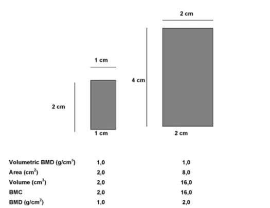
or of bone geometry nor does it distinguish between cortical and trabecular bone. Although bone size and geometry can be adjusted for mathematically, these are only estimates of these parameters. Because this is a 2-dimensional measurement and not a true volumetric density, measurements using DXA are often referred to as areal BMD (aBMD). Measurements of aBMD are influenced by bone size, with larger bones having artificially inflated aBMD measurements (figure 1). This is an important problem in pediatric bone assess-ment because of the large differences in body size and bone size within and across different ages. Studies show that aBMD by DXA increases with age, but com-puted tomography evaluations indicate that true volu-metric BMD (vBMD) is relatively constant during childhood until puberty, at which time there is a large increase in vBMD (37). BMC increases with age, and the increase in aBMD that is observed is likely the result of greater bone size.
Although adult aBMD has been shown to be predictive of future fracture risk in longitudinal epi-demiologic studies (Data on the Bone Densitometry Chapter), there is no evidence indicating this in chil-dren. The aBMD results are often presented as T- and Z-scores. Because T-scores compare the observed aBMD with that of young adults, they are not appro-priate for growing children and should never be used.
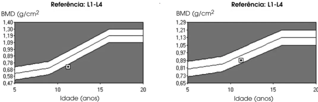
Z-scores, defined as the SD score based on age and gender-specific norms, must be used to determine how a child’s aBMD compares with other children’s (figures 2 and 3). This is a more appropriate method of com-parison of aBMD in pediatrics. As previously described, however, aBMD is highly correlated to body and bone size, and in children with chronic diseases in whom weight or height for age may be severely affected, the comparison of aBMD measurements to age-matched norms is difficult to interpret.
Total body and lumbar spine (L1-L4) DXA scans are reported in pediatric studies. Total body is a pre-dominantly cortical bone measurement while lumbar spine is mainly trabecular bone. Thus different skeletal sites will be affected by different factors. Dietary calcium intake has been shown to affect primarily appendicular bone sites that are predominantly cortical bone (38), whereas hypogonadism and steroid use affect primarily axial bone sites or the ends of long bones, which are predominantly trabecular bones (39,40).
Although regional DXA scans can measure BMD and BMC at sites that are predominantly tra-becular or cortical bone, it is not possible to obtain separate cortical and trabecular BMD results using DXA. The aBMD assessed by DXA is a function of both the amount of bone within the periosteal enve-lope and the size of the bone.
BONE MASS MEASUREMENT GENERAL GUIDELINES
There are currently no standard recommendations by either a U.S. pediatric or bone organization regard-ing who should have bone measurements for clinical purposes. The British Paediatric and Adolescent Bone Group recently published pediatric guidelines for the clinical use of DXA (41). They suggested that children with conditions that may increase their risk of low bone density and fracture should be consid-ered for a DXA scan if they also present low trauma or recurrent fractures, back pain, spinal deformity or loss of height, change in mobility status, or malnutri-tion. The list of conditions that place children at increased risk is given in table 1, along with some of the more rare conditions that also may be associated with decreased BMD. Because of the lack of pediatric reference databases, the variation between machines, and the different software analyses that are per-formed, it is important that clinicians consult with
pediatric bone specialists before using DXA diagnos-tically or prescribing treatment on the basis of DXA methods.
The International Society of Clinical Densitom-etry recently published an official position paper on recommendations for performance and clinical appli-cation of bone density testing, which included recom-mendations specific for diagnosis in children (table 2).
INTERPRETING PEDIATRIC DUAL-ENERGY X-RAY
Interpreting bone mineral measurements is far more complex in children than in adults and goes beyond cal-culating a Z-score (34,36,43,44). Unlike the adult whose bone dimensions are stable with time, children and adolescents are moving targets whose bone size, geometry, and mineral content are changing. These processes evolve at varying rates in different regions of the skeleton, with appendicular growth preceding spinal mineral acquisition (44). Furthermore, within a given
Figure 2.Shows the different graphs for the interpretation of bone densitometry. Pediatric bone densi-ty is compared for patients of the same age, the Z-score. Adult bone densidensi-ty is read comparing young adults, 20 to 40 years old, density, and T-score.
Figure 3.Spine and total body scan of a 11-year-old girl, 40,1 kg, 142,0 cm height, referred to evalua-tion for previous use of anticonvulsants for five years. Her BMI is 20, at the 80 th percentile for age, according to CDC Growth Charts, and Tanner Stage B2P2, no menarche. The Z-score for total skele-tal is -0,6 and for lumbar spine (L1L4) is -1,4. The diagnosis is "adequate bone mass density for chrono-logical age" since the Z-score values are less than -2,0 within the normal pediatric range.
Referência: L1-L4
BMD (g/cm2
Idade (anos)
Referência: L1-L4
BMD (g/cm2
Idade (anos)
Referência: L1-L4
BMD (g/cm2
Idade (anos)
Referência: L1-L4
BMD (g/cm2 YA T-Score
region of interest, trabecular and cortical compartments respond variably to sex steroids, calcium intake, and mechanical loading. The tempo of mineral accrual is linked more closely to pubertal and skeletal maturation than to chronologic age, and these processes vary with gender and ethnicity (43,44,45,49,50). For this reason, the influence of bone size and maturation must be con-sidered in evaluating DXA results.
In fact it is very important to correlate bone acquisition in young subjects not only with chrono-logical age or sex, as it is done in commercial soft-wares, but also with anthropometrical parameters, par-ticularly in longitudinal evaluations. On the basis of many studies published on literature (45-48), the pubertal development and weight are the most impor-tant parameters in monitoring bone mass in adoles-cents. When children have delayed growth or puberty and altered body composition, these factors must be considered in interpreting BMC and BMD. For chil-dren with delayed growth and maturation, it is also reasonable to adjust for pubertal stage rather than for chronological age. Unfortunately, only a few studies have reported normative data by pubertal stage (16,44,51). Alternatively, a bone age can be obtained and BMD data compared with norms for the patient’s skeletal rather than chronological age.
Additionally, BMC and BMD are strongly influenced by bone size; BMD corrects only for the area of bone studied but not the thickness of bone. For this reason, true (volumetric) bone density may be underestimated in patients with smaller bones and overestimated in larger children. Several methods have been proposed to adjust bone mass for the influence of bone size or lean body mass (52-55). Estimates of vol-umetric bone density at the spine and femoral neck
divide BMC by the estimated volume of bone in the region; total body BMC is corrected for relative height (55). None of these correction models has been estab-lished as best by the gold standard of predicting child-hood fracture. Furthermore, given two bones of equal density, the larger bone will be more resistant to frac-ture than the smaller one. Nonetheless, it is possible to estimate how much reduced BMD can be attributed to smaller bone size by calculating volumetric BMD. Limited pediatric norms for volumetric BMD have been published (49,55-57). An example of a pediatric DXA interpretation is provided in figure 2.
INTERPRETING LOW BONE MASS
The fracture risk associated with low BMD is far less certain in children and young adults. Patients with mild forms of osteogenesis imperfecta (OI), for exam-ple, have very low BMD but do not suffer spontaneous fractures. The International Society for Clinical Den-sitometry has determined that the diagnosis of osteo-porosis in a young patient “should not be made on the basis of densitometric criteria alone”. The WHO crite-ria for osteopenia and osteoporosis are not appropcrite-riate for use in children, adolescents, and young adults. Terms such as “low bone density for chronologic age” may be used if the Z-score is less than -2.0; the term “osteoporosis” must be avoided since it is better con-ditions of loss of bone. By implication, the diagnosis of “low bone density for chronologic age” in a child requires additional clinical findings such as a history of low impact fracture.
Finding low bone mass on a pediatric DXA does not necessarily imply bone loss. Low bone mass
Table 2.Position of the International Society for Clinical Densitometry (ISCD) on the Use of DXA in Diagnosis in Chil-dren (males or females less than 20 years of age) (32).
• The WHO classification (for defining osteopenia and osteoporosis) should not be used. • Z-scores should be used instead of T-scores in children.
• T-scores should not appear in reports or on DXA printouts for children.
• The diagnosis of osteoporosis in children should not be made on the basis of densitometric criteria alone. • Terminology such as "low bone density for chronological age" may be used if the Z-score is below -2.0. • Z-scores must be interpreted in light of the best available pediatric databases of age-matched controls. The
reference database should be cited in the report.
• Preferred skeletal sites for measurement are spine and total body.
• The value of BMD to predict fractures in children is not clearly demonstrated.
• Standards for adjusting BMD or bone mineral content (BMC) for factors such as bone size, pubertal stage, skele-tal maturity, or body composition have not been agreed upon. Clearly state any adjustments in the report. • Successive BMD studies should be done using the same machine, scanning mode, software, and analysis when
appropriate. Changes may be required with growth of the child.
in a child can result from inadequate gains of bone mineral, bone loss, or a combination of the two (8,58,59). Understanding this is critical, because most drugs used to treat osteoporosis in adults are anticata-bolic agents that reduce bone loss. Children who fail to gain adequate bone mineral may require therapy that is anabolic or bone building. It is beyond the scope of this article to review the current therapies for pediatric low bone mass (5,6). At the very minimum, however, the finding of low bone mass should prompt a search for possible cause(s), including a review of overall nutrition, calcium intake, vitamin D stores (serum 25 hydroxyvitamin D), hormonal status, phys-ical activity, and underlying disease status. All risk fac-tors should be addressed.
CONCLUSIONS
Bone acquisition early in life is considered an important predictor of osteoporosis risk later in life. DXA is the most common method for assessing bone health in pedi-atric populations. There are, however, several problems with interpreting DXA scans in children that need to be considered by clinicians before therapeutic interventions are implemented on the basis of DXA results. PQCT is a promising method that is currently being used in pedi-atric bone research that may find its way into clinical use for assessing bone strength and fracture risk. Further research is needed to determine whether QUS could be used as a radiation-free alternative for assessing bone development clinically and in epidemiologic studies.
REFERENCES
1. Heaney RP, Abrams S, Dawson-Hughes B. Peak bone mass. Osteoporos Int 2000;11:985-1009.
2. Mora S, Gilsanz V. Establishment of peak bone mass.
Endocrinol Metab Clin North Am 2003;32:39-63.
3. Bachrach LK. Acquisition of optimal bone mass in child-hood and adolescence. Trends Endocrinol Metab
2001;12:22-8.
4. Hui SL, Slemenda CW, Johnston CC. The contribution of bone loss to postmenopausal osteoporosis. Osteoporos
Int 1990;1:30-4.
5. Soyka LA, Fairfield WP, Klibanski A. Hormonal determi-nants and disorders of peak bone mass in children [Clin-ical review]. J Clin Endocrinol Metab 2000;86:3951-63.
6. Ward LM, Glorieux FH. The spectrum of pediatric osteo-porosis. In: Glorieux FH, Pettifor JM, Juppner H (eds).
Pediatric bone: biology and diseases. San Diego: Acad-emic Press, 2003. pp. 401-42.
7. Seeman E, Karlsson MK, Duan Y. On exposure to anorex-ia nervosa, the temporal varanorex-iation in axanorex-ial and appen-dicular skeletal development predisposes to site-specif-ic defsite-specif-icits in bone size and density: a cross-sectional study. J Bone Miner Res 2000;15:2259-65.
8. Soyka LA, Misra M, Frenchman A. Abnormal bone min-eral accrual in adolescent girls with anorexia nervosa. J
Clin Endocrinol Metab 2002;87:4177-85.
9. Warren MP. Health issues for women athletes: exercise-induced amenorrhea. J Clin Endocrinol Metab
1999;84:1892-6.
10. Mora S, Barera G, Beccio S. A prospective, longitudinal study of the long-term effect of treatment on bone den-sity in children with celiac disease. J Pediatr
2001;139:516-21.
11. Bhudhikanok GS, Lim J, Marcus R. Correlates of osteope-nia in patients with cystic fibrosis. Pediatrics 1996;97:103-11.
12. Heap J, Murray MA, Miller SC, Jalili T. Alterations in bone characteristics associated with glycemic control in ado-lescents with type l diabetes mellitus. J Pediatr
2004;144:56-62.
13. Tiosano D, Hochberg Z. Endocrine complications of tha-lassemia. J Endocrinol Invest 2001;24:716-23.
14. Vestergaard P. Bone loss associated with gastrointestinal disease: prevalence and pathogenesis. Eur J
Gastroen-terol Hepatol 2003;15:851-6.
15. Arikosko P, Komulainen J, Riikonen P. Alterations in bone turnover and impaired development of bone mineral density in newly diagnosed children with cancer: A 1-year prospective study. J Clin Endocrinol Metab
1999;84:3174-81.
16. van der Sluis IM, van den Heuvel-Eibrink MM, Hahlen K. Altered bone mineral density and body composition, and increased fracture risk in childhood acute lym-phoblastic leukemia. J Pediatr 2002;141:204-10.
17. Rose SR. Endocrinopathies in childhood cancer sur-vivors. Endocrinologist 2003;13:488-95.
18. Daniels MW, Wilson DM, Paguntalan HG. Bone mineral density in pediatric transplant recipients. Transplantation
2003;6:673-8.
19. Boot AM, Nauta J, de Jong M. Bone mineral density, bone metabolism and body composition of children with chronic renal failure, with and without growth hor-mone treatment. Clin Endocrinol (Oxf) 1998;49:665-72.
20. Kotaniemi A, Savolainen A, Kroger H. Development of bone mineral density at the lumbar spine and femoral neck in juvenile chronic arthritis — a prospective one-year follow-up study. J Rheumatol 1998;25:2450-5.
21. Leong GM, Center JR, Henderson NK. Glucocorticoid-induced osteoporosis. In: Marcus R, Feldman D, Kelsey J (eds). Osteoporosis. San Diego: Academic Press, 2001. pp. 169-93.
22. Baroncelli GI, Bertelloni S, Sondini F. Lumbar bone miner-al density at finminer-al height and prevminer-alence of fractures in treated children with GH deficiency. J Clin Endocrinol
Metab 2002;87:3624-31.
24. Lucidarme N, Ruiz JC, Czernichow P, Leger J. Reduced bone mineral density at diagnosis and bone mineral recovery during treatment in children with Graves’ dis-ease. J Pediatr 2000;137:56-62.
25. Riggs LB, Khosla S, Melton LJ. Sex steroids and the con-struction and conservation of the adult skeleton. Endocr
Rev 2002;23:279-302.
26. Miller KK, Klibanski A. Amenorrheic bone loss. J Clin
Endocrinol Metab 1999;84:1775-83.
27. Henderson RC, Lin PP, Greene WB. Bone-mineral density in children and adolescents who have spastic cerebral palsy. J Bone Joint Surg 1995;77A:1671-81.
28. Bianchi ML, Mazzanti A, Galbiati E. Bone mineral density and bone metabolism in Duchenne muscular dystrophy.
Osteoporos Int 2003;14:761-7.
29. Krassas GE. Idiopathic juvenile osteoporosis. Ann NY
Acad Sci 2000;900:409-12.
30. Cheng JCY, Qin L, Cheung CSK. Generalized low areal and volumetric bone mineral density in adolescent idio-pathic scoliosis. J Bone Miner Res 2000;15:1587-95.
31. Weiler HA, Janzen L, Green K, Grabowski J, Seshia MM, Yuen KC. Percent body fat and bone mass in healthy Canadian females 10 to 19 years of age. Bone 2000;27:203-7.
32. Specker BL, Johannsen N, Binkley T, Finn K. Total body bone mineral content and tibial cortical bone measures in preschool children. J Bone Miner Res 2001;16:2298-305.
33. Khosla S, Melton LJ III, Dekutoski MB, Achenbach SJ, Oberg AL, Riggs BL. Incidence of childhood distal fore-arm fractures over 30 years. JAMA 2003;290:1479-85.
34. Rauch F, Schoenau E. Changes in bone density during childhood and adolescence: An approach based upon bone’s biological organization. J Bone Miner Res 2001;16:597-604.
35. Heaney RP. Is the paradigm shifting? Bone 2003;33:457-65.
36. Mora S, Bachrach L, Gilsanz V. Noninvasive techniques for bone mass measurement. In: Glorieux FH, Pettifor JM, Juppner H (eds). Pediatric bone: biology and diseases. San Diego: Academic Press, 2003. pp. 303-24.
37. Gilsanz V, Roe TF, Mora S, Costin G, Goodman WG. Changes in vertebral bone density in black girls and white girls during childhood and puberty. N Engl J Med
1991;325:1597-600.
38. Wosje KS, Specker BL. Role of calcium in bone health during childhood. Nutrition Rev 2000;58:253-68.
39. Bianchi ML. Glucocorticoids and bone: some general remarks and some special observations in pediatric patients. Calcif Tissue Int 2002;70:384-90.
40. Hendrickson BA, Gokhale R, Cho JH. Clinical aspects and pathophysiology of inflammatory bowel disease.
Clin Microbiol Rev 2002;15:79-94.
41. Fewtrell MS. Bone densitometry in children assessed by dual x-ray absorptiometry: uses and pitfalls. Arch Dis
Child 2003;88:795-8.
42. Whyte MP. Osteogenesis imperfecta. In: Favus M (ed).
Primer on metabolic diseases and disorders of mineral metabolism. Washington: American Society for Bone and Mineral Research, 2003. pp. 470-3.
43. Bachrach LK. Dual-energy X-ray absorptiometry (DEXA) measurements of bone density and body composition: promise and pitfalls. J Pediatr Endocrinol Metab 2000; 13:983-8.
44. Seeman E. From density to structure: growing up and growing old on the surfaces of bone. J Bone Miner Res
1997;12:509-21.
45. Bonjour JP, Theintz G, Buchs B. Critical years and stages of puberty for spinal and femoral bone mass accumula-tion during adolescence. J Clin Endocrinol Metab
1991;73:555-63.
46. Lu PW, Briody JN, Ogle GD, Morley K, Humphries IRJ, Allen J, et al. Bone mineral density of total body, spine and femoral neck in children and young adults: a cross-sectional and longitudinal study. J Bone Miner Res 1994;9:1451-8.
47. Sabatier J-P, Guaydier-Souquières G, Laroche D, Beu-malek A, Fournier L, Guillon-Metz F, et al. Bone mineral acquisition during adolescence and early adulthood: Studying 574 healthy females 10–24 years of age.
Osteo-poros Int 1996;6:141-8.
48. Theintz G, Buchs B, Rizzoli R, Slosman D, Clavieu H, Sizo-nenko PC, et al. Longitudinal monitoring of bone mass accumulation in healthy adolescents – Evidence for a marked reduction after 16 years of age at the levels of lumbar spine and femoral neck in female subjects. J Clin
Endocrinol Metab 1992;75:1060-5.
49. Bachrach LK, Hastie T, Wang MC. Bone mineral acquisi-tion in healthy Asian, Hispanic, Black and Caucasian youth: a longitudinal study. J Clin Endocrinol Metab
1999;84:4702-12.
50. Bailey DA, McKay HA, Mirwald RL. A six-year longitudinal study of the relationship of physical activity to bone min-eral accrual in growing children: The University of Saskatchewan Bone Mineral Accrual Study. J Bone Miner Res 1999;14:1672-9.
51. Zanchetta JR, Plotkin H, Filgueira MLA. Bone mass in chil-dren: normative values for the 2–20-year-old population.
Bone 1995;16:393S-9.
52. Lu PW, Cowell CT, Lloyd-Jones SA. Volumetric bone min-eral density in normal subjects, aged 5–27 years. J Clin
Endocrinol Metab 1996;81:1586-90.
53. Hogler W, Briody J, Woodhead HJ. Importance of lean mass in the interpretation of total body densitometry in children and adolescents. J Pediatr 2003;143:81-8.
54. Carter DR, Bouxsein ML, Marcus R. New approaches for interpreting projected bone densitometry data. J Bone
Miner Res 1992;7:137-45.
55. Molgaard C, Thomsen GL, Prentice A. Whole body bone mineral content in healthy children and adolescents.
Arch Dis Child 1997;76:9-15.
56. van der Sluis IM, de Ridder MA, Boot AM. Reference data for bone density and body composition measured with dual-energy X-ray absorptiometry in white children and young adults. Arch Dis Child 2002;87:341-7.
58. Bhudhikanok GS, Wang M-C, Marcus R. Bone acquisition and loss in children and adults with cystic fibrosis: a lon-gitudinal study. J Pediatr 1998;133:18-27.
59. Bachrach LK, Katzman DK, Litt IF. Recovery from osteopenia in adolescent girls with anorexia nervosa. J
Clin Endocrinol Metab 1991;72:602-6.
60. Bachrach LK. Osteoporosis and measurement of bone mass in children and adolescents. Endocrinol Metab Clin
North Am 2005;34(3):521-35.
Address for correspondence:
João Lindolfo C. Borges
Clínica de Endocrinologia e Metabologia Centro Clínico do Lago, sala 305
SHIS QI 09, Brasília, DF