494
URINARY TRACT CHOLESTEATOMA Case Report
International Braz J Urol
Official Journal of the Brazilian Society of Urology
Vol. 30 (6): 494-495, November - December, 2004
CHOLESTEATOMA OF THE UPPER URINARY TRACT
DANIEL X. LIMA, ELI A. S. RABELO, PAULO G. O. SALLES
General Hospital, School of Medicine, Federal University of Minas Gerais, UFMG, Belo Horizonte, Minas Gerais, Brazil
ABSTRACT
We report the case of a 57-year old patient with complex cystic image in right kidney. Fol-lowing radical nephrectomy, the pathological study established the diagnosis of renal cholesteatoma. We discuss the frequency, pathogenesis, clinical presentation, propedeutics, histological findings and proposes for intervention observed in the literature.
Key words: kidney; cholesteatoma; cyst; disease, urinary tract Int Braz J Urol. 2004; 30: 494-5
INTRODUCTION
Cholesteatoma (leukoplakia) of the upper urinary tract is a rare benign condition, with approxi-mately 80 cases being described in the literature (1). The characteristic histopathological finding is squa-mous metaplasia of the urothelium associated with exuberant keratinization and desquamation of kerati-nized layers. The urinary elimination of horny mate-rial can lead to intermittent obstruction of the collec-tor system and flank pain, which are the main clini-cal manifestations of the disease. Classiclini-cally, the con-dition is treated by nephrectomy, though recently its malignant and recurrence potential has been ques-tioned, warranting conservative approaches.
CASE REPORT
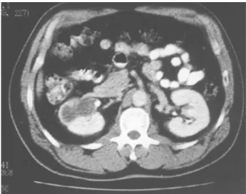
Male, 57-yar old patient, previously healthy, reported infrequent episodes of right lumbar colic for approximately 6 months. The physical examination did not present significant alterations, as well as the exam of urinary sediment and urine culture. The ul-trasound showed a pyelocaliceal cyst in right kidney,
with calcification on its inferior wall. On the com-puterized tomography (Figure-1), the cyst showed to be hypodense, with heterogeneous content, septate, with captation of contrast medium and calcification in its wall (Bosniak III). Considering the finding of complex renal cyst, right radical nephrectomy was
495
URINARY TRACT CHOLESTEATOMA
performed, due to the possibility of neoplasia. Surgi-cal procedure evolved without intercurrences, as well as postoperative outcome.
The pathological examination showed a kid-ney measuring 11.6 x 5.8 cm and weighting 305 g, with a pyelocaliceal cyst measuring 4.6 cm in its larger diameter, with smooth and regular walls, filled by whitish, semi-solid and somewhat friable material, which compressed the adjacent renal parenchyma (Figure-2). On microscopy, the cyst was covered by urothelium with extensive squamous metaplasia, abundant keratinization and corneal-lamellar content, compatible with the diagnosis of cholesteatoma (leu-koplakia) of the upper urinary tract. There was fibrosing chronic inflammation on the periphery of the cyst, focal chronic pyelonephritis and hyaline vascular nephrosclerosis. No neoplasia was observed.
COMMENTS
Desquamative keratinizing squamous meta-plasia of the upper urinary tract, or cholesteatomatous leukoplakia most often is located in renal pelvis and adjacent calices, where sometimes it assumes cystic form. It shows a clear predominance in adult popula-tion (97.5% of described cases) and is slightly more common in males than in females (3:2 ratio). The process is more commonly considered as a reactive phenomenon related to chronic urothelial
inflamma-Figure 2 – Surgical specimen from nephrectomy presenting cyst with semi-solid content located in renal pelvis and adja-cent calices. Insert: histological study of the cyst wall, where we can observe lining epithelium (keratinized squamous epi-thelium). HE, X100.
tion, though hypotheses of embryological anomaly or even spontaneous transformation of urothelium into squamous epithelium cannot be ruled out.
It is inconsistently correlated with squamous cell carcinoma, since the progression from metaplasia to neoplasia was never demonstrated (1). Conservative approaches, by percutaneous and transureteroscopic route, or even clinical follow-up have already been de-scribed (2,3). In the case of this patient, there was clini-cal and radiologiclini-cal suspicion of a malignant renal cyst, warranting radical surgery.
The most characteristic sign in renal choleste-atoma is the elimination of flake-like keratinized material in the urine, which becomes opaque. Such alteration is not always present, making pre-opera-tive diagnosis difficult for this rare condition. Imag-ing exams such as excretory urography and retrograde pyelography can be helpful in suspected cases of re-nal cholesteatoma, though urothelial tumor must be always considered in differential diagnosis (1).
REFERENCES
1. Hertle L, Androulakakis P: Keratinizing desquamative squamous metaplasia of the upper urinary tract: leu-koplakia-cholesteatoma. J Urol. 1982; 127: 631-5. 2. Houston W: Renal cholesteatoma. Br J Urol. 1983;
55: 239-40.
3. Neerhut G, Politis G, Alpert L, Griffith DP: Choleste-atoma of the renal pelvis: endoscopic management. J Urol. 1988; 139: 1032-4.
Received: June 23, 2004 Accepted after revision: July 27, 2004
Correspondence address: Dr. Eli Armando S. Rabelo
Section of Nephrology and Urology
General Hospital, School of Medicine, UFMG Rua Piauí 933 / 501