ADRIAMYCIN-INDUCED FETAL HYDRONEPHROSIS
ANDERSON GONÇALVES, WILLY G. FRANÇA, SUZANA G. MORAES, LUIS A.V. PEREIRA,
LOURENÇO SBRAGIA
Institute of Biology and School of Medicine, State University of Campinas, UNICAMP, Campinas, São Paulo, Brazil
ABSTRACT
Introduction: At the end of pregnancy, the amniotic fluid (AF) depends basically on renal function, corresponding to fetal urine. Changes in AF, especially oligohydramnios, are reported in association with fetal hydronephrosis (FH). The experimental model using adriamycin in pregnant female rats has a teratogenic effect and has been classically employed to study esophageal atresia. Nevertheless, adriamycin promotes FH with high frequency as well. In the present study, using this animal model, we tried to identify the incidence and microscopic changes of FH, as well as its corre-lation with AF weight.
Materials and Methods: Eight Spreague-Dawley pregnant female rats received adriamycin 2.2 mg/kg on the 8th and 9th gestational days (considering term gestation = 22 days). Those fetuses that received adriamycin (Adriamycin Group) were compared with fetuses from 2 female rats (Con-trol Group), which received 0.9% saline solution. On the 21.5 gestational day, the fetuses were col-lected by cesarean incision, sacrificed, and examined for macro and microscopic changes in kidneys and ureters. Fetuses with bilateral hydronephrosis formed the Hydronephrosis Group. AF weight was determined as well.
Results: Hydronephrosis occurred in 70 (95%) of the 74 fetuses in the adriamycin group against none of the 21 fetuses from the control group. The amniotic fluid weight was increased in the adriamycin group in relation to the control group (p <0.001). The histomorphometric study revealed dilation of the renal pelvis and reduction of renal parenchyma in the hydronephrosis group in relation to the control group. Severe cortical atrophy, cortical tubular atrophy and medullar atrophy were observed in the hydronephrosis group.
Conclusions: Slight renal lesions were in agreement with changes in AF weight, since they suggest that there was production of urine with the maintenance of AF.
Key words: rats; amniotic fluid; fetus; adriamycin; hydronephrosis
Int Braz J Urol. 2004; 30: 508-13
INTRODUCTION
The amniotic fluid (AF) at the end of preg-nancy depends basically on renal function, corre-sponding to the fetal urine (1). Changes in AF con-cerning volume, osmolarity and solute partition are reported in association with fetal hydronephrosis (FH) (2-4).
a predictive value on the prognosis of the newborn (7,8).
The experimental model using adriamycin in pregnant female rats has a teratogenic effect and has been classically employed to study esophageal atresia. However, adriamycin promotes other fetal morphologic changes, with FH being the urinary anomaly that occurs with higher frequency (9-11). Much has been described about gastrointestinal changes in the adriamycin model (12), however, little has been studied about renal changes and their cor-relation with AF.
In the present study, we tried to identify the feasibility of using adriamycin for the microscopic study of FH, aiming to correlate renal microscopic changes with AF weight (AFw).
MATERIALS AND METHODS
Eight Spreague-Dawley pregnant female rats, weighting between 250 and 300 g, received intra-peri-toneal adriamycin 2.2 mg/kg on the 8th and 9th ges-tational days (term = 22 days). The fetuses that re-ceived adriamycin (Adriamycin Group) were com-pared with fetuses from 2 female rats (Control Group) that had received 0.9% saline solution on the same gestational days.
On the day 21.5 of pregnancy, the rats under-went a cesarean incision and the amniotic sac was integrally extracted and weighted (ASw) in a preci-sion balance. The amniotic sac was then excised and the fetus (Fw), the placenta and the amniochorionic membranes (PMw) were weighted separately. The AFw was obtained through the formula: AFw = ASw - (Fw + PMw) in grams.
Then the fetuses were collected, sacrificed and examined for macroscopic changes in kidneys and ureters, bladder changes, and presence of proxi-mal digestive atresias. Fetuses from the adriamycin group with bilateral hydronephrosis formed the Hy-dronephrosis Group.
The abdominal region with the retroperito-neal cavity of fetuses, containing kidneys and ure-ters, was fixed in formaldehyde 4% and included in paraffin. Semi-serial coronal histological sections were obtained, measuring 5 µm, equidistant in 20 µm
between the anterior and posterior renal limits. Sec-tions were stained with hematoxylin and eosin.
Histomorphometric Study and Qualitative
Microscopic Analysis
The fetal kidneys from the hydronephrosis group and the control group were compared under light microscopy. Images obtained by microscopy were transmitted to the computer via digital camera. Subsequently, they were dimensioned using Image-Pro Plus 4.1 software (Media Cybernetics 1999), which allows the gauging of linear measures and area after manually defining 2 points and the perimeter, respectively. The micrometric scale was previously defined by a calibration file, according to the microscope’s objective lens.
With a X20 magnification, the 3 consecutive sections of each left kidney were determined, where the diameters of the ureteropelvic junction were larg-est. In these sections, the diameter of the ureteropel-vic junction, the mean parenchymal thickness, the area of the renal pelvis, and the area of renal parenchyma were measured, and the relationships between paren-chyma and pelvis were established (Figure-1).
The histological changes of pelvic epithelium, proximal ureter and kidney (epithelial, mesenchymal, epithelial-mesenchymal, obstructive and inflamma-tory) were qualitatively analyzed according to what is described in the literature for the model of obstruc-tive hydronephrosis (13,14).
Statistical Analysis
The statistical analysis of weights, with com-parisons between the adriamycin and control groups, and the histomorphometric measures, with compari-sons between the hydronephrosis and control groups, was performed through Mann-Whitney non-paramet-ric test, considering the difference as significant for p < 0.05 and highly significant for p < 0.001.
RESULTS
adriamycin group, of which 74 (91%) were alive and 7 (9%) dead (4 were reabsorbed and 3 were hydro-pic). In the control group, 21 fetuses were obtained, all alive.
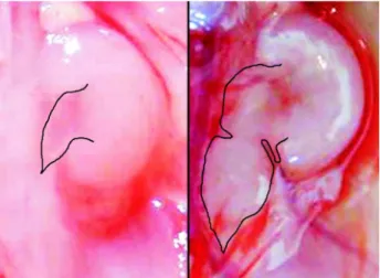
In the adriamycin group, hydronephrosis (Fig-ure-2) was observed in 70 out of 74 fetuses (95%), in addition to other malformations. In the 21 fetuses from the control group, the malformations under study were not observed (Table-1).
ASw, Fw and PMw were decreased and AFw was increased in the adriamycin group in relation to the control group (p <0.001) (Table-2).
The histomorphometric study revealed pel-vic dilation and reduction of renal parenchyma in the hydronephrosis group when compared with the con-trol group (p <0.001) (Table-3).
The qualitative microscopic analysis of the kidneys in both groups demonstrated the capacity of distinguishing between medullar and cortical in a lower magnification (X20) and the existence of neph-rogenic zone, composed by immature glomeruli (Fig-ure-3). In the hydronephrosis group, we observed se-Figure 1 – Scheme used for performing the histomorphometric
examination. 2-headed arrows indicate linear measurements. A1 = area of renal parenchyma and A2 = area of renal pelvis.
Table 1 – Frequency of malformations in the adriamycin
group.
Malformation
Hydronephrosis
- Bilateral - Unilateral
Duodenal Atresia Esophageal Atresia Bladder Hypoplasia
70 (95) 64 (87) 6 (8) 68 (92) 67 (91) 62 (84)
Frequency (%)
Figure 2 – Macroscopic comparison between a normal kidney from the control group (left) with hydronephrotic kidney from the adriamycin group (right). The ureters are delimited, showing dilation in the adriamycin group.
*p < 0.001; ASw = weight of amniotic sac; Fw = fetal weight; PMw = weight of placenta and amniotic membranes; AFw = weight
of amniotic fluid.
Table 2 – Comparison of weights. Mean, standard deviation and interval.
Weights
ASw (g) Fw (g) PMw (g) AFw(g)
Adriamycin Group
5.23* (± 0.56); 0.38 – 6,51
3.39* (± 0.43); 2.12 – 4.36
0.95* (± 0.16); 0.35 – 1.62
0.90* (± 0.33); 0.08 – 2.34
Control Group
vere cortical atrophy and tubular cortical atrophy; as well as moderate caliceal dilation, moderate to se-vere cystic tubular change and slight mesangial hy-perplasia, whereas in the control group such changes were not observed (Figure-4).
The search for microscopic changes in pel-vis and ureter revealed severe pelvic dilation with flat-tening of the epithelial cells, and dilation and tortu-osity of the proximal ureter in the hydronephrosis group. On the other hand, no changes were observed in the ureteral epithelium, which was similar to the control group.
COMMENTS
Adriamycin acts on the S-phase of the cell cycle, inhibiting topoisomerase II and consequently
Figure 4 – Microscopic comparison between a normal kidney from the control group (left) with hydronephrotic kidney from the hydronephrosis group (right). Note mature (arrows) and imma-ture (arrowheads) glomeruli in the control group. In the hydro-nephrosis group, we can see medullar and cortical atrophy and cystic tubular dilation (arrow), (HE, X200).
Figure 3 – Microscopic comparison between a normal kidney from the control group (left) with hydronephrotic kidney from the hydronephrosis group (right). The cortical-medullar limit is shown (arrowheads). Note the atrophy in the hydronephrosis group, (HE, X20).
DNA synthesis (15). This inhibition induces cell apoptosis, and is the potential molecular cause of the malformations detected, deriving mostly from a fail-ure in the embryologic development of mesoderma (16).
The decrease in ASw, Fw and PMw in the adriamycin group in relation to the control group is due to the drug’s deleterious effects, either by its pri-mary action, or by the repercussions from the mal-formations it induces (9,10).
The frequency of detected malformations was similar to those found by other authors who used adriamycin (9-11,17).
Fetuses with serious renal defects leading to intra-uterine urinary retention evolve with
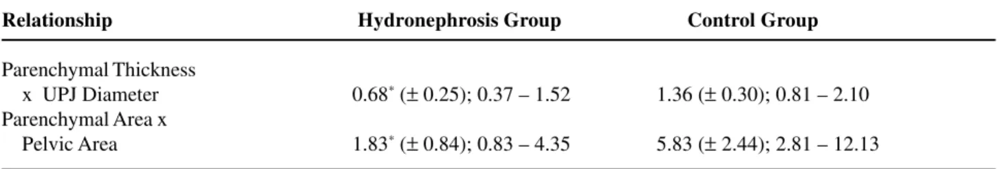
oligohy-Table 3 – Comparison of histomorphometric measures. Mean, standard deviation and interval.
Relationship Hydronephrosis Group Control Group
Parenchymal Thickness
x UPJ Diameter 0.68* (± 0.25); 0.37 – 1.52 1.36 (± 0.30); 0.81 – 2.10
Parenchymal Area x
Pelvic Area 1.83* (± 0.84); 0.83 – 4.35 5.83 (± 2.44); 2.81 – 12.13
dramnios, due to the relation between the production of fetal urine and a proper amount of AF (3,7). De-spite bilateral FH occurring in 87% of fetuses in the adriamycin group, there was an increase in AFw in relation to the control group (p < 0.0001). However, a bilateral FH occurred in association with 100% of digestive atresias. Such atresias, especially proximal ones, impair the deglutition of AF, as well as its ab-sorption in the gastrointestinal tract, and may lead to polyhydramnios (18).
The association between these anomalies, intestinal atresias and FH, which have a contrary ef-fect on AFw, and other potential causes of oligohy-dramnios can make results controversial. However, the present study showed that the ability of forming urine, and consequently AF by the fetal kidney was preserved, since there was no decrease in the AFw. Hence, it is not important to say that AFw increased, but that it did not decrease, since little AF would in-dicate low production of fetal urine.
Merei et al. (2001) studied urinary malfor-mations in the adriamycin model and concluded that the ureters would present a blind end, producing hy-dronephrotic fetal kidneys and altering the normal development of urinary bladder (11). In order to ex-plain the increase in AF that occurs concomitantly in blind-ending ureters, extra-renal compensatory mechanisms would be required.
Liu & Hutson (2000), while studying urogeni-tal malformations in the same model, concluded that the ureters might communicate with the urethra in male fetuses, and with the urogenital sinus in female fetuses, through an uretero-ureteral or uretero-uro-genital fistula, respectively (19). Thus, urinary flow would occur, which would allow for a proper mainte-nance of AF amount. The occurrence of FH non-as-sociated with oligohydramnios is suitable with the explanations proposed by Liu & Hutson (2000), since it may be the indirect result of passage of urine to the amniotic cavity (19).
In relation to bladder development, the oc-currence of 84% of bladder hypoplasia observed in the present study could be explained both by the lack of filling due to fistula between ureter and urethra or urogenital sinus and by the presence of blind-ending ureter (11,19).
Liu et al. (1999) studied bladder development, using adriamycin and verified that the 7th gestational day is the critical time when the drug administration causes bladder agenesis. Bladder hypoplasia observed in the present study, considering that the drug was administered on the 8th and 9th days, must be conse-quent to ureteral defects or represent an effectively distinct anomaly from that observed by the same au-thor (20).
The histomorphometric study revealed in-crease in the ureteropelvic junction (UPJ) diameter and in pelvic area, and decrease of parenchymal thick-ness and parenchymal area in the hydronephrosis group when compared with the control group. The parenchyma/pelvis relationships were decreased to half in the hydronephrosis group in relation to the control group, when we used linear measures de-creased to one third, when area measurements were used. These results objectively confirm the pelvic dilation and decrease in renal parenchyma observed in kidneys with hydronephrosis.
In models of surgically created hydroneph-rosis, we detected epithelial, epithelial-mesenchymal, obstructive and inflammatory changes, including at-rophy, dysplasia, fibrosis and changes in the renal de-velopment (3,13,14).
Microscopic findings in the present study are milder than the ones reported in those works, being restricted to the mechanical repercussion of urine retention, which is characterized by dilation of ureter, pelvis and ducts, and to secondary mass changes, such as atrophy of cortex and medulla. Moreover, no inflammatory or developmental changes were observed.
Other changes were expected in addition to dilation, since urine retention can be associated with renal damage and impair its normal differentiation (6,14). However, the microscopic findings are in agreement with other findings relative to AFw, which indirectly revealed production of fetal urine.
CONCLUSION
changes, despite being milder than the lesions de-scribed for surgically produced fetal hydronephrosis, were in agreement with amniotic fluid weight, since they enabled the production of urine with maintenance of the amniotic fluid.
Anderson Gonçalves received scholarship for Scientific Initiation from CNPq, and Lourenço Sbragia Neto from FAPESP
REFERENCES
1. Brace RA: Physiology of amniotic fluid volume regu-lation. Clin Obstet Gynecol. 1997; 40: 280-9. 2. Harrison MR, Nakayama DK, Noall R, de Lorimier
AA: Correction of congenital hydronephrosis in utero II. Decompression reverses the effects of obstruction on the fetal lung and urinary tract. J Pediatr Surg. 1982; 17: 965-74.
3. Chevalier RL, Thornhill BA, Chang AY: Unilateral ure-teral obstruction in neonatal rats leads to renal insuffi-ciency in adulthood. Kidney Int. 2000; 58: 1987-95. 4. Seseke F, Thelen P, Hemmerlein B, Kliese D, Zoller
G, Ringert RH: Histologic and molecular evidence of obstructive uropathy in rats with hereditary congeni-tal hydronephrosis. Urol Res. 2000; 28: 104-9. 5. Freedman AL, Bukowski TP, Smith CA, Evans MI,
Johnson MP, Gonzalez R: Fetal therapy for obstruc-tive uropathy: diagnosis specific outcomes [corrected]. J Urol. 1996; 156 (2 Pt 2): 720-3; discussion 723-4; Erratum in: J Urol. 1996; 156: 1786.
6. Bernstein J, Risdon RA, Gilbert-Barness E: Renal System. In: Gilbert-Barness E (ed.), Potter’s, Pathol-ogy of the Fetus and Infant. St Louis, Mosby. 1997; pp. 863-903.
7. Bastide A, Manning F, Harman C, Lange I, Morrison I: Ultrasound evaluation of amniotic fluid: outcome of pregnancies with severe oligohydramnios. Am J Obstet Gynecol. 1986; 154: 895-900.
8. Reddy PP, Mandell J: Prenatal diagnosis. Therapeutic implications. Urol Clin North Amer. 1998; 25: 171-80. 9. Beasley SW, Diez Pardo J, Qi BQ, Tovar JA, Xia HM: The contribution of the adriamycin-induced rat model of the VATER association to our understanding of con-genital abnormalities and their embryogenesis. Pediatr Surg Int. 2000; 16: 465-72.
10. Merei J, Hasthorpe S, Farmer P, Hutson JM: Visceral anomalies in prenatally adriamycin-exposed rat fetuses:
a model for the VATER association. Pediatr Surg Int. 1999; 15: 11-6.
11. Merei J, Batiha A, Hani IB, El-Qudah M: Renal anoma-lies in the VATER animal model. J Pediatr Surg. 2001; 36: 1693-7.
12. Franca WM, Goncalves A, Moraes SG, Pereira LA, Sbragia L: Esophageal atresia and other visceral anomalies in a modified Adriamycin rat model and their correlations with amniotic fluid volume variations. Pediatr Surg Int. 2004; 20: 602-8.
13. Steinhardt GF, Liapis H, Phillips B, Vogler G, Nag M, Yoon KW: Insulin-like growth factor improves renal architecture of fetal kidneys with complete ureteral obstruction. J Urol. 1995; 154: 690-3.
14. Steinhardt GF, Salinas-Madrigal L, deMello D, Farber R, Phillips B, Vogler G: Experimental ureteral obstruc-tion in the fetal Opossum: histologic assessment. J Urol. 1994; 152: 2133-8.
15. Tewey KM, Rowe TC, Yang L, Halligan BD, Liu LF: Adriamycin-induced DNA damage mediated by mam-malian DNA topoisomerase II. Science. 1984; 226 (4673): 466-8.
16. Orford JE, Cass DT: Dose response relationship be-tween adriamycin and birth defects in a rat model of VATER association. J Pediatr Surg. 1999; 34: 392-8. 17. Beasley SW, Diez Pardo J, Qi BQ, Tovar JA, Xia HM: The contribution of the adriamycin-induced rat model of the VATER association to our understanding of con-genital abnormalities and their embryogenesis. Pediatr Surg Int. 2000; 16: 465-72.
18. Kimble RM, Harding JE, Kolbe A: Does gut atresia cause polyhydramnios? Pediatr Surg Int. 1998; 13: 115-7. 19. Liu MI, Hutson JM: Cloacal and urogenital
malfor-mations in adriamycin-exposed rat fetuses. BJU Int. 2000; 86: 107-12.
20. Liu MI, Hutson JM, Zhou B: Critical timing of blad-der embryogenesis in an adriamycin-exposed rat fetal model: a clue to the origin of the bladder. J Pediatr Surg. 1999; 34: 1647-51.
Received: August 23, 2004 Accepted after revision: November 17, 2004
Correspondence address:
Dr. Lourenço Sbragia Neto
Discipline of Pediatric Surgery, FCM, Unicamp Rua Alexander Fleming, 181, Barão Geraldo Campinas, SP, 13084-970, Brazil
Fax: + 55 19 3788-9473