Identification of mechanisms involved in the relaxation
of rabbit cavernous smooth muscle by a new nitric oxide
donor ruthenium compound
João Batista Gadelha de Cerqueira, Lúcio Flávio Gonzaga-Silva, Francisco Ordelei Nascimento da Silva, João
Victor Medeiros de Cerqueira, Ricardo Reges Maia Oliveira, Maria Elisabete Amaral de Moraes, Nilberto
Robson Falcão do Nascimento
Departamento de Cirurgia (JBGC, LFGS, RRMO), Departamento de Química Orgânica e Inorgânica (FONS), Departamento de Fisiologia e Farmacologia (MEAM) Universidade Federal do Ceará, Ceará, Brazil and Instituto Superior de Ciências Biomédicas- Universidade Estadual do Ceará (JVMC, NRFN), Ceará, Brazil
ABSTRACT ARTICLE INFO
_________________________________________________________ ___________________
Key words:
Nitric oxide; Nitrosyl-ruthenium complex; Endothelium,
Vascular; Potassium Channel
Int Braz J Urol. 2012; 38: 687-94
__________________
Submitted for publication: March 08, 2012
__________________
Accepted after revision: July 27, 2012
Purpose: The aim of this study was to evaluate the relaxation in vitro of cavernous smooth muscle induced by a new NO donor of the complex nitrosil-ruthenium, named trans-[Ru(NH3)4(caffeine)(NO)]C13 (Rut-Caf) and sodium nitroprusside (SNP).
Materials and Methods: The tissues, immersed in isolated bath systems, were pre-con-tracted with phenilephrine (PE) (1 µM) and then concentration-response curves (10-12 - 10-4 M) were obtained. To clarify the mechanism of action involved, it was added to the baths ODQ (10 µM, 30 µM), oxyhemoglobin (10 µM), L-cysteine (100 µM), hydro-xicobalamine (100 µM), glibenclamide, iberotoxin and apamine. Tissue samples were frozen in liquid nitrogen to measure the amount of cGMP and cAMP produced.
Results: The substances provoked significant relaxation of the cavernous smooth mus-cle. Both Rut-Caf and SNP determined dose-dependent relaxation with similar potency (pEC50) and maximum effect (Emax). The substances showed activity through activation of the soluble guanylyl cyclase (sGC), because the relaxations were inhibited by ODQ. Oxyhemoglobin significantly diminished the relaxation effect of the substances. L--cysteine failed to modify the relaxations caused by the agents. Hydroxicobalamine significantly diminished the relaxation effect of Rut-Caf. Glibenclamide significantly increased the efficacy of Rut-Caf (pEC50 4.09 x 7.09). There were no alterations of po-tency or maximum effect of the substances with the addition of the other ion channel blockers. Rut-Caf induced production of significant amounts of cGMP and cAMP du-ring the relaxation process.
Conclusions: In conclusion, Rut-Caf causes relaxation of smooth muscle of corpus cavernosum by means of activation of sGC with intracellular production of cGMP and cAMP; and also by release of NO in the intracellular environment. Rut-Caf releases the NO free radical and it does not act directly on the potassium ion channels.
INTRODUCTION
Human corpus cavernosum contains vas-cular smooth muscle, kept under tonic
neu-rotransmitter that mediates penile erection in all animals (2). NO activates soluble guanylate cyclase (sGC), which induces production of cyclic guano-sine monophosphate (cGMP) from guanoguano-sine tri-phosphate (GTP). The cGMP acts in intracellular effectors, like protein kinase G (PKG), which cause diminishment of intracellular calcium and disas-sociation of actin and myosin fibers, ultimately leading to relaxation of the smooth muscle (2).
NO donors are substances that release NO, either in vivo or in vitro. One of these donors, sodium nitroprusside (SNP), is a powerful vasodi-lator utilized in patients with hypertensive crisis. Nevertheless, it is extremely labile, induces toler-ance and releases cyanide, which is toxic to the endothelium (3).
NO is a potent vasodilator synthesized by neurons, endothelial cells, leucocytes and platelets among others. It is released by the autonomous nerve terminals, and by the vascular and sinusoidal endothelium when stimulated by acetylcholine (4).
Synthesis of NO is catalyzed by NOS, which converts l-arginine and oxygen to l-citrul-ine and NO. NOS exists as three isoforms in mam-mals: nNOS and eNOS are preferentially expressed in neurons and endothelial cells, respectively, and iNOS in virtually all cell types. All three NOS iso-forms have been identified in the corpus caver-nosum, with nNOS and eNOS being considered responsible for initiating and sustaining erection, respectively (5). Down-regulation of nNOS expres-sion has been found in the corpus cavernosum of aging rats (6), a model in which corpus cavernous smooth muscle relaxation is impaired (7).
Endothelial dysfunction is present in a large group of patients with erectile dysfunction (ED) and co-morbidities like hypertension and diabetes. This syndrome is characterized by an endogenous NO production deficiency (8). About 56% of patients in this group show resistance to current ED treatment with phosphodiesterase 5 (PDE-5) inhibitors (9). Research for new drugs that increase bioavailability of endogenous NO is a permanent challenge.
Recently, new NO donor compounds with higher stability and less toxicity have been sub-ject of research. One group of such substances, S-nitroso-glutathione (GSNO) and
S-nitroso-N-acetylcysteine-etylester (SNACET), was utilized in studies with strips of human corpus cavernosum mounted in isolated bath systems, proving to be potentially useful for tissue relaxation (10).
SNP has already been utilized intracavern-ously in vivo, in both human and animals, to in-duce penile erection. In dogs and monkeys, SNP induced dose-related erection, without causing hypotension (11). In patients with ED, SNP pro-voked erection of shorter duration than did pa-paverine, without side effects - like hypotension, injection site pain, or priapism (11,12).
Ruthenium compounds, which are NO do-nors, have been tested in vitro and showed a simi-lar relaxation to SNP on rat aorta smooth muscle (13). These compounds theoretically have the po-tential to be better than SNP because they do not release cyanide nor they react with the superox-ide anion, forming peroxynitrite. However, further experimental studies are necessary to confirm this theory. Furthermore, they are stable at physiologic pH and soluble in water (13).
The aim of this study was to evaluated the relaxation in vitro of cavernous smooth muscle induced by a new NO donor of the complex nitro-syl-ruthenium, named trans-[Ru(NH3)4(caffeine) (NO)]C13 (Rut-Caf) and sodium nitroprusside (SNP).
MATERIALS AND METHODS
The study was approved by the local Com-mittee of Ethics on Animal Use in Research. Adult male New Zealand rabbits, weight 2 - 3 kg, were used. After anesthesia, the penis was removed entirely, and placed in Krebs-Henseleit solution. Cavernous tissue was dissected free of connective tissue and albuginea, providing two corpus caver-nosum strips (1 cm) from each penis.
transducer, while the other end was attached to a mobile unit that allowed tension adjustment.
The tissues rested for one hour. Tension was calibrated and the solution renewed every 15 minutes. Alterations on tension were registered on a polygraph (Gemini 7070, Ugo-Basile, Italy). Eight different experimental protocols were per-formed, as outlined next.
Experiment 1: After pre-contraction with phenilephrine (1 µM), rising concentrations (10-12
- 10-3 M) of SNP (as a control) and Rut-Caf were
administered to the baths and relaxation concen-tration-response curves were obtained.
Experiment 2: To evaluate the NO libera-tion profile for Rut-Caf, oxyhemoglobin (10 µM) was added to the baths, 30 minutes before pre-contraction with phenilephrine (1 µM).
Experiment 3: To evaluate possible anion nitroxyl (NO-) liberation by Rut-Caf during the re-laxation process, L-cysteine (100 µM), a specific NO- remover was added to the baths, 30 minutes before pre-contraction with phenilephrine (1 µM).
Experiment 4: To evaluate the contribu-tion of metabolically activated potassium chan-nels (KATP) on the relaxation process, one KATP blocker - glibenclamide (1 µM) - was added to the baths, 30 minutes before pre-contraction with phenilephrine (1 µM).
Experiment 5: To evaluate the contribu-tion of high, medium and low conductivity calci-um-activated potassium channels (KCA) on the re-laxation, two KCA blockers - iberotoxin (1 µM) and apamine (0.1 µM) - were added to the baths, 30 minutes before pre-contraction with phenileph-rine (1 µM).
Experiment 6: To evaluate the way NO is involved in the relaxation, hydroxocobalamin (0.1 mM), a NO remover, was added to the baths, 30 minutes before pre-contraction with phenileph-rine (1 µM).
Experiment 7: To determine the activa-tion of sGC by Rut-Caf, the agent 1H-[1,2,4] oxadiazole[4,3-α]quinoxalin-1-one (ODQ) - a sGC blocker - was added to the baths, 30 minutes be-fore pre-contraction with phenilephrine (1 µM).
In all the experiments, relaxation concen-tration-response curves were obtained according to rising concentrations (10-12 - 10-3 M) of Rut-Caf.
Experiment 8: The RbCC strips were frozen in liquid nitrogen for dosage of cGMP and cAMP. The method of non-acetylation was utilized and samples were separated for dosage of proteins ac-cording to the Bradford method (1976) (15). Tri-chloroacetic acid (TCA) was added to the tissue macerate, resulting in a final TCA concentration of 10%. After centrifugation, the supernatant was washed with water saturated diethylic ether. This process was repeated six times. After the wash-ings, the samples were dried in a nitrogen atmo-sphere at 60ºC, and then suspended again in the assay buffer of the immunoenzymatic kit for dos-age of cGMP and cAMP.
The relaxant effect of the substances was measured from the maximal contraction plateau induced by phenilephrine, and expressed as per-centages of contraction diminishment. The maxi-mal effect (Emax) was considered as the maximal amplitude response induced by the relaxant agents on the concentration-response curves. The drug concentrations that induced 50% maximal relax-ation (pEC50) were determined after logarithmic transformation of concentration-response normal curves, and expressed as the negative logarithmic of values for each tissue (pEC0). Percentages of contraction diminishment superior to 50% were considered significant.
The data were expressed as averages ± stan-dard deviation. Statistical analysis was performed with ANOVA, followed by the Tukey-Kramer test. Values of p < 0.05 were considered significant.
RESULTS
The SNP Emax value was 100%, and its pEC50 was 6.9 ± 0.2. For Rut-Caf, Emax value was 72.6 ± 6.6%, and pEC50 was 6.8 ± 0.2. There was no significant difference in potency between the two substances (p = 0.851).
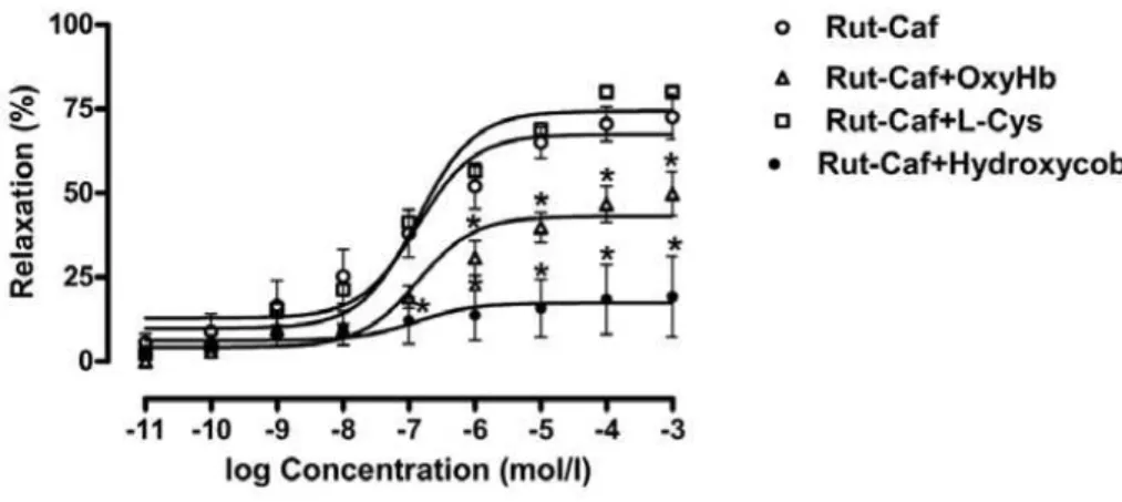
The relaxation curve induced by Rut-Caf was dislocated below with oxyhemoglobin, while the maximal response for Rut-Caf, in this setting, was 49.8 ± 6.5%.
0.915). The Emax value for Rut-Caf was 77% and for Rut-Caf + L-cysteine was 80% (Figure-1).
Glibenclamide modified potency and effi-cacy of Rut-Caf, as reflected by pEC50 values for Rut-Caf of 5.2 ± 0.2 and Rut-Caf + glibenclamide of 7.3 ± 0.2 (p = 0.016). Emax value increased from 68 ± 8.9% to 92 ± 7% (p = 0.060).
Tissue incubation with ibero-apamin did not interfere with Rut-Caf relaxations. The pEC50 for Rut-Caf was 4.7 ± 0.2, compared to 4.8 ± 0.3 for Rut-Caf + ibero-apamin (p = 0.779) (Figure-2).
Hydroxocobalamin almost abolished the relaxant effects of Rut-Caf. The pEC50 for Rut-Caf was 4.9 ± 0.1, while pEC50 for Rut-Caf +
hydroxo-mg. Rut-Caf generated 143.52 pmoL/mg (p < 0.01) and SNP 43.50 pmoL/mg (p < 0.05) (Figure-4).
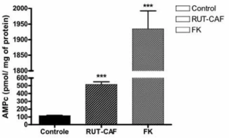
Finally, Rut-Caf (100 µM) produced cAMP in the corpus cavernosum in quantity similar to that of forskolin (10 µM) and significantly supe-rior to the control amount (p < 0.001) (Figure-5).
DISCUSSION
This study evaluated a new ruthenium compound and its capability to promote relax-ation of cavernous smooth muscle.
The substance Rut-Caf provoked sig-nificant relaxation of cavernous smooth muscle
Figure 1 - Effect of Rut-Caf upon rabbit corpus cavernosum strips with and without pretreatment with 10 µM/L oxyhemo-globin, 1 mmoL/L L-cysteine or 0.1 mmoL/L hydroxocobalamin. Concentration/response curves were plotted for graded concentrations (10-12 to 10-3 M) of Rut-Caf. Results were expressed as average ± standard error based on seven experi-ments. The statistical significance was verified with ANOVA followed by Buonferroni’s test.
cobalamin was 5.5 ± 0.2 (p = 0.713). As to Emax, values were respectively 85.3 ± 9.4% and 21.3 ± 2.2% (p < 0.001).
The relaxation induced by Rut-Caf was sig-nificantly inhibited by ODQ. This is evidenced by the Emax values for Rut-Caf - 58 ± 9.2% – and Rut-Caf + ODQ - 34.6 ± 3.7% (p = 0.038) (Figure-3).
Both SNP and Rut-Caf produced cGMP in the cavernous tissue in amounts significantly higher than the basal control value of 8.26 pmoL/
(Emax = 80%) with potency similar to SNP; how-ever, with lower maximum effect. The most prob-able hypothesis is that SNP acts on relaxation of vascular smooth muscle activating the sGC and as a hyperpolarizing agent through direct activa-tion of potassium ion channels and Rut-Caf acts mainly on activation of sGC (16).
The soluble isoform sGC plays a pivotal role in erectile function because it provides the link between NO and cGMP, which represent the *p<0.05 vs. Rut-Caf without oxyhemoblin pretreatment.
extracellular and intracellular signaling mole-cules, respectively, in physiologic erection (17).
Bonaventura et al., studying substances that also belong to the nitrosyl-ruthenium com-plex, in rabbit aorta rings, demonstrated a maxi-mal relaxation effect of 102% with pEC50 of 6.61 ± 0.09. These data confirm, as showed in this study, that ruthenium compounds are powerful vasodilators (13).
In vivo, NO arranges with hemoglobin creating a stable iron-nitrosyl complex that does not release NO. Bonaventura et al., evaluating the effects of ruthenium compounds in rabbit aorta rings in the presence of oxyhemoglobin, observed a decreased potency without significantly
modif-ing its maximum effect, due to intracellular re-lease of NO.
In this in vitro study, the addition of oxy-hemoglobin to the organ baths reduced the maxi-mum effect of Rut-Caf, but could not abolish it. Therefore, the relaxant effect of this substance is also probably due to the intracellular release of NO (13).
McDonald and Murad demonstrated that NO acts on the relaxation of vascular smooth muscle through activation of soluble guanylate cyclase, increasing the synthesis and bioavailabil-ity of intracellular cGMP (18). In this study, tis-sue incubation with a specific inhibitor of soluble guanylate cyclase (ODQ; 10 µM) abolished the re-Figure 2 - A. Relaxation induced by Rut-Caf in rabbit corpus cavernosum strips pre-contracted with 1 µM phenylephrine with and without glibenclamide pretreatment. Results were expressed as average ± standard error based on seven experiments. B. Relaxation induced by Rut-Caf in rabbit corpus cavernosum strips pre-contracted with 1 µM phenylephrine with and without iberiotoxin/apamin pretreatment. Results were expressed as average ± standard error based on seven experiments.
A
laxation response provoked by Rut-Caf. This dem-onstrates the action of the substance studied on the NO-cGMP cell signaling system.
According to the studies with compounds of the group nitrosyl-ruthenium, it was observed that these substances act by means of donation of free NO and nitroxyl anion (NO-) (13). Utiliz-ing a specific nitroxyl anion remover, L-cysteine, the potential of relaxation remained unchanged. However, the addition of the NO free radical
re-mover, hydroxocobalamin, to the baths signifi-cantly decreased the relaxation induced by Rut-Caf. In contrast with Bonaventura et al. findings, Rut-Caf does not release the nitroxyl anion dur-ing relaxation. In consequence, probably its re-laxation effect is related to release of the NO free radical.
Smooth muscle has neither a T-tubule system nor a well-developed sarcoplasmatic re-ticulum. Therefore extracellular calcium plays an Figure 3 - Concentration/response curves showing relaxation induced by Rut-Caf in rabbit corpus cavernosum strips with and without pre-treatment with 10 µM ODQ or 30 µM ODQ. Results were expressed as average ± standard error based on seven experiments.
Figure 4 - cGMP dosage of rabbit corpus cavernosum strips exposed to saline solution (control), 100 µM Rut-Caf or 3 µM SNP (positive control).
Figure 5 - cAMP dosage of rabbit corpus cavernosum strips exposed to saline solution (control), 100 µM Rut-Caf or 10 µM forscolin (positive control).
important role, and calcium must enter the cyto-plasm through the cyto-plasma membrane during an action potential.
A direct hyperpolarizing action of the Rut-Caf was not observed. The addition of calcium ac-tivated potassium ion channels of high, medium and low conductivity inhibitors did not modify the relaxation effect of Rut-Caf. When glibenclamide, which is an ATP dependent potassium channel blocker, was added to the baths, an increase on the efficacy of Rut-Caf was observed (pEC50 4.04 x 7.69, p < 0.05).
The results obtained in this study were similar to the findings of Lee and Kang in human corpus cavernosum (19). These researchers, study-ing the effect of relaxation of a NO donor - SIN-1, could observe an increase in the probability of opening of calcium activated potassium channels in an indirect way, through activation of soluble guanylate cyclase and activation of the cGMP protein kinase. However, it was not demonstrated direct action of SIN-1 on the ion channels studied.
The best explanation for the efficacy of Rut-Caf would be the action of glibenclamide as a reducing agent. Bates et al. showed that the bioactivation of NO in biological medium needs the presence of a reducing agent. In consequence, glibenclamide acting as a reducing agent increase the efficacy of Rut-Caf (3).
This hypothesis was supported by the spectroscopic profile. The product formed (UV-Vis) is compatible with the formation of the spe-cies Rut-Caf-H2O. Initially, there is a reduction of NO0 by glibenclamide with posterior liberation of the metal coordination sphere. In conclusion, this suggests that amplification in the presence of glib-enclamide occurs due to reduction of NO+ present in the metal coordination sphere, increasing the concentration of NO0 in the reactive environment.
Although it is known that ion channels ef-fectively act on the control of the basal tone of the cavernous smooth muscle (1), they do not partici-pate in the relaxation process induced by Rut-Caf.
The production of intracellular cGMP from activation of sGC by NO in different tissues was initially demonstrated by Arnold et al. (20). The production of cGMP in corpus cavernosum of animals and humans by NO donors, during the
relaxation process, was demonstrated in studies (10,21).
Similar to these studies, it was demon-strated that the production of cGMP induced by Rut-Caf in the relaxation of cavernous smooth muscle was significantly higher than that induced by saline solution and like that produced by SNP.
It was also observed production of cAMP induced by Rut-Caf during the relaxation pro-cess. Uckert et al. demonstrated the interaction of cGMP-cAMP cell signaling systems in the re-laxation of cavernous smooth muscle (22). These authors demonstrated the presence of cAMP-spe-cific phosphodiesterases, PDE-3 and PDE-5 in the cavernous endothelium. They also noted that the cAMP specific kinase PKA inhibited the relaxation induced by PDE-5 inhibitors, specific to cGMP, characterizing the integration of the cGMP-cAMP cell signaling systems on the cavernous smooth muscle relaxation. Although the mechanisms for that interaction are not clear, the authors have proposed that the increase in cAMP would be sec-ondary to the drop in the level of cGMP, which would diminish the activity of PDE-3, increasing the level of cAMP. So, the regulation of cAMP-specific phosphodiesterases by cGMP could ex-plain the increase of cAMP observed in this study, as a direct effect of Rut-Caf in the relaxation of cavernous smooth muscle.
Lindaman et al. studied the gallbladder smooth muscle in vitro. They noted that caffeine acting as an unspecific phosphodiesterases inhibi-tor 1 to 5, increased the production of cGMP and cAMP in the relaxation process (23). Similarly, the presence of caffeine in the structure of Rut-Caf could contribute to the increase of cAMP pro-duced by that substance.
It should be emphasized that the results of this study were obtained in in vitro experimental model and future studies in vivo and clinical stud-ies are necessary in order to confirm them.
CONCLUSIONS
in the intra-cellular environment. Rut-Caf releases the NO free radical and it does not act directly on the potassium ion channels.
CONFLICT OF INTEREST
None declared.
REFERENCES
1. Andersson KE: Pharmacology of penile erection. Pharmacol Rev. 2001; 53: 417-50.
2. Lohse MJ, Förstermann U, Schmidt HH: Pharmacology of NO:cGMP signal transduction. Naunyn Schmiedebergs Arch Pharmacol. 1998; 358: 111-2.
3. Bates JN, Baker MT, Guerra R Jr, Harrison DG: Nitric oxide generation from nitroprusside by vascular tissue. Evidence that reduction of the nitroprusside anion and cyanide loss are required. Biochem Pharmacol. 1991; 42 (Suppl:) S157-65.
4. Ignarro LJ: The unique role of nitric oxide as a signaling molecule in the cardiovascular system. Ital Heart J. 2000; 1 (Suppl 3): S28-9.
5. Hurt KJ, Musicki B, Palese MA, Crone JK, Becker RE, Moriar-ity JL, et al.: Akt-dependent phosphorylation of endothelial nitric-oxide synthase mediates penile erection. Proc Natl Acad Sci U S A. 2002; 99: 4061-6.
6. Carrier S, Nagaraju P, Morgan DM, Baba K, Nunes L, Lue TF: Age decreases nitric oxide synthase-containing nerve fibers in the rat penis. J Urol. 1997; 157: 1088-92.
7. Cartledge J, Minhas S, Eardley I: The role of nitric oxide in penile erection. Expert Opin Pharmacother. 2001; 2: 95-107. 8. Feldman HA, Goldstein I, Hatzichristou DG, Krane RJ,
McKinlay JB: Impotence and its medical and psychosocial correlates: results of the Massachusetts Male Aging Study. J Urol. 1994; 151: 54-61.
9. Rendell MS, Rajfer J, Wicker PA, Smith MD: Sildenafil for treatment of erectile dysfunction in men with diabetes: a randomized controlled trial. Sildenafil Diabetes Study Group. JAMA. 1999; 281: 421-6.
10. Seidler M, Uckert S, Waldkirch E, Stief CG, Oelke M, Tsikas D, et al.: In vitro effects of a novel class of nitric oxide (NO) donating compounds on isolated human erectile tissue. Eur Urol. 2002; 42: 523-8.
11. Trigo-Rocha F, Martinez-Piñeiro L, Donatucci CF, Hsu GL, Lue TF, Tanagho EA: Sodium nitroprusside: physiologic ef-fects as a nitric oxide donor in three species. Int J Impot Res. 1995; 7: 49-56.
12. Shamloul R, Atteya A, Elnashaar A, Gadallah A, Zohdy W, Abdelsalam W: Intracavernous sodium nitroprusside (SNP) versus papaverine/phentolamine in erectile dysfunction: a comparative study of short-term efficacy and side-effects. J Sex Med. 2005; 2: 117-20.
13. Bonaventura D, de Lima RG, Vercesi JA, da Silva RS, Bend-hack LM: Comparison of the mechanisms underlying the relaxation induced by two nitric oxide donors: sodium nitro-prusside and a new ruthenium complex. Vascul Pharmacol. 2007; 46: 215-22.
14. Palea S, Barras M: Comparison of the relaxant effects of alfu-zosin, phentolamine and sildenafil on rabbit isolated corpus cavernosum. BJU Int. 2003; 91: 873-7.
15. Bradford MM: A rapid and sensitive method for the quantita-tion of microgram quantities of protein utilizing the principle of protein-dye binding. Anal Biochem. 1976; 72: 248-54. 16. Bolotina VM, Najibi S, Palacino JJ, Pagano PJ, Cohen RA:
Nitric oxide directly activates calcium-dependent potassium channels in vascular smooth muscle. Nature. 1994; 368: 850-3.
17. Andersson KE: Neurophysiology/pharmacology of erection. Int J Impot Res. 2001; 13 (Suppl 3): S8-S17.
18. McDonald LJ, Murad F: Nitric oxide and cyclic GMP signal-ing. Proc Soc Exp Biol Med. 1996; 211: 1-6.
19. Lee SW, Kang TM: Effects of nitric oxide on the Ca2+-activat-ed potassium channels in smooth muscle cells of the human corpus cavernosum. Urol Res. 2001; 29: 359-65.
20. Arnold WP, Mittal CK, Katsuki S, Murad F: Nitric oxide acti-vates guanylate cyclase and increases guanosine 3’:5’-cyclic monophosphate levels in various tissue preparations. Proc Natl Acad Sci U S A. 1977; 74: 3203-7.
21. Kim NN, Huang YH, Goldstein I, Bischoff E, Traish AM: In-hibition of cyclic GMP hydrolysis in human corpus caver-nosum smooth muscle cells by vardenafil, a novel, selec-tive phosphodiesterase type 5 inhibitor. Life Sci. 2001; 69: 2249-56.
22. Uckert S, Hedlund P, Waldkirch E, Sohn M, Jonas U, Anders-son KE, et al.: Interactions between cGMP- and cAMP-path-ways are involved in the regulation of penile smooth muscle tone. World J Urol. 2004; 22: 261-6.
23. Lindaman BA, Hinkhouse MM, Conklin JL, Cullen JJ: The ef-fect of phosphodiesterase inhibition on gallbladder motility in vitro. J Surg Res. 2002; 105: 102-8.