DOI: 10.5935/2359-4802.20170036
181 International Journal of Cardiovascular Sciences. 2017;30(2):181-184
CASE REPORT
Mailing Address: Sabrina Weiss Sties
Rua Pascoal Simone, 358, Coqueiros. Postal Code: 88080-350, Florianópolis, SC – Brazil E-mail [email protected]
Differential Diagnosis of Marfan Syndrome in a Teenage Volleyball Athlete
Fabrissio Portelinha Graffunder,1 Sabrina Weiss Sties,2 Ana Inês Gonzáles,1 Tales de Carvalho3Universidade Federal de Santa Catarina (UFSC);1 Faculdade Avantis;2 Universidade do Estado de Santa Catarina (UDESC),3 Florianópolis, SC – Brazil
Manuscript received August 08, 2016, revised manuscript January 23, 2017, accepted January 31, 2017. Marfan Syndrome; Aortic Rupture; Cardiovascular
Diseases; Athletes; Exercise; Aortic Diseases.
Keywords
Introduction
Marfan syndrome (MFS) is an autosomal dominant hereditary disease mainly caused by mutations in the fibrillin-1 (FBN1) gene. It is characterized by the occurrence of thoracic aortic aneurysm and/or dissection,
ectopia lentis, and systemic abnormalities.1
The highest risk of death associated with the syndrome is attributed to cardiovascular abnormalities, in particular, progressive aortic root aneurysm, leading to aortic dissection and rupture if not corrected surgically.2 The clinical diagnosis of MFS may be established by the revised Ghent nosology,3 although this can be challenging, since many characteristics of this disease are dependent on the age of the patient, while others are frequently seen in the general population, with substantial phenotypic variability. In addition, MFS has considerable overlap with other connective tissue diseases.2,4
Even though MFS is a rare condition (1:5,000),1 its prevalence is speculated to be much higher among individuals participating in sports, especially those sports in which tall stature and long limbs are advantageous. One such example is volleyball,5 which is classified as a moderate dynamic and low static sport.6
Studies on the practice of competitive sports by individuals with borderline or evident aortic root dilation are limited. Therefore, it becomes necessary to evaluate the differential diagnosis of MFS or any evident systemic disease.
In this context, the present study describes the case of a volleyball athlete with a possible diagnosis of MFS.
The athlete and his guardian signed both agreement and informed consent forms. The research was approved by the Research Ethics Committee at Universidade do Estado de Santa Catarina (UDESC).
Case Report
We report the case of a 16-year-old black male athlete with a height of 2.08 meters and weighing 80.9 kg. He was summoned to the Brazilian National Junior Volleyball Team in October 2013. During pre-joining assessment procedures, the patient was asymptomatic. He underwent clinical examination, ophthalmologic evaluation by a specialist, electrocardiography at rest, cardiopulmonary testing, and laboratory analysis, which showed no significant findings. While undergoing unidimensional and bidimensional echocardiography with color Doppler, a 45-mm diameter dilatation was observed on the thoracic ascendant aorta involving the sinuses of Valsalva. The athlete was removed from practice and underwent magnetic resonance angiography of the thoracic aorta, which evidenced a dilatation at the valvular plane with maximum diameters measured at this topography of 48 X 46 mm.
182
Graffunder et al
Differential Diagnosis of Marfan Syndrome
Int J Cardiovasc Sci. 2017;30(2):181-184 Case Report
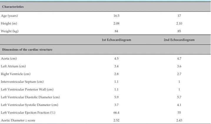
Table 1 – Patient characteristics and echocardiographic results
Characteristics
Age (years) 16.5 17
Height (m) 2.08 2.10
Weight (kg) 84 85
1st Echocardiogram 2nd Echocardiogram
Dimensions of the cardiac structure
Aorta (cm) 4.5 4.7
Left Atrium (cm) 3.4 3.6
Right Ventricle (cm) 2.8 2.7
Interventricular Septum (cm) 1.1 1
Left Ventricular Posterior Wall (cm) 1.1 1
Left Ventricular Diastolic Diameter (cm) 5.9 5.7
Left Ventricular Systolic Diameter (cm) 3.7 4.1
Left Ventricular Ejection Fraction (%) 66.4 55
Aortic Diameter z score 2.52 2.43
Six months after being summoned, his height was 2.10 m, and his weight was 85.5 kg. He continued to participate in volleyball training at his base team, but remained out from the Brazilian National Team. On April 2014, the athlete underwent a new echocardiography, in which no significant increase in the aortic dimensions was observed (Table 1).
His aortic diameter z score was > 2 standard deviations (SD) of the mean for his body surface area (2.43 m2). An aortic root diameter greater than 2 SD, in addition to a positive genetic test or ectopia lentis, confirms the diagnosis of MFS regardless of systemic criteria. However, an ophthalmologic evaluation, conducted by a specialist, demonstrated that the patient’s lenses were typical and within normal parameters, without any shape abnormality. A genetic test to investigate MFS was not performed, but the athlete had no family history of the syndrome, therefore, did not reach the score to confirm MFS.
The occurrence of MFS signs was assessed with the revised Ghent nosology, which totaled only 2 points (skin
striae, as well as reduced upper segment/lower segment ratio and increased arm span/height and absence of severe scoliosis), as shown in Table 2.
Discussion
The uncertainty regarding the diagnosis of MFS in this athlete was due to the fact that although his aortic diameter z score was 2 SDs above the mean for his body surface area, his phenotypic expression was incomplete on physical exam. In order to confirm
the diagnosis of MFS, the systemic score must be ≥ 7.
It is well established in the literature that the main concern in individuals with MFS is their increased risk of death due to aortic dissection.7
In this context, the recommendations of the 36th Bethesda Conference are used to regulate the participation of athletes in competitions and establish that individuals with aortic
diameters ≥ 4.0 cm or > 2 SD of the mean for body surface
area should only participate in low-to-medium intensity athletic competitions. However, these cutoff values are derived from the general population.
183 Graffunder et al
Differential Diagnosis of Marfan Syndrome Int J Cardiovasc Sci. 2017;30(2):181-184
Case Report
diagnosis is uncertain due to incomplete phenotypic expression in young athletes, and/or ineffective or negative genetic testing, the ESC allows the athlete to continue participating in sports, provided they undergo a periodic clinical reassessment.
According to the guidelines of the Brazilian Society of Cardiology and Brazilian Society of Sports Medicine, individuals with a complete MFS phenotype may participate in low-to-moderate dynamic and low static sports, and should be assessed by echocardiography every 6 months, provided they do not present the following conditions: dilation of the aortic root greater than 2 SDs, moderate to severe mitral regurgitation, and family history of aortic dissection or sudden death.8
Due to the characteristics presented by the athlete in this report, MASS phenotype was included in his differential diagnosis. This phenotype includes involvement of the skeletal muscle, striae atrophica,
Table 2 – Systemic scores for Marfan syndrome presented by the athlete
Characteristics Points
Wrist and thumb sign*,**
Pectus carinatum
or pectus excavatum or chest asymmetry
Hindfoot deformity
or plain pes planus
Pneumothorax
Dural ectasia
Protrusio acetabuli
Reduced upper segment/lower segment ratio and increased arm span/height and absence of severe scoliosis 1
Scoliosis or thoracolumbar kyphosis
Reduced elbow extension
Facial features (three out of five)***
Skin striae 1
Myopia > 3 diopters
Mitral valve prolapse
Total 2
*Wrist sign: This signal is positive when one hand wraps the contralateral wrist and the tip of the thumb of the hand covers the distal phalanx of the 5th
finger in the same hand. **Thumb sign: positive when during thumb adduction transversely across the hand, the distal phalanx of the thumb protrudes beyond the ulnar border. ***Facial characteristics: includes dolichocephaly, enophthalmos, downslanting palpebral fissures, malar hypoplasia, retrognathia.
borderline but nonprogressive aortic dilatation, and mitral valve prolapse. Only the latter was not observed in the athlete. However, the MASS phenotype only applies if the aortic diameter z score is < 2, the systemic score is
≥ 5, and the patient is at least 20 years old.3 The adolescent in the present study did not fit these criteria.
A cohort study by Joundeau et al.9 including individuals with confirmed MFS concluded that the risk of sudden death or aortic dissection is low in individuals with aortic diameter between 4.5 and 4.9 cm. For these authors, the diameter of 5 cm was deemed a reasonable threshold for prophylactic surgery.
In addition to the aortic dilation, an aortic growth at a rate greater than 0.5 cm per year is considered a concern in terms of dissection.2
184
1. Pepe G, Giusti B, Sticchi E, Abbate R, Gensini GF, Nistri S. Marfan syndrome: current perspectives. Appl Clin Genet. 2016;9:55-65.
2. Romaniello F, Mazzaglia D, Pellegrino A, Grego S, Fiorito R, Ferlosio A, et al. Aortopathy in Marfan Syndrome: an update. Cardiovasc Pathol. 2014;23(5):261-6.
3. Loeys BL, Dietz HC, Braverman AC, Callewaert BL, De Backer J,
Devereux RB, et al.The revised Ghent nosology for the Marfan
Syndrome. J Med Genet. 2010;47(7):476-85.
4. Tsang AK, Taverne A, Holcombe T. Marfan syndrome: a review of the literature and case report. Spec Care Dentist. 2013;33(5):248-54.
5. D’Andrea A. Cocchia R, Riegler L, Scarafile R, Salerno G, Gravino
R, et al.Aortic root dimensions in elite athletes. Am J Cardiol.
2010;105(11):1629-34.
6. Mitchell JH, Haskell W, Snell P, Van Camp SP.Task force 8: classification of sports. J Am Coll Cardiol. 2005;45(8):1364-7.
7. Castellano JM, Silvay G, Castillo JG. Marfan Syndrome: clinical, surgical, and anesthetic considerations. Semin Cardiothorac Vasc Anesth. 2014;18(3):260-71.
8. Ghorayeb N, Costa RV, Castro I, Daher DJ, Oliveira Filho JA, Oliveira MA, et al; Sociedade Brasileira de Cardiologia. [Guidelines on exercise and sports cardiology from the Brazilian Society of Cardiology and the Brazilian Society of Sports Medicine]. Arq Bras Cardiol. 2013;100(1 Suppl. 2):1-41.
9. Joundeau G, Detaint D, Tubach F, Arnoult F, Milleron O, Raoux F, et al. Aortic event rate in the Marfan Population: a cohort study. Circulation. 2012;125(2):226-32.
10. Pelliccia A, DI Paolo FM, Quatrinni FM. Aortic root dilatation in athletic population. Prog Cardiovasc Dis. 2012;54(5):432-7.
References
Graffunder et alDifferential Diagnosis of Marfan Syndrome
Int J Cardiovasc Sci. 2017;30(2):181-184 Case Report
without cardiovascular disease, including men and women participating in Olympic Games and world championships, and competitors in different sports at national and regional levels. The authors found that the 99th percentile value for the aortic root diameter was 40 mm in men and 34 mm in women. Overall, 17 male athletes showed larger aortic diameters than the set threshold, including young athletes practicing rowing, basketball or volleyball who were tall and had a large body surface area, but none satisfied the revised Ghent criteria for the diagnosis of MFS. They were allowed to practice sports and were followed up every 6 months. A significant increase in the size of the aorta was observed, which in two of these athletes exceeded 5 cm. However, they had no cardiovascular complications, remained without any Ghent criterion, and had a negative test for genetic FBN1 mutations.
Conclusion
Changes in the aortic diameter pattern should be taken into account the individual’s body surface area and progressive aortic dilation.
There is a paucity of information on the practice of competitive sports by individuals with borderline or obvious aortic root dilation but without diagnostic criteria for MFS or any evident systemic disease.
The athlete in this report had a possible differential diagnosis of MFS that could be confirmed after periodic
evaluations over time. It is important to highlight that trainers in sports in which tall stature is advantageous and, thus common, should become acquainted with MFS, in order to identify through known signs potential athletes who have the disease and help them seek appropriate guidance.
Author contributions
Conception and design of the research: Graffunder FP, Sties SW, Gonzáles AI, Carvalho T. Acquisition of data: Graffunder FP, Sties SW, Gonzáles AI, Carvalho T. Analysis and interpretation of the data: Graffunder FP, Sties SW, Gonzáles AI, Carvalho T. Writing of the manuscript: Graffunder FP, Sties SW, Gonzáles AI, Carvalho T. Critical revision of the manuscript for intellectual content: Graffunder FP, Sties SW, Gonzáles AI, Carvalho T.
Potential Conflict of Interest
No potential conflict of interest relevant to this article was reported.
Sources of Funding
There were no external funding sources for this study.
Study Association