Executive Summary – Guidelines for Mechanical Circulatory Support
of the Brazilian Society of Cardiology
Silvia Moreira Ayub-Ferreira
Instituto do Coração do Hospital das Clínicas da Faculdade de Medicina da Universidade de São Paulo, São Paulo, SP - Brazil Hospital Sírio-Libanês, São Paulo, SP - Brazil
Keywords
Heart Failure/complications, Heart Failure/therapy; Myocardial Ischemia/complications; Assisted Circulation/ instrumentation; Contraindicators; Risk Assessment. Mailing Address: Silvia Moreira Ayub-Ferreira •
Rua Enéas Carvalho de Aguiar, 44. CEP 05403-000, São Paulo, SP - Brazil E-mail: [email protected]
Manuscript received September 20, 2017, revised manuscript March 20, 2018, accepted June 13, 2018
DOI: 10.5935/abc.20180126
Evaluation of candidates for mechanical
circulatory support devices
In advanced heart failure (HF), the Interagency Registry for Mechanically Assisted Circulatory Support (INTERMACS) proposed seven clinical profiles (and modifiers) for a convenient, easy classification of disease status, risk of implantation of mechanical circulatory support devices (MCSDs) and adequate time for intervention (Chart 1).1
One of the main determinant factors for a successful MCSD implantation is patient eligibility. Correct selection of patients involves – (1) patients with advanced HF to which the risk of MCSD implantation surpasses mortality risk for current disease (making it a beneficial intervention); (2) patients with moderately advanced HF, i.e., implantation of MCSD would not increase patient’s morbidity and mortality due to increased complication rate; (3) no contraindications for MCSD implantation.2,3
Perioperative renal failure, pre-existing right HF, liver dysfunction, mechanical ventilation in the pre-operative period, low weight or overweight and reoperation have been related to worse clinical outcomes after MCSD implantation.3-5
The main scores for risk prediction in MCSD implantation are described in Chart 2.
Echocardiography
Evaluation of patients candidates for MCDS should include a transthoracic echocardiogram (TEE) complemented by a transesophageal echocardiography (TEE).
The effects of MCDS on right ventricular function depend on the balance between the benefits of decompression of the left chambers (reduction of the left ventricular afterload) and greater volumetric load to the right atrium (RA; increase of the right ventricular preload). Decompression of left chambers also cause changes in the geometry of the right chambers, such as leftward shift of interatrial (IAS) and interventricular septum (IVS), structural changes of tricuspid annulus, which can aggravate a pre-existing tricuspid insufficiency (TI) and right ventricular overload.10
Considering that right ventricular cardiac output determines left ventricular preload, a significant reduction in right ventricular function results in decreased output by the MCSD. It is estimated that approximately 30% of patients with left ventricular assist device develop limiting right ventricular dysfunction. For these reasons, a careful evaluation of right ventricular function is mandatory before MCDS implantation. In the presence of moderate-to-severe dysfunction, the requirement of a permanent biventricular support cannot be ruled out.11
In the assessment of right ventricular function before MCSD implantation, it is recommended the measurement of the right ventricle, as well as a semiquantitative assessment of right ventricular longitudinal and radial contractility combined with quantitative parameters, including fractional area change (FAC; FAC < 20% are associated with increased risk of right ventricular dysfunction after MCSD implantation),12 tricuspid annular plane systolic excursion (TAPSE) determined by M mode, peak systolic velocity of lateral tricuspid ring, measured by tissue Doppler (s’), and right ventricular performance index.13,14
Predictors of right ventricular dysfunction before mechanical circulatory support device implantation
Right ventricular dysfunction is multifactorial and includes an increase in preload, ventricular ischemia and mechanical interdependence of ventricular geometry. It is one of the most severe complications of left ventricular assist device, observed in up to 30% of cases and associated with a six-fold increase in morbidity and mortality (increased risk in up to 67%).11,15
Risk factors and the main risk score for right ventricular dysfunction after MCSD implantation are described in Charts 3 and 4.
Implantation of a MCSD in the left ventricle should be performed with caution in patients with important right ventricular dilation, moderate-to-severe tricuspid insufficiency, tricuspid valve annulus > 45 mm and CVP > 15 mmHg. By this means, hemodynamic variables directly reflect a preload or afterload increase and right ventricular contractility reductions, whereas venous congestion and organ hypoperfusion, consequence of right ventricular dysfunction, indicate hepatic and renal dysfunctions15,21
Chart 1 – Interagency Registry for Mechanically Assisted Circulatory Support (INTERMACS) profiles
Profile Description Hemodynamic status Time frame for definitive intervention
1 Critical cardiogenic shock Persistent hypotension despite the use of inotropes and
intra‑aortic balloon pumps, associated with organic dysfunction Hours 2 Progressive decline, but inotrope dependent and lactate levels, despite use of inotropes in optimized dosesDeterioration of renal and hepatic function, nutritional status Days
3 Stable but inotrope dependent Clinical stability on continuous inotropic therapy, and history of
failure to wean from it Weeks – months 4 Frequent hospitalization Signs of water retention, symptoms at rest and frequent admissions to emergency departments Weeks – months
5 At home, exertion intolerant Intolerant to activity, comfortable at rest despite water retention Intervention emergency depends on nutritional status and organic dysfunction severity 6 Exertion limited Moderate limitation to activity; absence of signs of hypervolemia Intervention emergency depends on nutritional status and organic dysfunction severity 7 NYHA III Hemodynamic stability and absence of hypervolemia Intervention is not indicated
NYHA: New York Heart Association.
Chart 2 – Risk predictors for mechanical circulatory support device implantation
Risk score for destination therapy6 Risk score for bridge/destination
therapy (HMII score)7 Pre-operative risk score
8 Pre-operative risk score9
Risk of 90‑day in‑hospital mortality
(pulsatile flow) Ninety-day mortality (continuous flow) Mortality risk after MCSD implantation (mean of 84 days) Mortality risk after MCSD implantation (mean of 100 days)
Platelets < 148.000/µL OR: 7.7
Age (for 10 years)
OR: 1.32
Urine flow < 30 mL/hour
RR: 3.9
Respiratory failure /sepsis OR: 11,2 Albumin < 3.3 mg/dL
OR: 5.7
Albumin OR: 0.49
CVP > 16 mmHg RR: 3.1
Right heart failure OR: 3.2 INR > 1,1
OR: 5.4
Creatinine OR: 2.1
Mechanical ventilation RR: 3
Age > 65 years OR: 3.01 Use of vasodilator
OR: 5.2
INR OR: 3.11
Prothrombin time > 16 seconds RR: 2.4
Postcardiotomy acute ventricular failure OR: 1.8
Pulmonary artery medium pressure < 25 mmHg
OR: 4.1
Center volume < 15 implants OR: 2.24
Reoperation RR: 1.8
Acute myocardial infarction OR: 1.7 ALT > 45 U/mL
OR: 2.6
Leucocytes > 15.000 RR: 1.1 Hematocrit < 34%
OR: 3,0
Temperature > 101.5 F RR: 0 BUN > 51 U/dL
OR: 2.9
Intravenous inotropic support OR: 2.9
HMII: HeartmateII; OR: odds ratio; RR: relative risk; CVP: central venous pressure; INR: international normalized ratio; ALT: alanine transaminase; BUN: Blood Urea Nitrogen. MCSD: mechanical circulatory support device
Temporary devices
Selection of strategies for temporary mechanical circulatory support devices
Temporary MCSD can be used for hemodynamic and clinical stability restoration, aiming at improvement of cardiac function and transplantation. Three strategies (which may be overlapped) can be defined:
1. Bridge to decision: should be considered in severely ill patients, who requires immediate hemodynamic
support due to high risk of cardiac failure. It may occur in different situations – lack of neurological recovery, multiple organ failure, hemodynamic stabilization and requirement of other devices – in which the final strategy of therapy cannot be established during device implantation (e.g. after cardiorespiratory arrest).22 2. Bridge to recovery: situation in which support device
Chart 3 – Risk factors for right ventricular dysfunction after mechanical circulatory support device implantation (MCSD)16
Indication of MCDS Destination therapy
Sex Female
Pre‑implantation support Intra‑aortic balloon pump and vasopressor requirement
Organic dysfunctions
Respiratory: invasive ventilatory support
Hepatic: ALT ≥ 80 UI/L. bilirubin > 2.0 mg/dL Renal: serum creatinine ≥ 2.3 g/dL
History of kidney replacement therapy
Nutritional: albumin ≤ 3.0 g/dL
Coagulation: platelets < 120,000 Others: increased BNP. PCR. Procalcitonin
Right ventricular dysfunction Right ventricular diastolic diameter > 35 mm. FAC < 30%. Right atrium > 50 mm Hemodynamic measures CVP ≥ 15 mmHg or CVP/PCP ≥ 0.63. right ventricular work index ≤ 300 mmHg mL/m
2; low pulmonary artery pressures,
low cardiac index or increased pulmonary vascular resistance Others Non‑ischemic cardiomyopathy, reoperation, important TI, history of PTE
ALT: alanine transaminase; BNP: brain natriuretic peptide; CRP: C-reactive protein; FAC: fractional area change; CVP: central venous pressure; PCP pulmonary capillary pressure; TI: tricuspid insufficiency; PTE: pulmonary thromboembolism
Chart 4 – Main risk scores for right ventricular failure after left ventricular mechanical circulatory support device implantation
Score Variables Prediction
University of Michigan, RV Failure Risk Score,
Matthews et al.17
Vasopressor requirement: 4 points
TGP ≥ 80 IU/L: 2 points Bilirubin ≥ 2.0 mg/dL: 2.5 points Creatinine ≥ 2.3 mg/dL or hemodialysis: 3 points
Likelihood of right ventricular failure
• ≥ 5.5 points: 7.6
• 4.0‑5.0 points: 2.8
• ≤ 3.0 points: 0.49
Kormos et al.18
Pre‑operative predictors for early left ventricular dysfunction: CVP/PCP > 0.63
Ventilatory support BUN > 39 mg/dL
One‑year survival: • Absent right ventricular dysfunction: 78% • Early right ventricular dysfunction: 59% (p< 0.001)
University of Pennsylvania, RV Failure Risk Score,
Fitzpatrick et al.19
Cardiac index ≤ 2.2 L/min/m2: 18 points
SVRI ≤ 0.25 mmHg-L/m2: 18 points
Important right ventricular dysfunction: 17 points
Serum creatinine ≥ 1.9 mg/dL: 17 points
Previous cardiac surgery: 16 points
Systolic arterial pressure ≤ 96 mmHg: 13 points
< 30: 96%, isolated left ventricular assist device
≥ 65 points: 11%, isolated left ventricular assist device
CRITT score20
CVP > 15 mmHg: 1 point Severe right ventricular dysfunction: 1 point Pre‑operative mechanical ventilation: 1 point
Important tricuspid insufficiency: 1 point Tachycardia (> 100 bpm) = 1 point
1‑2 points: low risk for right ventricular dysfunction 2‑3 points: moderate risk for right ventricular dysfunction
4‑5 points: high risk for right ventricular dysfunction
ALT: alanine transaminase; CVP: central venous pressure; PCP pulmonary capillary pressure; BUN: Blood Urea Nitrogen; SVRI: systemic vascular resistance index
3. Bridge to transplantation: situations in which the patient is in progressive severity and heart transplantation cannot be performed in a short term. Support devices may provide hemodynamic support and clinical stability until transplantation is performed.
Types of temporary mechanical circulatory support devices Main characteristics of temporary MCSDs available in Brazil are described in Chart 5.24
Indications and contraindications
Although temporary MCSDs are primarily indicated for patients INTERMACS levels 1 and 2, INTERMACS
level 3 patients, dependent of high doses of inotropes or at high risk of hemodynamic instability may also be considered eligible.
Contraindications for temporary MCDS include limiting clinical situations that require individualized approach and involvement of other professionals (e.g. oncologist for establishment of cancer prognosis).
Intra-aortic balloon pump (IABP)
Chart 5 – Temporary mechanical circulatory support devices available in Brazil
Characteristics Intra-aortic balloon ECMO TandemHeart™
Impella 2.5® Impella CP® Impella 5.0®
CentriMag® EXCOR®
Mechanism Pneumatic Centrifugal Centrifugal Axial Centrifugal Pulsatile
Access Percutaneous Percutaneous /
thoracotomy Percutaneous
Percutaneous Percutaneous Dissection
Thoracotomy Thoracotomy
Cannulation 7‑9 F 15-22 F Outflow18-21 F Inflow 15-17 F Outflow21 F Inflow
12 F 14 F 21 F
24‑34 F
27‑48 F
Inflow 36-48 F Outflow
Insertion technique Descending aorta via femoral artery
Percutaneous:
- Inflow: right atrium
via femoral or jugular vein
- Outflow:
descending aorta via femoral artery Thoracotomy:
- Inflow: right atrium - Outflow:
pulmonary artery (left mechanical circulatory assist
device) or ascending
aorta (biventricular
assist device)
Inflow: left atrium
via femoral vein and transseptal puncture
Outflow:
femoral artery
Insertion into left ventricle via
femoral artery
ACM‑E:
- Inflow: left ventricle
(via left atrium or
apex of left ventricle) - Outflow:
ascending aorta ACM‑D:
- Inflow: right atrium - Outflow: pulmonary
artery
ACM‑E:
- Inflow: left ventricle (apex of left ventricle) - Outflow: ascending
aorta ACM‑D:
- Inflow: right atrium - Outflow: pulmonary
artery
Hemodynamic
support 0.5 L/min > 4.5 L/min 4 L/min
2.5 L/min 3.7 L/min 5.0 L/min
Up to 8‑10 L/min Up to 8 L/min
ECMO: Extracorporeal membrane oxygenation
Recommendations for intra-aortic balloon pump implantation
Recommendation Class Evidence level
Post‑AMI cardiogenic shock IIa B Post‑AMI mechanical complication with
cardiogenic shock IIa C Refractory angina after standard
therapy for acute coronary syndrome IIa C Cardiogenic shock in ischemic /
non‑ischemic chronic cardiomyopathy IIa C Intervention support for patients at high
cardiac risk IIb C
AMI: acute myocardial infarction
Although IABP is still used in the clinical practice especially in younger patients with less severe cardiogenic shock, the efficacy of the method should be carefully evaluated based on improvement of objective parameters of tissue microperfusion. Lack of improvement of these variables in a short time period (hours) justifies the selection of more invasive devices.
Percutaneous circulatory devices
Definition and benefits
Percutaneous circulatory devices enable active support without requiring a synchronism with the cardiac cycle. The main benefits are maintenance of tissue perfusion, improvement of coronary perfusion, and reduction of myocardial oxygen consumption, filling pressures and ventricular wall stress, providing a circulatory support in cardiogenic shock.25,26
Recommendations for percutaneous circulatory support
device implantation
Recommendation Class Evidence level
Post‑AMI cardiogenic shock IIa C Support for interventions in patients at
high cardiac risk IIb C
Recommendations for extracorporeal membrane oxygenation implantation
Recommendation Class Level of evidence
Bridge to decision or recovery I C Bridge to transplantation IIa C
Recommendations for implantation of paracorporeal circulatory pumps
Recommendation Class Level of evidence
Bridge to decision or recovery IIa C Bridge to transplantation IIa C
Recommendations for long-term mechanical circulatory support
devices as bridge to transplant
Recommendation Class Level of evidence
Systolic heart failure ‑ INTERMACS
levels 2 and 3 Class IIa C Systolic heart failure ‑ INTERMACS
level 4 Class IIb C Systolic heart failure ‑INTERMACS
levels 1, 5, 6 and 7 Class III C
Recommendations for long-term mechanical circulatory support
devices as destination therapy
Recommendation Class Level of evidence
Systolic heart failure ‑ INTERMACS 3
Class IIa B Systolic heart failure ‑ INTERMACS 2 C Systolic heart failure ‑ INTERMACS 4 Class IIb C Systolic heart failure ‑ INTERMACS 1,
5, 6 e 7 Class III C
Types of percutaneous circulatory devices
Impella®
Impella device is composed of a continuous axial flow pump, that aspirates blood directly from the left ventricle and directs it to the aorta (works in series with left ventricle). It allows the flow of 2.5 L/min (Impella® 2.5), 4.0 L/min (Impella® CP) or 5.0 L/min (Impella® 5.0). The model currently available in Brazil is Impella® CP.24,27
TandemHeart™
TandemHeart™ system is composed of a centrifugal extracorporeal pump, a femoral cannula, a transseptal cannula and a control console. It pumps blood from the left atrium through a transseptal cannula to the ileo-femoral arterial system. Both TandemHeart™ and the left ventricle work in parallel and contribute to aortic blood flow.24,27
Extracorporeal membrane oxygenation
Definition, types and benefits
Extracorporeal membrane oxygenation (ECMO) is an invasive temporary mechanical support that provides partial or total cardiopulmonary support for patients with cardiogenic shock and/or acute respiratory insufficiency. There are two types of ECMO – venoarterial and venovenous. With quick installation technology, ECMO promotes rapid reversal of circulatory failure and/or anoxia.
Other conventional centrifugal pumps may be used with the same purpose.
Paracorporeal circulatory support
Definition, types and benefits
Paracorporeal circulatory support devices are surgically implanted pumps that promote hemodynamic support in individuals with refractory cardiogenic shock with high mortality risk.
A CentriMag® is a continuous flow, magnetically levitated centrifugal blood pump. It provides up to 10 L/minute of blood flow and low shear stress, promoting low thrombogenicity, moderate anticoagulation levels and minimum hemolysis during support.24
Berlin Heart EXCOR® is a pulsatile‑flow pump that provides up to 8 L/min of blood flow, with batteries connected to a transport system, allowing an up to ten hours of patient’s mobility.
Long term devices
Types of long-term mechanical circulatory support devices Due to technological progress, advances in long-term MCSD models have occurred during the last years, regarding pumping system and flow type, enabling its reduction in size, greater efficiency and lower complication rates (Figure 1).
The long-term MCSDs available in Brazil are described in Chart 6.
Indications and contraindications
Figure 1 – Progress of long-term mechanical circulatory support devices.
1st generation 2nd generation 3rd generation
• Pulsatile flow • Valves • Mechanical rotator
• Continuous flow • Axial • Mechanical axis
• Continuous flow • Centrifugal • Non‑contact bearing
• Magnetic or Hydrodynamic levitation
Chart 6 – Long-term mechanical circulatory support devices available in Brazil
Name Company Type of pump Type of support Presence of bearing Anvisa Approval
HeartMate II® Thoratec Axial flow Left Yes Yes
INCOR® Berlin Heart Axial flow Left No (electromagnetic levitation) Yes
HeartWare® HeartWare Centrifugal flow Left No (electromagnetic levitation) Yes
Anvisa: Agência Nacional de Vigilância Sanitária (The Brazilian Health Regulatory Agency); NA: not applicable
Recommendations for long-term mechanical circulatory support
devices as bridge to decision
Recommendation Class Level of evidence
Systolic heart failure ‑
INTERMACS 2 and 3 Class IIa C Systolic heart failure ‑ INTERMACS 4 Class IIb C Systolic heart failure ‑ INTERMACS 1,
5, 6 and 7 Class III C
Patients eligible for MCSD should be evaluated for the presence of factors that may contraindicate or negatively influence patients’ survival after transplant. Main contraindications are listed in Chart 7.
Strategy for selection of long-term MCSDs
1. Bridge to decision: long-term MCSDs may be indicated for patients with clinical conditions that contraindicate heart transplantation, but if modified, patients may become eligible for transplant (for example: pulmonary hypertension and curable cancers).
2. Bridge to transplant: Situations in which MCSDs may provide hemodynamic support and clinical stability until heart transplant, in patients with progressive severity and when a short-term transplant is not possible.
3. Destination therapy: Situations in which MCSDs may provide hemodynamic support and clinical stability in patients with refractory heart failure with contraindication for cardiac transplant, promoting higher survival and better quality of life as compared with clinical treatment with drugs.
Optimization and management of right ventricular function Right ventricular failure is still one of the main factors that affect patients’ survival after MCSD implantation.28
Its diagnostic criteria are – signs and symptoms for persistent right ventricular dysfunction; CVP > 18 mmHg with cardiac index < 2,0 L/min.m2 in the absence of ventricular arrhythmias or pneumothorax; requirement of ventricular support devices; or requirement for inhaled nitric oxide or inotropic therapy for more than one week after device implantation.29
Implantation of a MCSD increases cardiac output and consequently causes an increment in venous return to the right ventricle. To counteract this preload increase, right ventricular compliance should improve with reduction of its afterload (decrease in left ventricular filling pressure and pulmonary arterial pressure). However, leftward shift of IVS may occur in case of excessive left ventricular emptying.29
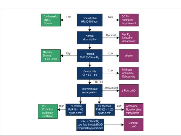
In addition to its contractility, optimization of right ventricular preload and afterload is crucial to prevent right ventricular failure in the perioperative period. CVP and systolic pulmonary pressure should be maintained lower than 16 mmHg and 65 mmHg , respectively. For maintenance of coronary perfusion, use of inotropes that cause pulmonary vasodilation (milrinone or dobutamine) and maintain adequate systemic pressure (adrenaline) is recommended. In addition, the use of specific pulmonary vasodilators, such as nitric oxide should be considered (Figure 2).30
Complications after long-term MCSD implantation
The main complications related to long-term MCSD
implantation are described in Chart 8.
Proposal of prioritization criteria for cardiac transplant in patients with MCSD
Figure 2 – Optimization and management of right ventricular function. MgSO4: magnesium sulfate; HR: heart rate; DC PM: dual-chamber pacemaker with right atrial
and ventricular stimulation and sensitivity; LVAD: Left ventricular assist device; CVP: central venous pressure; CI: cardiac index; TTE: transthoracic echocardiogram; TEE: transesophageal echocardiography; RV: right ventricular; PVR: pulmonary vascular resistance; LV: left ventricular; SVR: systemic vascular resistance; RVAD: right ventricular assist device; mAP: mean arterial pressure.
Cardioversion MgSO4
Digoxin
Fast
High
High
Slow
Abnormal
Low
Low
Low Leftward shift
Sinus rhythm HR 80‑100 bpm
Normal sinus rhythm
Preload CVP 10‑15 mmHg
Contractility CI > 2,0 – 4,0
Interventricular septal position
RV preload PVR 40 – 100
dynas.s.cm–5
LV afterload PVR 800 – 1200
dynas.s.cm–5
mAP < 50 mmHg Low flow through RVAD Peripheral hypoperfusion Diuresis
Dialysis ↓ Flow LVAD
iNO Protective pulmonary ventilation
DC PM Adrenaline Isoproterenol
MgSO4
Lidocaine Amiodarone
Volume
Milrinone Adrenaline Dobutamine
Adrenaline Noradrenaline Vasopressin ↓ Flow LVAD
Consider LVAD
TTE/TEE
Chart 7 – Contraindications for long-term mechanical circulatory support devices
Absolute
Coumarin intolerance Absence of trained caregivers
Severe psychiatric disorders or nonadherence to the staff instructions
Severe motor deficit or cognitive deficit related after stroke
Neoplastic disease with unfavorable prognosis
Vascular malformation of the small bowel that predisposes to bleeding Severe pulmonary obstructive disease
Severe hepatic dysfunction Active infection
Hematologic changes (platelets < 50,000 mm3 and thrombophilia)
Relative
Moderate‑to‑severe right ventricular dysfunction Dialytic therapy for renal failure
Difficult-to-control diabetes Partial motor deficit after stroke
Severe malnutrition
Chart 8 – Complications of long-term mechanical circulatory support devices (MCSDs)
Bleeding Pericardial effusion Respiratory insufficiency
Right ventricular dysfunction Hypertension Non‑neurological arterial thromboembolism Neurological events Arrhythmias Venous thromboembolism Infections Myocardial infarction Surgical wound dehiscence MCSD malfunction Hepatic dysfunction Psychiatric / behavioral change Hemolysis Renal dysfunction
Chart 9 – Proposal of prioritization criteria for cardiac transplant
Priority Criterium
1 Cardiogenic shock in patients using short/medium-term paracorporeal MCDS (including intra-aortic balloon) Long‑term MCDS with complications and substitution of device is not possible
2 Cardiogenic shock in patients using inotropes or vasopressors 3 Stable long‑term MCDS without complications
4 Outpatient management of advanced heart failure
MCDS: mechanical circulatory device support
1. Kirklin JK, Naftel DC, Kormos RL, Stevenson LW, Pagani FD, Miller MA, et
al. Third INTERMACS Annual Report: the evolution of destination therapy
in the United States. J Heart Lung Transplant. 2011;30(2):115‑23.
2. Moskowitz AJ, Rose EA, Gelijns AC. The cost of long‑term LVAD implantation.
Ann Thorac Surg. 2001;71(3 Suppl):S195-8.
3. Rose EA, Gelijns AC, Moskowitz AJ, Heitjan DF, Stevenson LW, Dembitsky
W, et al; Randomized Evaluation of Mechanical Assistance for the Treatment
of Congestive Heart Failure (REMATCH) Study Group. Long‑term use of
a left ventricular assist device for end-stage heart failure. N Engl J Med. 2001;345(20):1435-43.
4. Reedy JE, Swartz MT, Termuhlen DF, Pennington DG, McBride LR, Miller LW, et al. Bridge to heart transplantation: importance of patient selection. J
Heart Transplant. 1990;9(5):473-80.
5. Lietz K, Miller LW. Patient selection for left‑ventricular assist devices. Curr
Opin Cardiol. 2009;24(3):246-51.
6. Lietz K, Long JW, Kfoury AG, Slaughter MS, Silver MA, Milano CA, et al.
Outcomes of left ventricular assist device implantation as destination therapy in the post-REMATCH era: implications for patient selection. Circulation. 2007;116(5):497-505.
7. Cowger J, Sundareswaran K, Rogers JG, Park SJ, Pagani FD, Bhat G, et al. Predicting survival in patients receiving continuous flow left ventricular assist devices: the HeartMate II risk score. J Am Coll Cardiol. 2013;61(3):313-21.
8. Oz MC, Goldstein DJ, Pepino P, Weinberg AD, Thompson SM, Catanese KA, et al. Screening scale predicts patients successfully receiving long-term implantable left ventricular assist devices. Circulation. 1995;92(9 Suppl):II169-73.
9. Deng MC, Loebe M, El‑Banayosy A, Gronda E, Jansen PG, Vigano M, et al.
Mechanical circulatory support for advanced heart failure: effect of patient selection on outcome. Circulation. 2001;103(2):231-7.
10. Santamore WP, Gray LA Jr. Left ventricular contributions to right ventricular systolic function during LVAD support. Ann Thorac Surg. 1996;61(1):350‑6.
11. Loforte A, Stepanenko A, Potapov EV, Musumeci F, Dranishnikov N,
Schweiger M, et al. Temporary right ventricular mechanical support in
high-risk left ventricular assist device recipients versus permanent biventricular or total artificial heart support. Artif Organs. 2013;37(6):523-30.
12. Scalia GM, McCarthy PM, Savage RM, Smedira NG, Thomas JD. Clinical utility of echocardiography in the management of implantable ventricular assist devices. J Am Soc Echocardiogr. 2000;13(8):754-63.
13. Lang RM, Badano LP, Mor‑Avi V, Afilalo J, Armstrong A, Ernande L, et al.
Recommendations for cardiac chamber quantification by echocardiography in adults: an update from the American Society of Echocardiography and the European Association of Cardiovascular Imaging. Eur Heart J Cardiovasc Imaging. 2015;16(3):233-70.
14. Rudski LG, Lai WW, Afilalo J, Hua L, Handschumacher MD, Chandrasekaran
K, et al. Guidelines for the echocardiographic assessment of the right heart in adults: a report from the American Society of Echocardiography endorsed by the European Association of Echocardiography, a registered branch of the European Society of Cardiology, and the Canadian Society of Echocardiography. J Am Soc Echocardiogr. 2010;23(7):685-713.
15. Feldman D, Pamboukian SV, Teuteberg JJ, Birks E, Lietz K, Moore SA, et al; International Society for Heart and Lung Transplantation. The 2013 International Society for Heart and Lung Transplantation Guidelines for mechanical circulatory support: executive summary. J Heart Lung Transplant. 2013;32(2):157‑87.
16. Argiriou M, Kolokotron SM, Sakellaridis T, Argiriou O, Charitos C, Zarogoulidis P, et al. Right heart failure post left ventricular assist device implantation. J Thorac Dis. 2014 Mar;6 Suppl 1:S52-9.
17. Matthews JC, Koelling TM, Pagani FD, Aaronson KD. The right ventricular failure risk score a pre-operative tool for assessing the risk of right ventricular failure in left ventricular assist device candidates. J Am Coll Cardiol. 2008;51(22):2163-72.
18. Kormos RL, Teuteberg JJ, Pagani FD, Russell SD, John R, Miller LW, et al; HeartMate
II Clinical Investigators. Right ventricular failure in patients with the HeartMate II continuous-flow left ventricular assist device: incidence, risk factors, and effect on outcomes. J Thorac Cardiovasc Surg. 2010;139(5):1316-24.
19. Fitzpatrick JR 3rd, Frederick JR, Hsu VM, Kozin ED, O’Hara ML, Howell E,
et al. Risk score derived from pre-operative data analysis predicts the need
for biventricular mechanical circulatory support. J Heart Lung Transplant.
This is an open‑access article distributed under the terms of the Creative Commons Attribution License 20. Atluri P, Goldstone AB, Fairman AS, MacArthur JW, Shudo Y, Cohen JE, et
al. Predicting right ventricular failure in the modern, continuous flow left ventricular assist device era. Ann Thorac Surg. 2013;96(3):857-63.
21. Holman WL, Acharya D, Siric F, Loyaga‑Rendon RY. Assessment and
management of right ventricular failure in left ventricular assist device patients. Circ J. 2015;79(3):478-86.
22. Goldstein D, Neragi-Miandoab S. Mechanical bridge to decision: what are the options for the management of acute refractory cardiogenic shock? Curr Heart Fail Rep. 2011;8(1):51-8.
23. Kar B, Basra SS, Shah NR, Loyalka P. Percutaneous circulatory support
in cardiogenic shock: interventional bridge to recovery. Circulation. 2012;125(14):1809-17.
24. Gilotra NA, Stevens GR. Temporary mechanical circulatory support: a review of the options, indications, and outcomes. Clin Med Insights Cardiol. 2014;8(Suppl 1):75-85.
25. Thiele H, Lauer B, Hambrecht R, Boudriot E, Cohen HA, Schuler G. Reversal
of cardiogenic shock by percutaneous left atrial-to-femoral arterial bypass assistance. Circulation. 2001;104(24):2917-22.
26. Raess DH, Weber DM. Impella 2.5. J Cardiovasc Transl Res. 2009;2(2):168-72.
27. Rihal CS, Naidu SS, Givertz MM, Szeto WY, Burke JA, Kapur NK, et al. Society for Cardiovascular Angiography and Interventions (SCAI); Heart Failure Society of America (HFSA); Society of Thoracic Surgeons (STS); American Heart Association (AHA), and American College of Cardiology (ACC). 2015 SCAI/ ACC/HFSA/STS Clinical Expert Consensus Statement on the Use of Percutaneous Mechanical Circulatory Support Devices in Cardiovascular Care: Endorsed by the American Heart Association, the Cardiological Society of India, and Sociedad
Latino Americana de Cardiologia Intervencion; Affirmation of Value by the
Canadian Association of Interventional Cardiology-Association Canadienne de Cardiologie d’intervention. J Am Coll Cardiol. 2015;65(19):e7-e26.
28. Kirklin JK, Naftel DC, Pagani FD, Kormos RL, Stevenson LW, Blume ED, et al.
Seventh INTERMACS annual report: 15,000 patients and counting. J Heart
Lung Transplant. 2015;34(12):1495‑504.
29. Patlolla B, Beygui R, Haddad F. Right-ventricular failure following left ventricle assist device implantation. Curr Opin Cardiol. 2013;28(2):223-33.
30. Meineri M, Van Rensburg AE, Vegas A. Right ventricular failure after LVAD