470 Maciel R, Melo AC, Carvalho EB
J Bras Pneumol. 2005; 31(4):470-3
Yellow nail syndrome*
RENATO MACIEL1, ANDRÉIA CRISTINA DE MELO2, EMANUELLA BRAGA CARVALHO2
ABSTRACT
Yellow nail syndrome is a rare disorder characterized by three main features: discoloration of the nails, together with dystrophic alterations; lymphedema; and pleural effusion. It is often accompanied by bronchiectasis and chronic rhinosinusitis. Herein, we report a case of the complete syndrome with bilateral pleural effusion in a patient under treatment for pulmonary tuberculosis for nine months. There was a family history of two similar cases in siblings.
Keywords: Nail diseases; Lymphedema; Pleural effusion; Antitubercular; Syndrome; Case report
* Study carried out at the Governador Israel Pinheiro Hospital, Belo Horizonte, Minas Gerais, Brazil
1. Pulmonologist and Chief Resident in Clinical Medicine of the Governador Israel Pinheiro Hospital, Belo Horizonte, Minas Gerais, Brazil
2. Resident in Clinical Medicine of the Governador Israel Pinheiro Hospital, Belo Horizonte, Minas Gerais, Brazil Correspondence to: Renato Maciel. Rua Tomé de Souza, 300, apto 1.202, Belo Horizonte, MG CEP: 30140130. Phone.: 55 31 3223-0484. E-mail: [email protected]
Submitted: 17 February 2005. Accepted, after review: 15 April 2005.
INTRODUCTION
Yellow nail syndrome (YNS) is a rare disorder of unknown etiology and characterized by yellow nails, lymphedema and pleural effusion. It is often accompanied by other features, mainly bronchiectasis and chronic rhinosinusitis.
We report herein the case of a female patient presenting typical YNS and developing cavitary pulmonary tuberculosis. Through etiological investigation of the pleural effusion, pleural tuberculosis and chylothorax were ruled out.
CASE REPORT
In March of 2004, a 61-year-old female was hospitalized in the Governador Israel Pinheiro Hospital with a clinical profile that included dyspnea, fever and a considerable amount of yellowish expectoration, as well as postnasal drip.
Case Report
Physical examination revealed yellowing of the fingernails (Figure 1) and toenails, both of which also presented areas that were discretely darker, with longitudinal and transversal overcurvature, as well as loss of the cuticle and lunula. Onycholysis and the transverse ridging characteristic of hyperkeratosis were also seen. The patient presented a nonpitting edema of the lower limbs, more pronounced and with mild erythema in the left leg (Figure 2). Upon auscultation, diffuse ronchi and sparse wheezing, as well as crackles, were heard in the middle third of the right hemithorax. In the right lung base, crackles and decreased vocal fremitus were heard. The remaining systems presented no alterations.
J Bras Pneumol. 2005;31(4):470-3
Síndrome da unha amarela 4 7 1
with pulmonary tuberculosis. Treatment with rifampin was started. On the ninth day of medication, the patient developed drug-induced hepatitis, which was treated with isoniazid and ethambutol, and the end of the treatment was scheduled for July 2004. The patient reported recent ungual alterations and stated that she had had edema of the lower limbs since adolescence.
Regarding family history, the patient reported that one brother had presented clinical profile with ungual alterations, edema of lower limbs and pleural effusion, and that he had been submitted to multiple thoracenteses. She also reported that a sister had died from a disease similar to hers.
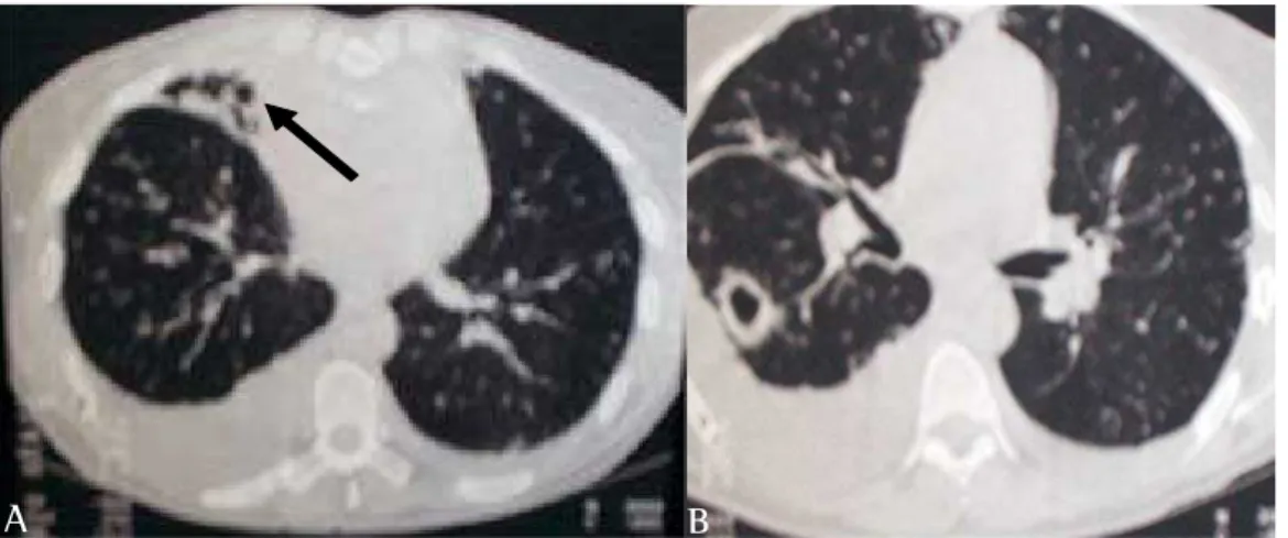
The patients herself provided an abdominal ultrasound, showing mild nonspecific hepatomegaly, bilateral pleural effusion and microlithiasis in the gallbladder, as well as a high-resolution computed tomography scan, revealing bilateral pleural effusion, bronchial cystic dilations in the middle lobe and evidence of cavitation in the apical segment of the lower right lobe (Figures 3a and 3b).
Due to the fever and abundant purulent expectoration in this patient, diagnosed (via tomography) with bronchiectasis of the middle lobe, treatment with intravenous ceftriaxone (2 g every 24 hours) was initiated, and complementary exams were requested, the results of which are described herein. The chest X-ray revealed bilateral pleural effusion, more pronounced on the right side. Blood tests were conducted. The blood count showed a leukocyte count of 6380 (N68.5 - L20.1 - M6.1 - E4.0 - B1.2), and hemoglobin was 12.8. The platelet count was 351,000, triglycerides were 100 mg/dL, total cholesterol was 158 mg/dL, HDL was 47 mg/dL, VLDL was 20 mg/dL, and LDL was 91 mg/dL. Prothrombin activity was 88.9%, RNI was 1.06, PCR was negative; albumin was 3.1 g/dL, and globulin was 3.8 g/dL. Three samples were collected for the blood culture, and there was no growth after seven days.
In order to thoroughly elucidate the etiological diagnosis of the pleural effusion, we opted for thoracentesis with pleural biopsy, from which the information described herein was gleaned. A pleural fluid sample was collected. After being centrifuged, the pleural fluid was yellowish and transparent, with hematogenous deposition. The cytometry showed 1100 cells, polymorphonuclear counts at 11%, mononuclear counts at 89% and innumerable erythrocytes, as well as, rarely, histiocytes and mesothelial cells. In the culture, there was no growth of germs after 72 hours. Gram staining revealed no bacteria, a few pus cells per field. The glucose level was 92 mg/dL, total cholesterol was 75 mg/dL, triglycerides were 28 mg/dL, LDH was 166 IU/L, total protein was 5.0 g/dL, and there were no residual lipoproteins. Cytological analysis of the pleural fluid revealed a dark-reddish liquid, and smear of hemorrhagic nature containing rare isolated mesothelial cells, by interposition, among various typical lymphocytes, and no malignant cells. The pleural biopsy revealed moderate mononuclear inflammatory infiltrate and dense fibrosis, permeated by cells of a fusiform pattern. Neither granulomas Figure 1 - Image showing yellow, dystrophic nails
472 Maciel R, Melo AC, Carvalho EB
J Bras Pneumol. 2005; 31(4):470-3 nor specific agents were identified.
These findings led us to the diagnosis of pleural exudate unrelated to tuberculosis, confirming the YNS etiology and providing no evidence of chylothorax.
Other complementary exams were carried out. A color Doppler echocardiogram revealed preserved global function of the left ventricle, an ejection fraction of 63%, a minor prolapse of the mitral valve (with minimal regurgitation), an absence of pericardial effusion, and mild dilation of the ascending aorta. Computed tomography of the sinuses revealed turbinate hypertrophy and opacity of the bilateral sinus, with an air-fluid level and opacification of the left ethmoid sinus.
During hospitalization, the patient was submitted to two thoracenteses, with an interval of eleven days between the two. During each procedure, 500 mL and 300 mL of xanthochromic fluid, respectively, were removed. The patient presented favorable evolution, with a decrease in the volume, and alteration in the color of, the expectoration, improvement of dyspnea, and no further bouts of fever. She was discharged on 03/29/04 and became an outpatient.
DISCUSSION
The YNS is more common among females (1:1.6) and occurs mainly in middle age, although it has been described in patients ranging from infancy to the eighth decade of life.(1) A family history of the disease is extremely rare.
The syndrome was initially described in 1964, in a study of thirteen patients with yellow nails and lymphedema.(2)
In 1966, three cases of individuals with lymphedema and pleural effusion were reported. Two of those patients presented yellow nails, and it was suggested that all these alterations could be due to inefficient lymphatic drainage, either of the pleural cavity or of the fingertips. In those three patients, the study of the pleural fluid revealed a high protein concentration, above 4.0 g/dL, and a predominance of lymphocytes.(3)
In 1969, bronchiectasis was suggested as the fourth component of YNS.(4)
A study involving 12 patients treated at the Mayo Clinic was published in 1972, in which various combinations of the syndrome were observed: 5 had the three findings, 4 presented lymphedema and pleural effusion, 1 had yellow nails and lymphedema, and 2 only presented ungual alterations.(5) The order of appearance of the several findings in the natural history of these patients varied, but pleural effusion was typically the last to appear. The time it takes the entire triad to appear may be prolonged, and the authors therefore included two cases that presented only ungual alterations. Both of those patients had bronchiectasis.
In a review carried out in 1986, it was reported that, to date, 62 cases of YNS had been described, and only 17 presented the complete triad of yellow nails, lymphedema and pleural effusion.(6)
The syndrome has been associated with respiratory diseases such as bronchiectasis(4-5) and
A B
Figure 3 - A) Images suggestive of cysts in the middle lobe with signs of bronchiectasis-related retraction (arrow) and bilateral pleural effusion, more pronounced on the right; B) Thick wall cavitation in the lower right lobe and bilateral pleural effusion.
J Bras Pneumol. 2005;31(4):470-3
Síndrome da unha amarela 4 7 3
chronic sinusitis.(5,7) In 12 cases described in one study, bronchiectasis was present in 5 and chronic sinusitis in 4.(5)
Ungual alterations seem to result from inefficient lymphatic drainage of the fingertips, leading to problems with the fingernails, including retarded growth, thickening, loss of the cuticle, loss of the lunula, and onycholysis of varying degrees.
The lymphedema may appear in the lower and upper limbs, is frequently asymmetric and is sometimes quite modest.
Pleural effusion is an exudate, characterized by a predominance of lymphocytes of different sizes. It may be unilateral or bilateral and is generally oligosymptomatic. After the relief drainage, fluid may re-accumulate within a few days or months. In case of early recurrence, it may be necessary to use pleurodesis (chemical or surgical).(8)
In 1990, the case of a 65-year-old male patient was reported, presenting the complete triad, with significant left-sided pleural effusion, who required multiple relief thoracenteses.(9) Because the patient refused to submit to pleurodesis, a pleuroperitoneal shunt was inserted, and there was a favorable response to treatment after one year. According to the author, this was the first time such procedure had been adopted.
Pleural effusion seems to be a late manifestation of the disease.(10) Cases of spontaneous regression of the effusion have been reported.(11-12)
According to some authors, the theory of lymphatic obstruction, in isolation, does not suffice to explain all the clinical manifestations of YNS and the high protein contents found in studies of the pleural fluid of these patients.(13) They suggest microangiopathy as the probable physiopathological mechanism, with increased microvascular permeability.
In the Brazilian literature, two cases of YNS have been described. Both present the components of the syndrome, the first one being associated with pericardial effusion(14) and the second accompanied by bronchiectasis and sinusitis.(15)
There are aspects of the case presented herein that are worth mentioning. The triad was complete and was accompanied by chronic sinusitis and bronchiectasis, confirmed by high-resolution computed tomography. In addition, in July of 2003, the patient presented pulmonary tuberculosis with evidence of cavitation confirmed by computed chest tomography and tested positive for acid-fast bacilli
in sputum. Furthermore, she had had a sister who died with a similar clinical profile and had a brother, still living, who presented the complete triad. Moreover, chylothorax was ruled out as the cause of the pleural effusion, and no pericardial or peritoneal effusion was found.
The YNS is a rare disorder whose treatment is basically symptom based. However, a diagnosis of YNS should be considered in patients with pleural effusion of unknown cause, especially when it involves bilateral effusion or chylothorax, or in patients with lymphedema.
REFERENCES
1. Nordkild P, Kromann-Andersen H, Struve-Christensen E. Yellow nail syndrome - the triad of yellow nails , lymphedema and pleural effusion. A review of the literature and a case report. Acta Med Scand. 1986;219(2):221-7. 2. Samman PD, White WF. The "yellow nail" syndrome. Br
J Dermatol. 1964;76:153-7.
3. Emerson PA. Yellow nails, lymphoedema and pleural effusions. Thorax. 1966;21(3):247-53.
4. Bowers D. Unequal breasts, yellow nails, bronchiectasis and lymphedema. Can Med Assoc J. 1969;100(9):437-8. 5. Hiller E, Rosenow EC 3rd, Olsen AM. Pulmonary
manifestations of the yellow nail syndrome. Chest. 1972;61(5):452-8.
6. Gupta AK, Davies GM, Haberman HF. Yellow nail syndrome. Cutis. 1986;37(5):371-4.
7. Varney VA , Cumberworth V, Sudderick R, Durham SR, Mackay IS. Rhinitis sinusitis and the yellow nail syndrome: a review of symptoms and response to treatment in 17 patients. Clin Otolaryngol Allied Sci. 1994;19(3):237-40. 8. Glazer M, Berkman N, Lafair JS, Kramer MR. Successful talc slurry pleurodesis in patients with nonmalignant pleural effusion. Chest. 2000;117(5):1404-9. 9. Brofman JD, Hall JB, Scott W, Little AG. Yellow nails,
lymphedema and pleural effusion. Treatment of chronic pleural effusion with pleuroperitoneal shunting. Chest. 1990;97(3):743-5.
1 0 . Christu AK, Pastaka C, Papadopoulos D, Klimi E, Gourgoulianis KI. Yellow nail syndrome or diffuse lymphatic network disease. Acta Medica (Hradec Kralove). 2002;45(4):181-2.
11. Riedel M. Multiple effusions and lymphedema in the yellow nail syndrome. Circulation. 2002;105(3):E25-6. 1 2 . Rigau NC, Daele JJ. The yellow nail syndrome. Acta
Otorhinolaryngol Belg. 2003;57(3):221-4.
1 3 . D'Alessandro A, Muzi G, Monaco A, Filiberto S, Barboni S, Abbritti G. Yellow nail syndrome: does protein leakage play a role? Eur Respir J. 2001;17(1):149-52. 14. Bessler R, Spector N. Síndrome da unha amarela. Arq
Bras Med. 1988;62(6):437-8.