an important role.(3) Most patients are under 40 years of age and were previously healthy. The male/female ratio is as high as 1:4 in some studies,(1) although other studies have shown little gender bias.(4)
The onset of the disease is acute or subacute, progressing over the course of two to three weeks.(5) The most common form of presentation is lymphadenopathy, primary cervical (74% to 90% of the cases) or supraclavicular. Adenopathies
Introduction
Kikuchi-Fujimoto disease (KFD), also known as Kikuchi’s disease or necrotizing granulomatous lymphadenitis, is rare, has a benign course and is of unknown cause. The disease was initially described in 1972, in Japan, in young females affected by fever and cervical lymphadenopathy.(2) Although its pathogenesis remains little understood, it is thought to be a hyperimmune reaction induced by different antigenic stimuli or an autoimmune process in which apoptosis play
Rogério Gastal Xavier1, Denise Rossato Silva2, Mauro Waldemar Keiserman3, Maria Francisca Torres Lopes4
Abstract
Kikuchi-Fujimoto disease is characterized by fever and lymphadenopathy, usually localized in the cervical region. This disease princi-pally affects young females. It can be confused with lymphoma, adenocarcinoma metastasis and tuberculosis. We report two cases of Kikuchi- Fujimoto disease. In the first case, a 28-year-old female had been treated for tuberculosis one year prior and presented with a clinical and histological profile consistent with Kikuchi-Fujimoto disease. The second patient, a 58-year-old female, initially received treat-ment for Wegener’s granulomatosis and, subsequently, for tuberculosis. Histopathological examination followed by immunohistochemical analysis confirmed the diagnosis of Kikuchi-Fujimoto disease in both cases. After the definitive diagnosis had been made, both patients were treated symptomatically, and both presented clinical improvement within one month. Subsequently, the latter patient developed systemic lupus erythematosus.
Keywords: Fever; Lymph nodes; Rare diseases; Tuberculosis, lymph node; Histiocytic necrotizing lymphadenitis; Lupus erythematosus, systemic.
Resumo
A doença de Kikuchi-Fujimoto é caracterizada por febre e linfadenopatia, geralmente cervical. Esta doença acomete principalmente mulheres jovens. Pode ser confundida com linfoma, metástase de adenocarcinoma e tuberculose. Relatamos dois casos da doença de Kikuchi- Fujimoto. No primeiro caso, uma paciente de 28 anos havia tratado tuberculose há um ano e apresentava quadro clínico e histológico compatível com a doença de Kikuchi-Fujimoto. A segunda paciente, de 58 anos, recebeu tratamento inicialmente para granulomatose de Wegener e, posteriormente, para tuberculose. O exame histopatológico com estudo imunohistoquímico permitiu estabelecer o diagnóstico da doença de Kikuchi-Fujimoto nos dois casos. Após o diagnóstico definitivo, ambas foram tratadas sintomaticamente e melhoraram clinicamente dentro de um mês. Posteriormente, a segunda paciente desenvolveu lúpus eritematoso sistêmico.
Descritores: Febre; Linfonodos; Doenças raras; Tuberculose dos linfonodos; Linfadenite histiocítica necrosante; Lúpus eritematoso sistêmico.
* Study carried out in the Department of Internal Medicine, Universidade Federal do Rio Grande do Sul – UFRGS, Federal University of Rio Grande do Sul – School of Medicine, Porto Alegre, Brazil.
1. Associate Professor. Department of Internal Medicine, Universidade Federal do Rio Grande do Sul – UFRGS, Federal University of Rio Grande do Sul – School of Medicine, Porto Alegre, Brazil.
2. Pulmonologist. Universidade Federal do Rio Grande do Sul – UFRGS, Federal University of Rio Grande do Sul – Porto Alegre, Brazil.
3. Professor. Department of Rheumatology, São Lucas Hospital, Pontifícia Universidade Católica do Rio Grande do Sul – PUC-RS, Pontifical Catholic University of Rio Grande do Sul – Porto Alegre, Brazil.
4. Pathologist. Anapat Pathology Laboratory, Moinhos de Vento Hospital, Porto Alegre, Brazil.
Correspondence to: Rogério Gastal Xavier. Avenida Soledade, 478, CEP 90470-340, Porto Alegre, RS, Brasil. Tel 55 51 3331-8664. E-mail: [email protected]
Financial support: None.
tuberculosis, treatment with the RHZ regimen was resumed, and, after the onset of skin rash, pyrazina-mide was replaced with ethambutol. The treatment was discontinued 30 days later due to the lymph node biopsy results: there was a xanthogranuloma-tous and necrotizing inflammatory process, without granuloma formation and with an extensive popu-lation of (CD68+) xanthomatous histiocytes; as well as negativity for AFB and fungi. The findings were not suggestive of tuberculosis, and, therefore, the possibility of KFD was considered. The patient was treated symptomatically, and there was improve-ment of arthralgia, together with resolution of the fever, within 30 days. After 18 months, the patient no longer presented clinical alterations or ANF.
Case 2
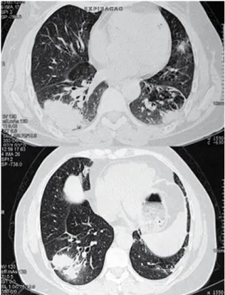
A 58-year-old female nonsmoker reported cough, moderate weight loss, fatigue and joint pain. The patient had a history of childhood asthma. After age 40, she experienced recurrence of the asthma—a fact that she attributed to contact with paints and solvents in the workplace. The patient used inhaled typically measure less than 3 cm in diameter, present
a firm consistency and are sometimes painful upon palpation. Generalized lymphadenopathy occurs in a minority (5%) of cases. Although extranodal involvement is rare, it has been described in the kidneys, liver, gastrointestinal tract, thyroid, parath-yroid glands, adrenal glands and bone marrow.(6) The diagnosis of KFD is made by lymph node biopsy, which should be performed to rule out more severe conditions with which KFD can be confused, such as lymphoma, tuberculosis and Kawasaki disease.(1)
Various case reports have been published in the international literature,(7-12) and some presentations on the subject have been made at national confer-ences.(13,14) However, there have been no published reports of cases of KFD in Brazil.
Case reports
Case 1
A 28-year-old female smoker with a history of tuberculosis treatment for one year sought medical treatment due to mild constitutional symptoms and pleuritic pain in the left hemithorax. A chest X-ray and a tomography scan of the chest revealed bilateral consolidations, with posterior cavitation of the consolidation in the posterior segment of the left upper lobe (LUL). Sputum smear microscopy results were negative. Bronchoscopic examina-tion demonstrated that the LUL bronchial mucosa was hyperemic and edematous, consistent with an inflammatory process. In the bronchoalveolar lavage fluid, acid-fast bacilli (AFB) staining was nega-tive, and culture was positive for Mycobacterium tuberculosis (although only two colonies were found to grow). The patient received a regimen of rifampin, isoniazid and pyrazinamide (RHZ) for 6 months, and presented clinical and radiological improvement. One year later, the patient again presented with fever (for three weeks), arthralgia and submandibular lymph node enlargement. Laboratory tests revealed the following: hematocrit, 38.7%; hemoglobin, 13.1 g/ dL; leukocytes, 2,180 (with 569 lymphocytes); erythrocyte sedimentation rate (ESR), 27 mm/h; antinuclear factor (ANF), present (1:320), with a homogeneous nuclear pattern; anti-DNA, nonreactive; and rheumatoid factor, nonreactive. A chest X-ray revealed alterations with a residual aspect in the LUL. Due to the history of
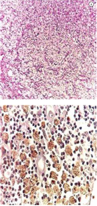
the following: hematocrit, 41,1%; hemoglobin, 13.7 g/dL; leukocytes, 8,970, with lympho-penia (1,076 lymphocytes); ANF, present (1:160), with a dotted nuclear pattern; ESR, 56 mm/h; and C-reactive protein, 27.60 mg/L. At the time, Wegener’s granulomatosis was suspected due to antineutrophil cytoplasmic antibody (ANCA) serum positivity. There was no upper airway involvement. Cyclophosphamide treatment was started. In the first month, there was an increase in the left anterior cervical ganglion. The lymph node biopsy identified chronic granulomatous lymphadenitis with exten-sive noncaseating necrosis (Figure 2). Testing for AFB was negative. Since tuberculosis was suspected, the patient received the RHZ regimen for 6 months, presenting clinical involvement. However, in the following two years, the patient continued to expe-rience fatigue, joint pain and well-defined episodes of pneumonia, as well as, during the final 6 months, oral lesion at the base of the tongue. She used systemic corticosteroids and antibiotics temporarily (5 to 6 times, for 10 to 14 days). With the worsening of the symptoms, the patient again sought treat-ment. Laboratory tests showed the following: ANF, 1:80 (homogeneous nuclear pattern); anti-DNA, nonreactive; lymphopenia; and ESR, 65 mm/h. A review of the anatomopathological diagnosis confirmed chronic granulomatous lymphadenitis with necrosis, numerous vacuolated histiocytes and giant Langerhans-type cells, as well as negativity for AFB and fungi. In addition, an immunohistochem-ical panel was performed, revealing CD68 (KP-1) positivity (xanthomatous histiocytes) and myeloper-oxidase negativity, which is suggestive of KFD. The patient was then diagnosed as presenting a combi-nation of KFD (which had gone undiagnosed for two years) and systemic lupus erythematosus (SLE). Her condition was controlled by the addition of oral azathioprine (100 mg/day) and prednisone (40 mg/day), the latter being tapered to discontinu-ation. Within 30 days, the patient presented clinical improvement, as well as improved laboratory test and tomography results.
Discussion
Here, we have described two cases of KFD. The actual incidence of this disease is estimated to range from 0.5% to 5% of all cases of aden-opathy analyzed histologically.(15) In a retrospective formoterol and budesonide (12/400 µg) daily,
achieving satisfactory symptom control. A chest X-ray and a tomography scan of the chest showed bilateral consolidations and bronchiectasis in the lower lobes (Figure 1). Laboratory tests revealed
a
b
vasculitis and Kawasaki disease.(19) Due to multisys-temic involvement and spontaneous resolution of lymphadenopathy, sarcoidosis can also be included in the differential diagnosis.(19)
One group of authors described the case of a patient with KFD and concomitant pulmonary tuberculosis.(20) Cases have also been reported in India. However, in countries with a high prevalence of tuberculosis, the concomitant diagnosis of tuber-culosis should be well investigated.(1,16)
Histologically, KFD is characterized by ganglia with foci of paracortical necrosis surrounded by aggregates of histiocytes.(6) Three histological profiles, which might correspond to evolutional or sequential phases, to different etiologic agents or even to differences in the inflammatory responses of patients, have been described: proliferative, necrotizing and xanthomatous.(18)
One group of authors described 25 cases of KFD. In that study, two patients had undergone chemo-therapy for the treatment of lymphoma, and two patients were treated with antituberculous drugs prior to the receipt of the biopsy results.(3) In the first case reported here, the patient received treatment for Wegener’s granulomatosis and for tuberculosis before the final diagnosis was made. Since empir-ical treatment is potentially toxic, it is important to consider KFD in the differential diagnosis of cervical lymphadenopathy in young females.
There is no effective treatment. The signs and symptoms usually resolve within 4 months.(5) Patients with more severe or persistent symptoms have been treated with corticosteroids. Long-term follow-up evaluation should be performed due to disease recurrence (in up to 3% of the cases) and to the possibility of SLE development.(1,18) Our second patient developed SLE during a two-year follow-up period.
The presence of fever and cervical lymphaden-opathy, especially in young females, should raise the suspicion of KFD.
References
1. Richards MJ. Kikuchi’s disease. UpToDate [cited 2007 Aug, version 15.3]. Available from: http://www.uptodate.com/ patients/content/topic.do;jsessionid=C5D9967BC119B2367 198967022B688F4.1102?topicKey=~S7j/7LBd.4veB5o&sele ctedTitle=4~6&source=search_result
2. Cho KJ, Lee SS, Khang SK. Histiocytic necrotizing lymphadenitis. A clinico-pathologic study of 45 cases with study of 1,724 lymph node biopsy samples, KFD
was found in 36 cases.(16) Various infectious agents, such as Epstein-Barr virus, herpes virus 6, herpes virus 8, parvovirus B19, parainfluenza virus, Yersinia spp. and toxoplasma, have been proposed as antigenic stimuli.(1) In our report, both patients presented bilateral consolidations, consistent with an infectious or inflammatory process. Although no infectious germs were identified, it is possible that this was the antigenic stimulus for the develop-ment of KFD.
Typical cases usually occur in young females, as was the case of the first patient (28 years old). However, patient ages range from 6 to 80 years.(1)
Fever is the initial symptom in 30% to 50% of patients. Symptoms such as fatigue, joint pain, skin rash, arthritis and hepatosplenomegaly are also found. Leukopenia, increased ESR and anemia are some of the laboratory findings reported. Systemic symptoms are more prominent in cases of extranodal involvement.(1) The first patient developed skin rash, which was initially attributed to an adverse effect of the antituberculous drugs, although one cannot rule out the possibility that it was secondary to KFD.
There are some characteristics, such as gender and age, as well as histological alterations, that KFD and SLE have in common, some authors consid-ering the former to be a lupus-like syndrome.(1) It has been proposed that KFD is a manifestation of, or evolves to, SLE,(6) justifying the surveillance of KFD patients using immunological studies such as ANF testing. Patients can present positivity for ANF, anti-ribonucleoprotein, anti-DNA and lupus anticoagulant. (17) Our two patients presented ANF. The second patient presented ANCA positivity at the onset of the condition. We found no descriptions of cases of KFD with ANCA positivity. However, it is known that these antibodies can be present in various autoimmune diseases, such as SLE, and the second patient described here subsequently devel-oped SLE. Other autoimmune conditions, such as antiphospholipid antibody syndrome, polymyositis, Still’s disease, uveitis, scleroderma, thyroiditis, pulmonary hemorrhage and interstitial lung disease, can be associated with KFD.(5,17)
um caso. In: XXIV Congresso Brasileiro de Reumatologia; 2002 Sep 15-20; Goiânia, Brazil: Academia Brasileira de Reumatologia; p. S68.
13. Oliveira RCV, Muglia VF, Bellucci AD, Louzada P, Santos AC, Elias Jr J, et al. Doença de Kikuchi-Fujimoto: uma causa rara de síndrome do lobo médio. In: 33rd Jornada Paulista de Radiologia; 2003 May 1-4, São Paulo, Brazil: Sociedade Paulista de Radiologia e Diagnóstico por Imagem; 2003. 14. Parappil A, Rifaath AA, Doi SA, Pathan E, Surrun SK. Pyrexia
of unknown origin: Kikuchi-Fujimoto disease. Clin Infect Dis. 2004;39(1):138-43.
15. Infante MJ, Lovillo C, Santaella IO, Checa RM, González MR. Enfermedad de Kikuchi-Fujimoto como causa de linfadenopatías. An Pediatr (Barc). 2007;67(1):83-5. 16. Mohan A, Reddy MK, Phaneendra BV, Chandra A. Aetiology
of peripheral lymphadenopathy in adults: analysis of 1724 cases seen at a tertiary care teaching hospital in southern India. Natl Med J India. 2007;20(2):78-80.
17. Reichert A, Correia T, Freitas O, Almeida T, Rosado L. Doença de Kikuchi - Fujimoto. Acta Med Port. 2005;18(3):231-4. 18. Veja GE, Martín CV, Ruiz-Capillas JJ, Rojas MG, Calvet CL,
Lázaro FB, et al. Linfadenitis histiocítica necrotizante de Kikuchi. Rev Clin Esp. 2003;203(7):343-5.
19. Sharma OP. Unusual systemic disorders associated with interstitial lung disease. Curr Opin Pulm Med. 2001;7(5):291-4.
20. Malagón MJ, García JL, Cansino MD, Jiménez FR. Enfermedad de Kikuchi-Fujimoto y tuberculosis pulmonar, una asociación poco frecuente. Rev Clin Esp. 2003;203(10):507-11. in situ hybridization for Epstein-Barr virus and hepatitis B
virus. J Korean Med Sci. 1996;11(5):409-14.
3. Menasce LP, Banerjee SS, Edmondson D, Harris M. Histiocytic necrotizing lymphadenitis (Kikuchi-Fujimoto disease): continuing diagnostic difficulties. Histopathology. 1998;33(3):248-54.
4. Bosch X, Guilabert A. Kikuchi-Fujimoto disease. Orphanet J Rare Dis. 2006;1:18.
5. Bosch X, Guilabert A, Miquel R, Campo E. Enigmatic Kikuchi-Fujimoto disease: a comprehensive review. Am J Clin Pathol. 2004;122(1):141-52.
6. Calvo Romero JM. Enfermedad de Kikuchi-Fujimoto (linfadenitis necrotizante histiocitaria). Rev Clin Esp. 2002;202(2):94-95.
7. Famularo G, Giustiniani MC, Marasco A, Minisola G, Nicotra GC, De Simone C. Kikuchi Fujimoto lymphadenitis: case report and literature review. Am J Hematol. 2003;74(1):60-3. 8. Bennie MJ, Bowles KM, Rankin SC. Necrotizing cervical
lymphadenopathy caused by Kikuchi-Fujimoto disease. Br J Radiol. 2003;76(909):656-8.
9. Mahajan T, Merriman RC, Stone MJ. Kikuchi-Fujimoto disease (histiocytic necrotizing lymphadenitis): report of a case with other autoimmune manifestations. Proc (Bayl Univ Med Cent). 2007;20(2):149-51.
10. Rodríguez Martorell J, Martín MV, Báez JM, Gil JL. Kikuchi-Fujimoto necrotizing lymphadenitis associated with brucellosis [Article in Spanish]. Sangre (Barc). 1992;37(3):201-4. 11. Kucukardali Y, Solmazgul E, Kunter E, Oncul O, Yildirim S,
Kaplan M. Kikuchi-Fujimoto Disease: analysis of 244 cases. Clin Rheumatol. 2007;26(1):50-4.