Belén RETAMAL-VALDES(a) Geisla Mary SOARES(a) Bernal STEWART(b)
Luciene Cristina FIGUEIREDO(a) Marcelo FAVERI(a)
Steven MILLER(b) Yun Po ZHANG(b) Magda FERES(a)
(a) Universidade de Guarulhos, Department of Periodontology, Dental Research Division, Guarulhos, São Paulo, Brazil.
(b) Colgate-Palmolive Company, Piscataway, NJ, USA.
Effectiveness of a pre-procedural
mouthwash in reducing bacteria in
dental aerosols: randomized clinical trial
Abstract: The aim of this randomized, single blinded clinical trial was to evaluate the effect of a pre-procedural mouthwash containing
cetylpyridinium chloride (CPC), zinc lactate (Zn) and sodium luoride (F)
in the reduction of viable bacteria in oral aerosol after a dental prophylaxis with ultrasonic scaler. Sixty systemically healthy volunteers receiving dental prophylaxis were randomly assigned to one of the following experimental groups (15 per group): (i) rinsing with 0.075% CPC, 0.28%
Zn and 0.05% F (CPC+Zn+F), (ii) water or (iii) 0.12% chlorhexidine
digluconate (CHX), and (iv) no rinsing. Viable bacteria were collected
from different locations in the dental ofice on enriched TSA plates and
anaerobically incubated for 72 hours. The colonies were counted and
species were then identiied by Checkerboard DNA–DNA Hybridization. The total number of colony-forming units (CFUs) detected in the aerosols from volunteers who rinsed with CPC+Zn+F or CHX was statistically signiicantly (p<0.05) lower than of those subjects who did not rinse or
who rinsed with water. When all locations were considered together, the
aerosols from the CPC+Zn+F and CHX groups showed, respectively, 70% and 77% fewer CFUs than those from the No Rinsing group and 61% and
70% than those from the Water group. The mean proportions of bacterial
species from the orange complex were statistically signiicantly (p<0.05) lower in aerosols from the CPC+Zn+F and CHX groups compared
with the others two groups. In conclusion, the mouthwash containing
CPC+Zn+F, is effective in reducing viable bacteria in oral aerosol after a
dental prophylaxis with ultrasonic scaler.
Keywords: Cetylpyridinium; Chlorhexidine; Aerosols; Mouthwashes; Microbiology.
Introduction
The propagation of oral microorganisms in the dental ofice during
different oral procedures has been a concern. The use of certain equipment such as ultrasonic devices,1,2,3,4,5 highspeed dental handpieces6,7 or three-way
syringes8 may spread aerosols and splatters containing microorganisms in
the environment. These microorganisms may cause cross-infections in the
dental ofice, jeopardizing the health of patients and dental professionals.9
Different procedures, materials and antimicrobial agents have been proposed to minimize microbial cross-contamination in the dental ofice,
Declaration of Interests: Bernal Stewart, Steven Miller and Yun Po Zhang are currently employed by Colgate Palmolive Company. The other authors certify that they have no commercial or associative interest that represents a conflict of interest in connection with the manuscript.
Corresponding Author: Magda Feres
E-mail: [email protected]
https://doi.org/10.1590/1807-3107BOR-2017.vol31.0021
Submitted: Sep 26,2016
such as immunization of dental staff, decontamination of surfaces, sterilization of instruments, use of personal protective barriers and pre-procedural mouthwashes.9,10,11,12 Chlorhexidine (CHX) is considered
the gold standard substance in controlling oral bioilm
growth in the oral cavity or microbial spread by oral aerosols3,4,9,13,14,15 due to its broad antibacterial
spectrum14,16 and substantivity of 8 to 12 hours.14
However, other antiseptics have also been used as pre-procedural mouthwashes, such as essential oils17
and cetylpyridinium chloride (CPC).3,15 CPC has an
important antimicrobial activity18,19 and is considered
a safe product for marketing.20
The authors of this study have previously demonstrated the effectiveness of a pre-procedural mouthwash containing CPC in reducing viable bacteria in dental aerosol after a prophylaxis with an ultrasonic scaler. This study introduced CPC as a good alternative to CHX, due to its effective antibacterial action, fewer adverse effects and lower cost.3 In addition, a recent clinical study has indicated
that a higher concentration of CPC – 0.07%, is very
effective in controlling plaque accumulation and gingivitis;18 however, to date no studies have evaluated
the effectiveness of CPC at this higher concentration as a pre-procedural mouthwash.
The present trend has been to combine more than one active substance in mouthwash and toothpaste
formulations with the aim of increasing the eficacy
of the products, or treating more than one clinical problem, such as plaque accumulation and halitosis;21,22
caries23 or gingival inlammation.24 Following this
line of thought, zinc has been added to some oral hygiene products due to its good antimicrobial
and anti-inlammatory properties25 and its capacity
of neutralizing volatile compounds that play an important role in halitosis.21,26 Similarly, fluoride has been added to mouthwashes formulations due to its effect in caries prevention23 and in dental
remineralization.27 Based on this body of evidence,
the aim of this study was to evaluate the effectiveness of a mouthwash formulation containing 0.075%
CPC, 0.28% zinc lactate and 0.05% sodium luoride (CPC+Zn+F), in reducing viable bacteria present in
oral aerosol/splatter.
Methodology
Sample size calculation
The ideal sample size to assure adequate power for this study was calculated based on the data of
Feres et al.3 Considering a difference of at least 682
CFUs between groups CPC+Zn+F and Water and assuming a standard deviation of 435, it was deined that 15 subjects per group is adequate to provide a
95% power with an α of 0.05.
Subject population
Systemically healthy volunteers were selected from the population referred to the Center for Clinical
Trials at Guarulhos University (Guarulhos, SP, Brazil).
The medical and dental histories were obtained and a full-mouth periodontal examination was performed. The study protocol was explained to each volunteer and those who met the inclusion/exclusion criteria,
signed the term of Free and Informed Consent, illed
out a health questionnaire, and were enrolled in the study. The study protocol was previously approved
by the Guarulhos University Ethics Committee on Clinical Research (CAAE: 41244315.4.0000.5506). The ClinicalTrials.gov identiier of the present study is NCT02875769.
Inclusion criteria
The inclusion criteria were as follows: male or
female aged between 18–70 years, minimum of
20 natural teeth, at least 80% of the sites with visible supragingival plaque, fewer than 10% of sites with visible supragingival calculus, fewer than 30% of
sites with probing depth (PD) ≥ 5 mm.
Exclusion criteria
The exclusion criteria were as follows: presence of orthodontic bands; partial removable dentures; lesions of the soft or hard tissues of the oral cavity; carious lesions requiring immediate restorative treatment; history of allergy to CHX, CPC, zinc lactate or sodium
luoride; participation in any other clinical study
women; antibiotic therapy in the previous 6 months;
continuous use of oral mouthwashes and any systemic condition that may require prophylactic medication for dental treatment (e.g., mitral valve prolapse).
Clinical assessment
In order to select the volunteers for the study, one trained periodontist (G.M.S.) performed a clinical examination that included an oral tissue evaluation and periodontal assessment. The oral examination included an evaluation of the soft and hard palate; gingival mucosa; buccal mucosa; mucogingival fold areas; tongue, sublingual and submandibular areas; salivary glands, and the tonsilar and pharyngeal areas.
Visible plaque (0/1) and PD (mm) were measured at
six sites per tooth (mesiobuccal, buccal, distobuccal, mesiolingual, lingual and distolingual) in all teeth,
excluding third molars. The PD was recorded to the nearest millimeter using a North Carolina periodontal probe (Hu-Friedy, Chicago, IL, USA). The volunteers
have refrained from any oral hygiene procedure for twelve hours prior to the clinical examination, and
from eating, drinking or smoking for four hours
prior to the examination.
Experimental design, randomization, allocation concealment and treatment protocol
In this single blinded randomized clinical
trial (RCT), using simple randomization and a 1:1 allocation ratio, the study coordinator (L.C.F.) used
a computer-generated table to randomly assign the volunteers to the following groups (n = 15 per group):
1- CPC+Zn+F (test): mouthwash containing 0.075% CPC, 0.28% zinc lactate and 0.05% sodium luoride in an Alcohol-free base; 2- Water (negative control):
water from a three-way syringe; 3- CHX (positive
control): 0.12% CHX with 10% alcohol; 4- No Rinsing
(negative control): standard of care - no rinsing.
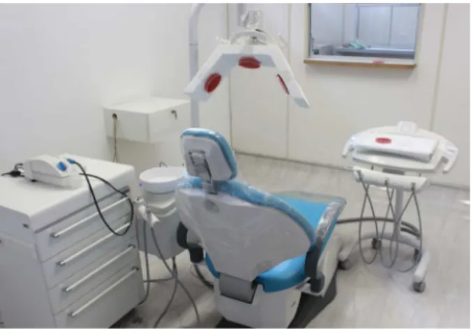
In an unoccupied dental ofice, previously cleaned
with detergent, alcohol and sodium hypochlorite, in which no regular patients had been treated on that
day, the study coordinator placed ive plates containing Tryptic Soy Agar with Yeast Extract enriched with 5% menadione, 5% sheep blood, and 1% N-Acetylmuramic acid (HNK plates) to sample microorganisms contained
in the unit atmosphere. Three plates were placed on
a support board attached to the relector, which had
been previously designed3 for this type of experimental
design, one on the bracket tray, and another on the ofice bench (Figure 1). The room was locked and after
30 minutes, the plates were closed and labeled properly and sent to the laboratory for incubation. Subsequently, the volunteers were invited to sit on the dental chair to
proceed with the irst phase of the study. A researcher,
who did not participate in any of the other phases
of the study (B.R.V.), checked the randomization list containing the names of the Water and No Rinsing groups, and codes for CPC+Zn+F and CHX groups, and
selected the solutions for the volunteers from groups
CPC+Zn+F, CHX and Water. CPC+Zn+F and CHX
mouthwashes were placed in identical opaque coded
lasks, which assured the allocation concelament for these two groups, but not for the Water and No Rinsing groups. The researcher entered the dental ofice and
handled one of three solutions to the volunteer, who rinsed for one minute with 20 ml of the solution and then expectorated the remaining liquid. The clinician (G.M.S.) did not participate on this phase of the study,
and only entered the ofice after the rinsing phase
had been completed. The volunteers were instructed to not mention to the clinician whether or not they had rinsed with a given solution. These procedures
assured the masking of the clinician (G.M.S.) and the
allocation concealment of the study.
After this phase, one volunteer at a time was reclined to a supine position and three new HNK
agar plates were placed on the support board, one on the volunteer’s chest (immediately in front of the volunteer’s mouth) and one on the clinician’s forehead. Subsequently, the study coordinator opened the plates and the clinician proceeded with the full mouth dental prophylaxis using an ultrasonic scaler
at a frequency of 25 kHz (Cavitron Select, Dentsply, York, Pa) at less than 50% power for 10 minutes. After the oral prophylaxis was completed, the ive HNK
agar plates were covered, labeled and sent to the laboratory for incubation. Volunteers were dismissed
from the dental room. The dental ofice was then
cleaned and disinfected with detergent, alcohol
and sodium hypochlorite and the next subject was
Microbiological procedures
a. Microbial quantiication: the HNK plates were incubated under anaerobic condition in jars
containing 10% CO2 at 37°C for 72 hours under
anaerobic conditions, using the BD GasPak EZ system (Becton Dickinson, Sparks, MD). Using a Lab Line Colony Counter (Phoenix model CP 600 N 302), a laboratory technician counted the colony-forming units (CFUs). For this purpose, the technician was calibrated using the kappa
test and a value of 90% was obtained and accepted. The technician was not aware of which plates were exposed to each treatment.
b. Microbial identiication: after counting, the
colonies were washed off the agar plates. The
technician added 1 ml of TE buffer (pH = 7.6)
to the plates and the bacterial colonies were
scraped off the surfaces using L-shaped
Pasteur pipettes. The suspensions were placed
in individual Eppendorf tubes and sonicated for 10 seconds (Analyzer model SP 2000 UV) and the optical density (OD) at 700 nm of each suspension was adjusted to a inal value of 1.0,
corresponding to approximately 109 bacterial
cells/ml. After this, 10 ul of each suspension was removed and placed in another Eppendorf tube and 140 ul of TE buffer and 100 ul of 0,5 M NaOH
were added. The samples were analyzed for 40
oral bacterial species using the Checkerboard DNA-DNA Hybridization technique.28,29
c. Checkerboard DNA-DNA hybridization: the
samples were boiled for 10 min. and neutralized using 0.8 ml of 5 M ammonium acetate. The
released DNA was then placed into the extended
slots of a Minislot 30 apparatus (Immunetics,
Cambridge, MA, USA), concentrated on a
15 x 15 cm positively charged nylon membrane
(Boehringer Mannheim, Indianapolis, IN, USA) and ixed to the membrane by baking it at
120°C for 20 min. The membrane was placed in a Miniblotter 45 (Immunetics, Cambridge,
MA, USA) with the lanes of DNA at 90° to the lanes of the device. Digoxigenin-labelled whole genomic DNA probes for 40 bacterial
species were hybridized in individual lanes
of the Miniblotter. After hybridization, the
membranes were washed at high stringency
and the DNA probes were detected using the antibody to digoxigenin conjugated with alkaline phosphatase and chemiluminescence detection using the scanner G:Box iChemi XL (Syngene/ USA). The last two lanes in each run
contained standards at concentrations of 105 and
106 cells of each species and the signals were converted to absolute counts by comparing them with the controls in each membrane. The
sensitivity of the assay was adjusted to allow
detection of 104 cells of each bacterial species.
Primary and secondary outcome variables
The primary outcome variable was the difference
between groups for the mean number of total CFUs in
all plates (all locations). Secondary outcome variables were differences among groups for the following
parameters: mean number of total CFUs in plates
positioned on the clinician forehead, volunteer’s chest and support board; mean proportion of the microbial complexes, as well as the mean percent
reduction in CFUs in the CPC+Zn+F and CHX groups in comparison with the Water and No Rinsing groups.
Statistical analysis
The total number of CFUs in the plates placed
on the support board, clinician and volunteer, as well as all locations grouped were computed for each volunteer and then averaged in each group. Figure 1. Microbial sampling technique used to evaluate
For the support board, the mean number of CFUs
of the three plates per volunteer was computed.
For the Checkerboard DNA-DNA hybridization
analysis, the mean levels of each bacterial species was averaged within each participant and then across participants in each of the four groups.
After this, the percentage of the total DNA probe
counts (proportions) was determined per volunteer, and averaged across volunteers within each group. In addition, the proportions of the species belonging to the six complexes and the “other”
group deined by Socransky et al.30 were added to
generate the proportion of each complex per group.
The signiicance of differences between groups for
the microbial data and for age were determined by
Kruskall-Wallis and Dunn tests. Chi-square test was
used to compare the differences in the frequency of
gender and smoking status. The level of signiicance
was set at 5%.
Results
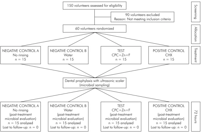
This RCT was conducted between February and April of 2015. Figure 2 presents the low diagram of
the study. Sixty volunteers participated in the study. Table 1 presents the demographic characteristics of the volunteers according to the different experimental
groups. No statistically signiicant differences were
observed among the four groups at the time of data
collection. No volunteer reported any adverse events
associated with the use of the mouthwashes evaluated.
No bacterial growth was detected in the agar plates
located in the dental office before the experiment
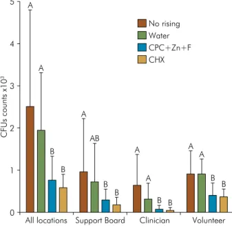
started (environmental contamination). The mean CFUs
in different locations according to the experimental
groups are presented in Figure 3. The aerosol/splatter from volunteers that rinsed with CPC+Zn+F or CHX harbored statistically signiicantly (p < 0.05) lower CFUs
counts than those who rinsed with water or who did
CPC: cetylpyridinium chloride; Zn: zinc lactate; F: sodium fluoride; CHX: chlorhexidine digluconate.
Figure 2. Flow chart of the study design.
150 volunteers assessed for eligibility
60 volunteers randomized
NEGATIVE CONTROL A No rinsing
n = 15
NEGATIVE CONTROL B Water n = 15
TEST CPC+Zn+F
n = 15
POSITIVE CONTROL CHX n = 15
NEGATIVE CONTROL A No rinsing (post-treatment microbial evaluation)
n = 15 analyzed Lost to follow-up: n = 0
NEGATIVE CONTROL B Water (post-treatment microbial evaluation)
n = 15 analyzed Lost to follow-up: n = 0
TEST CPC+Zn+F (post-treatment microbial evaluation)
n = 15 analyzed Lost to follow-up: n = 0
POSITIVE CONTROL CHX (post-treatment microbial evaluation)
n = 15 analyzed Lost to follow-up: n = 0 90 volunteers excluded
Reason: Not meeting inclusion criteria
Screening
Allocatio
nT
reatment
72 hours
not rinse. This occurred in the agar plates placed on the clinician’s forehead, volunteer’s chest and when all plates were evaluated together. However, when the plates placed on the support board were evaluated, this
difference was not statistically signiicant (p > 0.05)
between the Water group and the other two mouthwash
(CPC+Zn+F or CHX) groups.
Table 2 shows the mean decrease in CFUs in the CPC+Zn+F and CHX groups compared with No Rinsing and Water groups. When all locations were
considered together, the aerosols/splatters from the
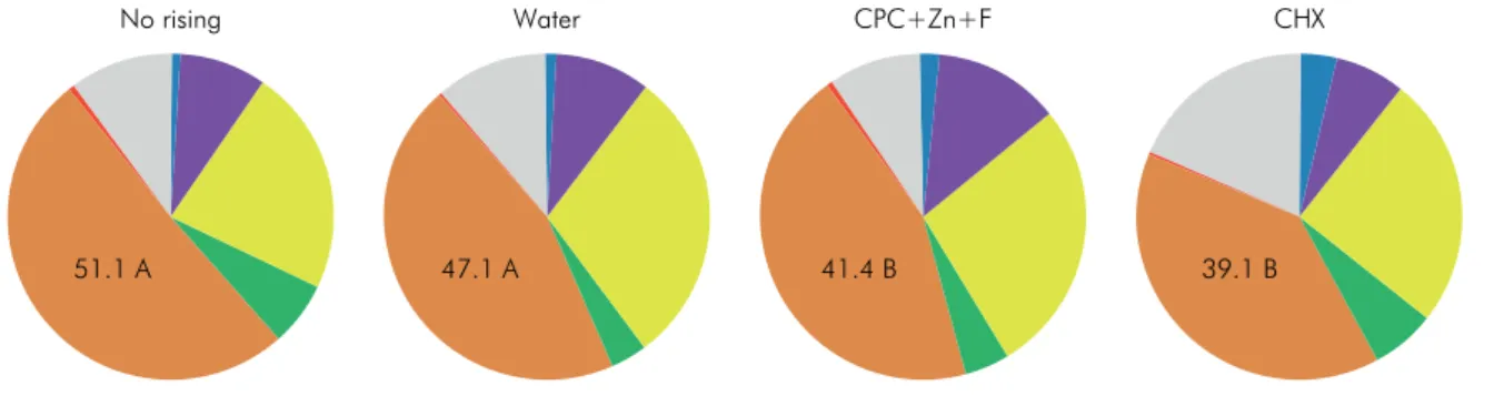
CPC+Zn+F and CHX groups showed, respectively, 70% and 77% fewer CFUs than the group that did not rinse, and 61% and 70% than those from the group that rinsed with water. Figure 4 presents the mean proportions of DNA probe counts of the 40 subgingival species evaluated by Checkerboard DNA-DNA Hybridization technique. No statistically signiicant (p > 0.05) differences between groups were
observed for any individual proportion of the species tested. When the bacterial species were grouped into
“Microbial Complexes” (Figure 5), the data showed that aerosols/splatters from CPC+Zn+F and CHX groups
had a lower proportion of bacterial species from the orange complex (41.4% and 39.1%, respectively) when
compared with aerosols/splatter from No rinsing
and Water groups (51.1% and 47.1%, respectively).
Discussion
The results of the present study showed that oral
aerosol/splatter from subjects who rinsed with a
mouthwash containing 0.075% CPC, 0.28% Zn and
0.05% F, before receiving an ultrasonic prophylaxis, harbored a signiicantly lower bacterial content that from subjects who did not rinse or rinsed with water.
This pre-procedural mouthwash was as effective as the CHX positive control mouthwash in reducing viable bacteria in dental aerosols/splatter. These results are in agreement with those of a previous study that evaluated a mouthwash containing CPC.3
According to our results, the clinician was the person who most beneited from the pre-procedural
rinsing. The blood agar plates positioned on the clinician’s forehead, from the volunteers who rinsed
with CPC+Zn+F or CHX, harbored, 89% and 94% fewer CFUs respectively, than the plates from the volunteers
Table 1. Demographic characteristics (n = 60).
Variable Experimental groups p-value
No rinsing (n = 15) Water (n = 15) CPC+Zn+F (n = 15) CHX (n = 15)
Age (mean years ± SD) 46.27 ± 6.63 36.13 ± 8.71 45.47 ± 11.79 41.40 ± 10.93 p > 0.05
Gender (female/male) 7/8 9/6 10/5 10/5 p > 0.05
Smokers (yes/not) 0/15 0/15 1/14 1/14 p > 0.05
The significance of differences among groups for gender and percentage of smokers was assessed using the Chi-square test and for mean age was assessed using Kruskal-Wallis test. CPC: cetylpyridinium chloride; Zn: zinc lactate; F: sodium fluoride; CHX: chlorhexidine digluconate; SD: standard deviation.
CPC: cetylpyridinium chloride; Zn: zinc lactate; F: sodium fluoride; CHX: chlorhexidine digluconate; CFUs: colony-forming units.
Figure 3. Bar chart of the mean total colony-forming units (CFUs) in the volunteer, clinician, support board and all locations according to experimental groups. The significance of differences among groups was assessed using Kruskal-Wallis and Dunn test (different capital letters indicate significant differences between test and control groups, p < 0.05).
CFUs counts x10
3
5
4
3 A
A
A
No rising Water CPC+Zn+F CHX
AB B
B
B B
B B
B B A
A
A A 2
1
0
Volunteer Clinician
who did not rinse. This represents important protection for the dentist and dental hygienists, who are the main targets of the microorganisms generated during oral procedures.6 It is important to emphasize that the
dental/surface barriers, the methods most commonly
used to minimize cross-infection in the dental ofice,
do not reduce the levels of microorganisms in the environment. Pre-procedural rinsing provides a viable
CPC: cetylpyridinium chloride; Zn: zinc lactate; F: sodium fluoride; CHX: chlorhexidine digluconate; %: percentage.
Figure 4. Mean percentage of DNA probes counts of 40 subgingival species in the four experimental groups. The species were
ordered according to the microbial complexes described by Socransky et al.30 The proportion that each species was determined
for each participant and then averaged in each experimental group. The significance of differences among the four groups was assessed using Kruskal-Wallis test (p < 0.05, adjusted for multiple comparisons).
DNA probe counts (%) 0 5.7 11.4 17.1 22.8
Actinomyces
Purple
Yellow
Green
Orange
Red
Other
A.gerencseriae A.israelii A.naeslundii A.oris A.odontolyticus V.parvulla S.gordonii S.intermedius S.mitis S.oralis S.sanguinis A.a C.gingivalis C.ochracea C.sputigena E.corrodens C.gracilis C.rectus C.showae E.nodatum F.nucleatum.ssp.nucleatum F.nucleatum.ssp.polymorphum F.nucleatum.ssp.vincentii F.periodonticum P.micra P.intermedia P.nigrescens S.constellatus T.forsythia P.gingivalis T.denticola E.saburreum G.morbillorum L.buccalis P.acnes P.melaninogenica N.mucosa S.anginosus S.noxia T.socranskii
No rising Water CPC+Zn+F CHX
CPC: cetylpyridinium chloride; Zn: zinc lactate; F: sodium fluoride; CHX: chlorhexidine digluconate.
Figure 5. Pie charts of the mean proportion of each microbial complex in the four experimental groups. The colors represent
different microbial complexes described by Socransky et al.30 The significance of differences among experimental groups was
assessed using the Kruskal-Wallis and Dunn tests (different capital letters indicate significant differences between groups, p < 0.05).
No rising Water CPC+Zn+F CHX
mean of protection because these barriers, such as
gloves, masks and glasses may have openings, smaller
pores or defects, through which bacteria can pass.
Furthermore, the aerosol particles may remain in the
environment for up to 4 hours after a procedure31
and normally, the clinician and patients remove the protective barriers shortly after completion of the
procedure. Therefore, the risk of airway contamination
by these microorganisms even after the completion of the appointment is high.1 Thus, minimizing the
quantity of microorganisms in the oral cavity before the aerosol/splatter is generated is essential to reduce
the risk of cross-infection in the dental environment.
In addition, the pre-procedural rinsing also
represented a major beneit for the patients. In the
present study, high bacterial counts, which reached
an average of ≈1.000 CFUs in the Water and No Rinsing groups, were observed on the blood agar
plates positioned on the volunteer’s chest. The plates positioned on the chest of the volunteers
who pre-rinsed with CPC+Zn+F showed 55% fewer
bacterial colonies than those who did not rinse or
who rinsed with water, suggesting a major effect of
this solution in reducing the levels of bacteria in the
patient’s oral cavity. A similar effect was observed for the CHX group, which showed 60% fewer bacterial colonies than the Water and No Rinsing groups.
These are relevant items of information for the
dental practitioner, as they may beneit the patients
in several ways during dental procedures, such as for example, by reducing the odds of bacterial spread to other areas of the body, such as the eyes through aerosol,11,32 or even into the lungs though inhalation.33
The previous studies that have evaluated the effects of different antiseptics in reducing aerosol contamination, have recommended CHX as the gold standard.3,4,13,34,35,36
CHX as a pre-procedural rinse has been compared with other antiseptics, such as essential oils,13 povidone
iodine and ozone,36 tea tree oil,35 a solution containing
different types of herb extracts4 and CPC.3 CHX was
shown to be more effective than most of these other antimicrobial agents. Only CPC has shown to be as effective as CHX in reducing the content of bacteria in dental aerosols,3 results that corroborate those reported
in the present study for 0.075% CPC+Zn+F. An additional
advantage for CPC compared with CHX is its lower cost37 and the reduced chance of patient intolerance
to the product.38 Therefore, the main strength of this
study is that it is the irst RCT to determine the eficacy
of a new formulation containing CPC in reducing oral bacterial load in the patient’s mouth and consequently, in the aerosols, as well as determining the microbial composition of these aerosols.
The analysis of the microbial composition showed statistically significantly higher proportions of putative pathogens from the orange complex30 in the
aerosols/splatters from the Water and No Rinsing groups in comparison with those from the CPC+Zn+F and
CHX groups, in agreement with a previous study.3 This
represents an additional beneicial effect of the pre-rinsing,
since certain species from the orange complex, such as
the Fusobacterium species, may be associated with the
etiology of ophthalmic and respiratory infections.32,33
One limitation of the present study is that it presents results for a single dental procedure, prophylaxis with an ultrasonic scaler that has been shown to have a great
potential for aerosol generation in the dental ofice.1,2,3,4,5,39
However, other dental procedures can also generate a large amount of aerosol with infectious components launched into the dental environment, such as the air turbine handpiece,6,7 air-water from a three-way syringe8 and sodium bicarbonate jet.39 Therefore, one
Table 2. Mean percentage of CFUs reduction (average values) in the CHX and CPC+Zn+F groups compared with the No Rinsing and Water groups considering all evaluated locations.
Variable Locations (% of CFUs reduction)
Clinician Volunteers Board All
CHX versus No rinsing 94 60 81 77
CPC+Zn+F versus No rinsing 89 55 70 70
CHX versus Water 87 60 75 70
CPC+Zn+F versus Water 78 55 59 61
could assume that rinsing with CPC would yield similar
beneits if used before the majority of dental procedures.
Conclusion
In conclusion, the results of this study showed that a mouthwash containing 0.075% CPC, 0.28% zinc
lactate and 0.05% sodium luoride as a pre-procedural
mouthwash was effective in reducing bacterial species present in viable oral aerosols during prophylaxis with ultrasonic instruments.
Acknowledgments
The research described in this article was supported
by Colgate Palmolive Company (Piscataway, NJ, USA) and Latin America Oral Health Association (LAOHA).
1. Rivera-Hidalgo F, Barnes JB, Harrel SK. Aerosol and
splatter production by focused spray and standard
ultrasonic inserts. J Periodontol. 1999;70(5):473-7. https://doi.org/10.1902/jop.1999.70.5.473
2. Timmerman MF, Menso L, Steinfort J,
Winkelhoff AJ, Weijden GA. Atmospheric
contamination during ultrasonic scaling.
J Clin Periodontol. 2004;31(6):458-62.
https://doi.org/10.1111/j.1600-051X.2004.00511.x
3. Feres M, Figueiredo LC, Faveri M, Stewart B, Vizio
W. The effectiveness of a preprocedural mouthrinse containing cetylpyridinium chloride in reducing bacteria
in the dental office. J Am Dent Assoc. 2010;141(4):415-22. https://doi.org/10.14219/jada.archive.2010.0193
4. Gupta G, Mitra D, Ashok KP, Gupta A, Soni S,
Ahmed S et al. Efficacy of preprocedural mouth rinsing in
reducing aerosol contamination produced by ultrasonic
scaler: a pilot study. J Periodontol. 2014;85(4):562-8. https://doi.org/10.1902/jop.2013.120616
5. Veena HR, Mahantesha S, Joseph PA, Patil SR, Patil SH.
Dissemination of aerosol and splatter during ultrasonic scaling: a pilot study. J Infect Public Health. 2015;8(3):260-5. https://doi.org/10.1016/j.jiph.2014.11.004
6. Bentley CD, Burkhart NW, Crawford JJ. Evaluating
spatter and aerosol contamination during dental
procedures. J Am Dent Assoc. 1994;125(5):579-84. https://doi.org/10.14219/jada.archive.1994.0093
7. Toroğlu MS, Haytaç MC, Köksal F. Evaluation of aerosol
contamination during debonding procedures. Angle Orthod. 2001;71(4):299-306. https://doi.org/10.1043/0003-3219(2001)071<0299:EOACDD>2.0.CO;2
8. Souza-Gugelmin MC, Lima CD, Lima SN,
Mian H, Ito IY. Microbial contamination in dental unit waterlines. Braz Dent J. 2003;14(1):55-7. https://doi.org/10.1590/S0103-64402003000100010
9. Leggat PA, Kedjarune U. Bacterial aerosols in the
dental clinic: a review. Int Dent J. 2001;51(1):39-44. https://doi.org/10.1002/j.1875-595X.2001.tb00816.x
10. Cochran MA, Miller CH, Sheldrake MA. The
efficacy of the rubber dam as a barrier to the spread of microorganisms during dental
treatment. J Am Dent Assoc. 1989;119(1):141-4. https://doi.org/10.14219/jada.archive.1989.0131
11. Nejatidanesh F, Khosravi Z, Goroohi H, Badrian H, Savabi O.
Risk of contamination of different areas of dentist’s face during dental practices. Int J Prev Med. 2013;4(5):611-5.
12. Kanjirath PP, Coplen AE, Chapman JC, Peters MC,
Inglehart MR. Effectiveness of gloves and infection control in dentistry: student and provider perspectives. J Dent Educ. 2009;73(5):571-80.
13. Logothetis DD, Martinez-Welles JM. Reducing bacterial
aerosol contamination with a chlorhexidine gluconate
pre-rinse. J Am Dent Assoc. 1995;126(12):1634-9. https://doi.org/10.14219/jada.archive.1995.0111
14. Jones CG. Chlorhexidine: is it still the gold
standard? Periodontol. 2000. 1997;15(1):55-62. https://doi.org/10.1111/j.1600-0757.1997.tb00105.x
15. Thomas E. Efficacy of two commonly available mouth
rinses used as preprocedural rinses in children.
J Indian Soc Pedod Prev Dent. 2011;29(2):113-6. https://doi.org/10.4103/0970-4388.84682
16. Varoni E, Tarce M, Lodi G, Carrassi A. Chlorhexidine
(CHX) in dentistry: state of the art. Minerva Stomatol.
2012;61(9):399-419.
17. Yengopal V. The use of essential oil mouthwashes
as preprocedural rinses for infection control. SADJ. 2004;59(6):247-8.
18. Costa X, Laguna E, Herrera D, Serrano J, Alonso B,
Sanz M. Efficacy of a new mouth rinse formulation
based on 0.07% cetylpyridinium chloride in the control
of plaque and gingivitis: a 6-month randomized clinical trial. J Clin Periodontol. 2013;40(11):1007-15. https://doi.org/10.1111/jcpe.12158
19. Pandit S, Cai JN, Jung JE, Lee YS, Jeon JG. Effect of
brief cetylpyridinium chloride treatments during early
and mature cariogenic biofilm formation. Oral Dis. 2015;21(5):565-71. https://doi.org/10.1111/odi.12312
20. Department of Health and Human Service (US).
Food and Drug Administration. 21 CFR Part 356.
Oral health care drug products for over-the-counter human use; anti-gingivitis/anti-plaque drug products;
establishment of a monograph; proposed rules. Federal Register 2003 May 29[cited 2016 Feb 1]. Available from:
https://federalregister.gov/a/03-12783
21. Kang JH, Jang YJ, Kim DJ, Park JW. Antimicrobial
effectiveness of cetylpyridinium chloride and zinc chloride-containing mouthrinses on bacteria of halitosis
and peri-implant disease. Int J Oral Maxillofac Implants. 2015;30(6):1341-7. https://doi.org/10.11607/jomi.3824
22. Mendes L, Coimbra J, Pereira AL, Resende M,
Pinto MG. Comparative effect of a new mouthrinse containing chlorhexidine, triclosan and zinc on volatile sulphur compounds: a randomized, crossover,
double-blind study. Int J Dent Hyg. 2016;14(3):202-8.
https://doi.org/10.1111/idh.12132
23. Giertsen E. Effects of mouthrinses with triclosan,
zinc ions, copolymer, and sodium lauryl sulphate combined with fluoride on acid formation by
dental plaque in vivo. Caries Res. 2004;38(5):430-5. https://doi.org/10.1159/000079623
24. Kjaerheim V, Skaare A, Barkvoll P, Rölla G. Antiplaque,
antibacterial, and anti-inflammatory properties of triclosan mouthrinses in combination with zinc citrate
or polyvinylmethylether maleic acid (PVM-MA) copolymer. Eur J Oral Sci. 1996;104(5-6):529-34. https://doi.org/10.1111/j.1600-0722.1996.tb00137.x
25. Lang C, Murgia C, Leong M, Tan LW,
Perozzi G, Knight D, et al. Anti-inflammatory
effects of zinc and alterations in zinc transporter
mRNA in mouse models of allergic inflammation. Am J Physiol Lung Cell Mol Physiol. 2007;292(2):L577-84. https://doi.org/10.1152/ajplung.00280.2006
26. Erovic Ademovski S, Lingström P, Renvert S.
The effect of different mouth rinse products on
intra-oral halitosis. Int J Dent Hyg. 2016;14(2):117-23.
https://doi.org/10.1111/idh.12148
27. Maggio B, Guibert RG, Mason SC, Karwal R,
Rees GD, Kelly S et al. Evaluation of mouthrinse
and dentifrice regimens in an in situ erosion
remineralisation model. J Dent. 2010;18 Supple 3:S37-44. https://doi.org/10.1016/S0300-5712(11)70007-0
28. Socransky SS, Smith C, Martin L, Paster BJ, Dewhirst FE,
Levin AE. “Checkerboard” DNA-DNA hybridization.
Biotechniques. 1994;17(4):788-92.
29. Mestnik MJ, Feres M, Figueiredo LC, Duarte PM,
Lira EA, Faveri M. Short-term benefits of the adjunctive use of metronidazole plus amoxicillin
in the microbial profile and in the clinical
parameters of subjects with generalized aggressive periodontitis. J Clin Periodontol. 2010;37(4):353-65. https://doi.org/10.1111/j.1600-051X.2010.01538.x
30. Socransky SS, Haffajee AD, Cugini MA, Smith C,
Kent RL, Jr. Microbial complexes in subgingival plaque. J Clin Periodontol. 1998;25(2):134-44. https://doi.org/10.1111/j.1600-051X.1998.tb02419.x
31. Grenier D. Quantitative analysis of bacterial aerosols in
two different dental clinic environments. Appl Environ Microbiol. 1995;61(8):3165-8.
32. Arat YO, Shetlar DJ, Rose JE. Blindness from septic
thrombophlebitis of the orbit and cavernous sinus
caused by Fusobacterium nucleatum. Arch Ophthalmol. 2004;122(4):652-4. https://doi.org/10.1001/archopht.122.4.652
33. Bhattacharya S, Livsey SA, Wiselka M,
Bukhari SS. Fusobacteriosis presenting as community acquired pneumonia. J Infect. 2005;50(3):236-9. https://doi.org/10.1016/j.jinf.2003.11.007
34. Reddy S, Prasad MG, Kaul S, Satish K, Kakarala S,
Bhowmik N. Efficacy of 0.2% tempered chlorhexidine as a pre-procedural mouth rinse: A clinical
study. J Indian Soc Periodontol. 2012;16(2):213-7. https://doi.org/10.4103/0972-124X.99264
35. Shetty SK, Sharath K, Shenoy S, Sreekumar C,
Shetty RN, Biju T. Compare the effcacy of two
commercially available mouthrinses in reducing viable bacterial count in dental aerosol produced during ultrasonic scaling when used as a preprocedural
rinse. J Contemp Dent Pract. 2013;14(5):848-51. https://doi.org/10.5005/jp-journals-10024-1414 36. Kaur R, Singh I, Vandana KL, Desai R. Effect of
chlorhexidine, povidone iodine, and ozone on microorganisms in dental aerosols: randomized
double-blind clinical trial. Indian J Dent Res.
2014;25(2):160-5. https://doi.org/10.4103/0970-9290.135910
37. Slots J. Low-cost periodontal therapy. Periodontol 2000.
2012;60(1):110-37. https://doi.org/10.1111/j.1600-0757.2011.00429.x
38. Keni NN, Aras MA, Chitre V. Chlorhexidine allergy due
to topical application. Indian J Dent Res. 2012;23(5):674-6.
https://doi.org/10.4103/0970-9290.107393
39. Legnani P, Checchi L, Pelliccioni GA, D’Achille C.