504
1 . Physician, cardiologist, Belo Horizonte, MG, Brazil.
2 . MD, PhD, Professor, Federal University of Minas Gerais, Belo Horizonte, MG, Brazil.
3 . PhD student; Postgraduate Program in Adult Health Federal University of Minas Gerais and Surgeon Cardiovascular. 4. Resident in the Department of Cardiology and Cardiovascular
Surgery. Hospital das Clinicas, Federal University of Minas Gerais. 5. Cardiologist and Professor, Federal University of Minas Gerais. Study conducted at Hospital das Clínicas, Federal University of Minas
Isabella Morais Martins
1, Júlia Medeiros Fernandes
1,
Cláudio Léo Gelape
2,
Renato Braulio
3, Vagner
de Campos Silva
4,
Maria do Carmo Pereira Nunes
5Rev Bras Cir Cardiovasc 2011;26(3):504-7 CASE REPORT
Grande cisto pericárdico manifestando-se com compressão das câmaras cardíacas direitas
A large pericardial cyst presenting with
compression of the right-side cardiac chambers
Abstract
Pericardial cysts are rare, usually congenital. Cysts frequently occur in the right cardiophrenic angle and their diagnosis is usually suspected after an abnormal chest X ray. The present case report shows a case of pericardial cyst with atypical radiographic aspect in an athletic patient who presented clinical with symptoms of right ventricular failure. The diagnosis was suggested by echocardiogram and subsequently was confirmed by pathologic examination.
Descriptors: Mediastinal cyst/surgery. Pericardium/
pathology. Cardiac surgical procedures.
Gerais, Belo Horizonte, MG, Brazil. Mailing Address:
Maria do Carmo Pereira Nunes. Departamento de Clínica Médica -UFMG.
Avenida Professor Alfredo Balena, 190 - Santa Efigênia - Belo Horizonte, MG, Brazil – Zip Code: 30130-100
E-mail: [email protected]
Article received on April 18th, 2010
Article accepted on August 15th, 2010
Resumo
Cistos pericárdicos são raros, comumente congênitos, com localização mais frequente no ângulo cardiofrênico direito. O diagnóstico é suspeitado pela radiografia de tórax anormal. O presente relato descreve um caso de cisto pericárdico com aspecto radiográfico atípico, em um paciente atleta, manifestando-se clinicamente com sintomas de insuficiência ventricular direita. O diagnóstico foi sugerido pelo ecocardiograma transesofágico e confirmado pelo estudo anatomopatológico.
Descritores: Cisto mediastínico/cirurgia. Pericárdio/
patologia. Procedimentos cirúrgicos cardiovasculares.
INTRODUCTION
The cysts of the heart and pericardium are rare [1]. They are mainly congenital, with an estimated incidence of 1:100,000, accounting for about 7.6% of all mediastinal masses described in the literature [2,3]. In most cases they are asymptomatic and are usually diagnosed incidentally by chest radiograph. Some cases present with symptoms of dyspnea and chest pain. In addition, complications such as cardiac tamponade can occur, justifying the need for diagnosis.
We report the case of a patient previously asymptomatic, athlete, presenting with signs of systemic congestion due to right heart compression by a large pericardial cyst after blunt chest trauma.
CASE REPORT
Male 40 years old, bodybuilder, with habits of lifting excessive weight and previous history of deep venous thrombosis in oral anticoagulation therapy. Previously asymptomatic when he developed dyspnea on exertion,
505 Martins IM, et al. - A large pericardial cyst presenting with compression
of the right-side cardiac chambers
Rev Bras Cir Cardiovasc 2011;26(3):504-7
jugular vein engorgement and dry coughing. Informing blunt trauma to the chest with a strong impact by a bar last month. He evolved with progressive worsening of symptoms when sought medical attention. Initially, we required transthoracic echocardiography, which did not show enough impact diagnosis, and subsequently, a chest CT scan was performed without contrast, which suggested the presence of pericardial effusion. Transesophageal echocardiogram showed extrinsic compression of right heart chambers and interventricular septum deviation to the left as well as significant thickening of the pericardium, with hemodynamic repercussions. The function and morphology of the left chambers were slightly reduced, and there were no significant changes in heart valves (Figure 1).
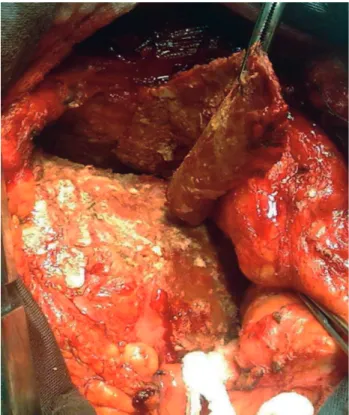
Fig. 3 - Median sternotomy, demonstrating the calcifications on the right chambers and the thickened wall of the pericardial cyst. Repair, and the upper right side of the cyst
Fig. 1 – Image of the transesophageal echocardiography demonstrating extrinsic compression of the right chambers, with deviation of the interventricular septum to the left as a result of pericardial cyst (arrows). AD: right atrium, VD: right ventricle, AE: left atrium, VE: left ventricle
The patient was admitted and that moment the patient was normal colored, hydrated, eupneic, acyanotic, with preserved peripheral capillary perfusion. Engorged jugular veins with normal cardiac auscultation.
The patient underwent median low ministernotomy with CPB, being found a large cystic structure with thick hemorrhagic fibrin content. The walls of the cyst were grossly calcified in the portion adhered to the epicardium in the right ventricle and right atrium from the superior vena cava to the lower right diaphragmatic wall (Figures 2 and 3) It was difficult for excision of the cyst, due to calcifications strongly adhered to the epicardium, increasing the risk of injury. The incision had to be expanded to the sternal notch (in L). The operation was uneventful.
After surgery, the patient recovered well and was discharged on the 5th postoperative day. Histopathologic
506
analysis confirmed the preoperative suspicion of a large pericardial cyst measuring 10 x 3 x 0.3 cm. Postoperative transthoracic echocardiogram showed a thickened pericardium, but normal right ventricle. In follow-up of 6 months, the patient had complete remission of symptoms.
DISCUSSION
Pericardial cysts are uncommon congenital anomalies that occur almost exclusively in adults in the fourth and fifth decades of life [4]. These cysts are caused by a defect in the development of the coelomic cavity and is consistently adhered to the pericardial leaflet, although communication with the pericardial cavity may occur [5]. They are classically located next to the right anterior cardiophrenic sinus, being its occurrence in other sites much less common [6].
The cysts vary in diameter from 2 to 5 cm or more and represent 6% of mediastinal masses and 33% of mediastinal cysts [7]. Most cases have no symptoms, diagnosed incidentally on chest radiography. Rarely calcify or rupture. Acquired cysts are extremely rare and may be associated with mediastinal neoplasia, parasitic infection, illness, trauma or cardiac surgery [8].
In this case, the patient is a man of 40, who belongs to the common age of manifestation of this disease. However, we emphasize the fact of being a bodybuilding patient with a history of recent chest trauma, causing an increase in previously asymptomatic cyst. Thus, the compression of the right heart chambers and heart failure associated may be present in an asymptomatic patient with pericardial cyst after chest trauma [9]. In fact, the patient reported onset of symptoms four weeks after the trauma. Added to this, we highlight the unusual size of the cyst and its pathological characteristics with hemorrhagic content.
The most common symptoms are chest pain, dyspnea, cough, and palpitations. Occasionally, they may alter the cardiovascular hemodynamics or lung expansion and produce signs and symptoms that mimic tricuspid stenosis, pulmonary stenosis or constrictive pericarditis [9]. In this study, symptoms of right ventricular failure were present secondary to compression of the right chambers, as evidenced by echocardiography.
The incidental diagnosis is based on the realization of a chest x-ray reveals a mass radiodense, homogeneous, rounded, showing the classic sign silhouette described by Felson & Felson [10]. Transthoracic echocardiography is usually sufficient to establish the diagnosis, and occasionally necessary to complement with transesophageal exam in cases of atypical location or difficult to visualize. Computerized tomography and magnetic resonance imaging also contribute mainly in the differential diagnosis of other mediastinal masses. Needle
REFERENCES
1. Engle DE, Tresch DD, Boncheck LI, Foley WD, Brooks HL. Misdiagnosis of pericardial cyst by echocardiography and computed tomography scanning. Arch Intern Med. 1983;143(2):351-2.
2. Davis RD Jr, Oldham HN Jr, Sabiston DC Jr. Primary cysts and neoplasms of the mediastinum: recent changes in clinical presentation, methods of diagnosis, management, and results. Ann Thorac Surg. 1987;44(3):229-37.
3. Cohen AJ, Thompson LN, Edwards FH, Bellamy RF. Primary cysts and tumors of the mediastinum. Ann Thorac Surg. 1991;51(3):378-84.
4. Saad R, Carvalho WR, Ximenes Neto M. Cirurgia torácica geral. 1ª ed. São Paulo:Atheneu;2005.
5. Gilbert-Barness E. Potter’s pathology of the fetus and infant.
4th ed. St. Louis: Mosby-Year Book;1997.
aspiration is an attractive alternative for being diagnostic and therapeutic and has low mortality. However, the only definitive diagnosis is confirmed by the findings of the pathology, as occurred in this case.
The differential diagnosis includes other solid tumors and cysts of the mediastinum, diaphragmatic hernia or tumors, aneurysms of the heart or great vessels. Among the complications include rupture, cardiac tamponade, obstruction of right main bronchus, obstruction of the outflow tract of the right ventricle and acute heart failure [11].
Spontaneous resolution of pericardial cyst was described in a few cases, probably due to rupture of the cyst [12]. Conservative treatment should be reserved for asymptomatic cases. The percutaneous aspiration and ethanol sclerosis are alternative therapies, reserving surgical removal of those symptomatic cases with cardiorespiratory repercussions [13]. The surgery was the best treatment option in our case, due to the size of the cyst and the presence of hemodynamic compromise by evident compression of the right chambers.
Martins IM, et al. - A large pericardial cyst presenting with compression of the right-side cardiac chambers
507
6. Wang ZJ, Reddy GP, Gotway MB, Yeh BM, Hetts SW, Higgins CB. CT and MR imaging of pericardial disease. Radiographics. 2003;23(Spec No):S167-80.
7. Maisch B, Seferovic PM, Ristic AD, Erbel R, Rienmüller R, Adler Y, et al. Guidelines on the diagnosis and management of pericardial diseases executive summary; The Task Force on the Diagnosis and Management of Pericardial Diseases of the European Society of Cardiology. Eur Heart J. 2004;25(7):587-610.
8. Borlaug BA, DeCamp MM, Gangadharan SP. Neoplastic pericardial disease. In: Basow DS, ed. UpToDate. Waltham:UpToDate;2008.
9. Temizkan V, Onan B, Inan K, Ucak A, Yilmaz AT. Hemorrhage into a pericardial cyst and associated right ventricular
compression after blunt chest trauma. Ann Thorac Surg. 2010;89(4):1292-5.
10. Felson B, Felson H. Localization of intrathoracic lesions by means of the postero-anterior roentgenogram; the silhouette sign. Radiology. 1950;55(3):363-74.
11. Patel J, Park C, Michaels J, Rosen S, Kort S. Pericardial cyst: case reports and a literature review. Echocardiography. 2004;21(3):269-72.
12. Abbey AM, Flores RM. Spontaneous resolution of a pericardial cyst. Ann Thorac Cardiovasc Surg. 2010;16(1):55-6.
13. Nina VJ, Manzano NC, Mendes VG, Salgado Filho N. Giant pericardial cyst: case report. Rev Bras Cir Cardiovasc. 2007;22(3):349-51.
Martins IM, et al. - A large pericardial cyst presenting with compression of the right-side cardiac chambers