663
www.scielo.br/rsbmt I www.rsbmt.org.br
Case Report
Case Report
Revista da Sociedade Brasileira de Medicina Tropical 47(5):663-665, Sep-Oct, 2014http://dx.doi.org/10.1590/0037-8682-0087-2014
INTRODUCTION
Address to: Dr. Nishanth Dev. Department of Medicine/ESIC Medical College.
PO Box 110001 Faridabad, India.
Phone: 91 97 1728-3133
e-mail: [email protected]
Received 17 April 2014
Accepted 17 July 2014
An unusual case of heart failure due to
Plasmodium vivax
infection with a favorable outcome
Nishanth Dev
[1], Adesh Kumar Gadpayle
[2],
Jhuma Sankar
[3]and Mona Choudhary
[4][1]. Department of Medicine, ESIC Medical College, Faridabad, India. [2]. Department of Medicine, Post Graduate Institute of Medical Education and Research, Dr Ram Manohar Lohia Hospital, New Delhi, India. [3]. Department of Pediatrics, AIIMS, New Delhi, India. [4]. Department of Psychiatry PGIMER, Dr RML Hospital, New Delhi, India.
ABSTRACT
Although malaria is one of the oldest types of parasitic infection, we have recently witnessed substantial changes in the outcome of malarial infections. Severe Plasmodium vivax infections have recently become more frequent, and are occasionally associated with fatal outcomes. Cardiac arrhythmia and myocardial failure have also been reported, typically in association with Plasmodium falciparum infections. We report a case of myocarditis and heart failure, due to Plasmodium vivax infection, along with the favorable outcome.
Keywords: Malaria. Plasmodium vivax. Myocarditis. Heart failure.
With its associated morbidity and mortality, malaria has threatened human beings since time immemorial. The infection is common in tropical and sub-tropical areas, as their environment favors the survival and multiplication of the malarial parasite.
According to the latest fi gures, released in December 2013 by
the World Health Organization, there were approximately 207 million cases of malaria in 2012 (uncertainty range, 135-287 million) and an estimated 627,000 deaths (uncertainty range, 473,000-789,000)1. Of the 5 species of the malaria parasite,
Plasmodium falciparum (P. falciparum)infection is most often associated with complicated malaria. However, over the past few years there has been an increasing trend in cases of complicated malaria associated with Plasmodium vivax (P. vivax). Once considered benign, P. vivax malaria is becoming hostile, creating fresh challenges for clinicians and epidemiologists. Although rare, cardiovascular complications are known to be associated with P. falciparum malaria2, while myocarditis associated with
P. vivax is extremely rare in the literature3. We report the case
of a 22-year-old male who presented with myocarditis and heart failure due to P. vivax malaria.
CASE REPORT
A 22-year-old male resident of India presented with high-grade fever associated with chills and rigor of 5 days duration, as well as breathlessness of 1 day duration. There was no
history of cough, chest pain, dysuria, loose stools, yellowish discoloration of the eyes or skin, bleeding, joint pain, or other
symptoms that might indicate previous signifi cant medical or
surgical illness. Upon physical examination, the patient was febrile with a temperature of 101°F. His respiratory rate was 30/min, pulse was 98/minute, and systolic blood pressure was 70mmHg. Examination of the cardiovascular system revealed no signs of murmur or gallop rhythm, and his heart sounds were normal. His spleen tip was palpable during an abdominal examination, and respiratory system examination revealed crepitations in the bilateral infrascapular regions. The rest of his systemic examination was normal, and there were no clinical
fi ndings suggestive of acute rheumatic fever.
The patient’s hemoglobin level was 12.3g/dL, with a total leucocyte count of 5.7 × 10⁹/L (68% polymorphs and
32% lymphocytes), and a platelet count of 270 × 109/L. His
erythrocyte sedimentation rate was 8mm/h, his C reactive protein (CRP) level was 1mg/L, and his anti-streptolysin O (ASO) titer was 32 units. Liver and kidney function tests were normal, and his serum calcium was 8.4mg/dL. A peripheral smear detected the ring and schizont stage of P. vivax, and the malaria antigen test was positive for P. vivax only. Blood and urine cultures were sterile, and typhoid immunoglobulin G and Leptospira slide macroagglutination tests were negative. Viral serology for adenovirus, cytomegalovirus, Coxsackie virus, and enteric cytopathic human orphan virus were also negative, and the
patient was non-reactive for the human immunodefi ciency virus.
Electrocardiography (ECG) detected sinus tachycardia with T-wave inversion in the inferior leads. Cardiac enzymes were within the normal range and the troponin-T test was negative. Chest radiography revealed cardiomegaly with bilateral lower
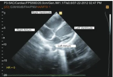
zone infi ltrates (Figure 1A). Echocardiogram revealed dilated cardiac chambers (Figure 2) with left ventricular systolic dysfunction (ejection fraction of 25-30%); no regional wall motion abnormalities were detected.
664 www.scielo.br/rsbmt I www.rsbmt.org.br
Dev N et al. - A case of heart failure due to Plasmodium vivax infection
DISCUSSION
Antimalarial treatment was provided as a combination of artemether and lumefantrine. Supportive care was administered
in the form of oxygen, intravenous fl uids, furosemide, and
antipyretics. The patient’s blood pressure normalized and he became afebrile in 3 days. Glucose-6-phosphate dehydrogenase levels also returned to the normal range, and the patient was treated with primaquine for 2 weeks to prevent relapse. Seven days later, follow-up ECG, radiography (Figure 1B), and echocardiography detected no cardiac abnormalities.
FIGURE 2 - Echocardiograph showing dilated cardiac chambers.
FIGURE 1 - A: Chest radiography image obtained at admission. B: Chest radiography obtained at discharge.
P3-5AC/Cardiac/FPS59D/20.0cm/Gen./MI1.1/TIs0.6/07-22-2012 02:47 PM
[2D]G38/90dB/FA4/P90/HAR/FSI 1
Right Ventricule
Right Atrium Left Ventricule
Left Atrium
0
5
10
15
HR = 0
20
A
B
The clinical course of P. vivax malaria, traditionally called benign tertian malaria, appears to be changing, given the recent reports of P. vivax-associated serious complications3,4.
Cardiovascular complications, such as bundle branch block, pericardial effusion, and cardiomyopathy, are more frequently
reported as complications of P. falciparum malaria, compared to P. vivax and experimental malaria5. In the present case,
we found evidence of myocarditis with heart failure, which improved following symptomatic treatment. In a study of 22 adult cases of P. falciparum malaria, ECG abnormalities were observed in 23% of cases, while pericardial effusion and global hypokinesia were observed in 9% and 4.5% of cases, respectively6. Myocarditis has also been reported as a
complication of P. falciparum malaria2, although it is rarely
reported following P. vivax malaria7,8.
Unfortunately, the mechanism of cardiac complications associated with malaria is not well understood. Various case reports and pathological studies have indicated that malaria might be a cause of toxic and ischemic myocarditis. As well, ischemic cardiomyopathy can develop following the blockage of capillaries by parasites and parasitized red blood cells, as shown by various autopsy studies9. Interestingly, the phenomenon of
cytoadherence, where red blood cells adhere to the walls of capillaries and are sequestered to the brain and heart, is typically caused by P. falciparum. However, as the parasite density in P. vivax malaria is low, these cases do not typically develop cytoadherence. Therefore, it appears that the toxic effects of cytokines, such as tumor necrosis factor alpha, interleukin-10, or other unknown factors, might play a role in cases of P. vivax malaria. Cytokine-mediated endothelial activation has been found to be associated with disease severity in both P. falciparum and complicated P. vivax malaria. In a case-control study from Pakistan, the authors compared 82 complicated cases and 100 uncomplicated cases, and observed that tumor necrosis factor alpha, interleukin-10, intercellular adhesion molecule-1, and vascular cell adhesion molecule-1 were individual predictors of complicated P. vivax malaria10.
665
www.scielo.br/rsbmt I www.rsbmt.org.br
Rev Soc Bras Med Trop 47(5):663-665, Sep-Oct, 2014
REFERENCES
troponin-T test was negative. Although they may not be specifi c
to ischemic cardiomyopathy, their absence also supported our exclusion of this condition. As well, echocardiography did not reveal any regional wall motion abnormalities, which are
generally helpful in differentiating cardiac diseases and defi ning
the nature of the myocardial dysfunction. Myocarditis is typically characterized by a decreased left ventricular ejection fraction and hypokinesia of the ventricular walls, and may be associated with pericardial effusion and valvular regurgitation11. Regarding other
diagnoses, we excluded acute viral myocarditis, as the serology results were negative for commonly implicated viruses. Acute rheumatic fever was also unlikely, as the ASO titer and CRP levels
were normal, and there were no other signifi cant clinical fi ndings.
Treatment for myocarditis is mainly supportive, and involves the management of shock, arrhythmia, and heart failure. Recovery is typically complete, pending resolution of the underlying cause. Few patients exhibit evidence of residual cardiac dysfunction on follow-up12, and in the present case,
the patient’s recovery was complete, with full resolution of all
clinical and echocardiographical fi ndings at the time discharge.
Myocarditis is an unusual complication of P. vivax malaria, although the presence of heart failure and shock during admission should arouse suspicion regarding underlying myocarditis. Echocardiography may help prove the diagnosis, and guide subsequent management of such cases.
1. World Health Organization. World Malaria Report. Washington, DC: WHO, 2013.
2. Mohsen AH, Green ST, West JN, McKendrick MW. Myocarditis associated with Plasmodium falciparum malaria: a case report and a review of the literature. J Travel Med 2001; 8:219-220.
3. Oh MD, Shin H, Shin D, Kim U, Lee S, Kim N, et al. Clinical features of vivax malaria. Am J Trop Med Hyg 2001; 65:143-146.
4. Choi HJ, Lee SY, Yang H, Bang JK. Retinal haemorrhage in vivax malaria. Trans R Soc Trop Med Hyg 2004; 98:387-389.
5. Günther A, Grobusch MP, Slevogt H, Abel W, Burchard GD. Myocardial damage in falciparum malaria detectable by cardiac troponin T is rare. Trop Med Int Health 2003;8:30-32
6. Franzen D, Curtius JM, Heitz W, Hopp HW, Diehl V, Hilger HH, et al. Cardiac involvement during and after malaria. Clin Invest 1992; 70:670-673.
7. Herrera JM. Cardiac lesions in vivax malaria. Study of a case with coronary and myocardial damage. Arch Inst CardiolMex 1960; 30: 26-36.
8. Kim SA, Kim ES, Rhee MY, Choi SI, Huh HJ, Chae SL. A Case of Myocarditis Associated With Plasmodium vivax Malaria. J Travel Med 2009; 16:138-140.
9. Clark IA, Chaudhri G, Cowden WB. Roles of tumour necrosis factor in the illness and pathology of malaria. Trans R Soc Trop Med Hyg 1989; 83:436-440.
10. Raza A, Ghanchi NK, Sarwar Zubairi Ab, Raheem A, Nizami S, Beg MA.
Tumor necrosis factor-α, interleukin-10, intercellular and vascular
adhesion molecules are possible biomarkers of disease severity in complicated Plasmodium vivax isolates from Pakistan. PLoS One 2013;8:e81363.
11. Lieberman EB, Hutchins GM, Herskowitz A, Rose NR, Baughman KL. Clinicopathologic description of myocarditis. J Am Coll Cardiol 1991; 18:1617-1626.
12. McCarthy RE 3rd, Boehmer JP, Hruban RH, Hutchins GM, Kasper EK,