Arq Neuropsiquiatr 2007;65(4-B):1220-1223
1220
BROCA’S APHEMIA
An illustrated account of its clinico-anatomic validity
Ricardo de Oliveira-Souza
1,2, Jorge Moll
2, Egas M.A. Caparelli-Dáquer
1,2ABSTRACT - Objective:To present the case of a 54-year-old man with loss of speech, but with preservation of voluntary facio-lingual motility, language and other cognitive abilities (Broca’s aphemia). Method: Ob-servation of patient oral communicative abilities and general behavior, neuropsychological assessment and cranial computed tomography. Results:Computed tomography showed a hyperdense lesion in the sub-cortex of the left precentral gyrus corresponding to Brodmann’s area 6 and 44. Neuropsychological assess-ment confirmed that the major cognitive domains were intact. Conclusion:Our patient reiterates the va-lidity of Broca’s aphemia as a clinico-anatomic entity allowing us to portray it for the first time in pictures. From a neurobehavioral perspective, aphemia is related to apraxia rather than to aphasia, a fact that may have hampered the full grasp of its far-reaching implications for neurology and aphasiology.
KEY WORDS: aphemia, Broca’s area, Broca’s aphasia, apraxia.
Afemia de Broca: um relato ilustrado sobre sua validade anátomo-clínica
RESUMO - Objetivo:Apresentar o caso de um paciente de 54 anos de idade com perda da fala, mas pre-servação da linguagem, das demais capacidades cognitivas, e da motilidade fácio-lingual voluntária (afe-mia de Broca). Método:Observação da capacidade de comunicação oral e do comportamento geral, exa-me neuropsicológico e tomografia computadorizada do crânio. Resultados:A tomografia computadori-zada revelou lesão hiperdensa no subcórtex do giro precentral esquerdo correspondendo às áreas 6 e 44 de Brodmann. O exame neuropsicológico confirmou que os principais domínios cognitivos se encontravam in-tactos. Conclusão:Nosso paciente reiterou a validade da afemia de Broca como entidade anátomo-clínico permitindo documentá-la em fotos pela primeira vez. Da perspectiva neurocomportamental, a afemia está vinculada às apraxias e não às afasias, o que pode ter prejudicado a apreensão plena do seu profundo sig-nificado para a neurologia e para a afasiologia.
PALAVRAS-CHAVE: afemia, área de Broca, afasia de Broca, apraxia.
1Hospital Universitário Gaffrée e Guinle, Rio de Janeiro RJ, Brazil; 2Cognitive and Behavioral Neuroscience Unit, LABS - D´Or Hos-pitals Network, Rio de Janeiro RJ, Brazil.
Received 27 July 2007. Accepted 22 September 2007.
Dr. Ricardo de Oliveira-Souza - Rua Conde de Bonfi m 232 / 304 - 20550-012 Rio de Janeiro RJ - Brasil. E-mail: rdeoliveira@gmail.com
Pierre Paul Broca (1824-1880) coined the term “aphemia” for the loss of speech without impair-ment of language in patients with left frontal lobe damage1. As shown in the passage below, Broca was
mostly impressed by retained ability of such patients to move the faciolingual territories employed in speech:
“There are cases where the general language fac-ulty persists unaltered, where the auditory appara-tus is intact, where all the muscles, not even except-ing those of the voice and those of articulation, obey the will, and yet where a cerebral lesion abolishes ar-ticulated language. This abolition of speech (…) con-stitutes a symptom so singular that it seems to me useful to designate it with a special name (…) aphe-mia (α,deprive;ϕηµι,I speak, pronounce); it is only
the faculty of articulating words that these patients lack. They hear and comprehend all that one says to them; they all have their intelligence; they emit vo-cal sounds with ease; they execute with their tongue and their lips movements much more extensive and energetic than those required for the articulation of sounds, and yet the perfectly sensible response that they would want to make is reduced to a very small number of articulated sounds, always the same and always performed in the same manner (…)”
We report on a case of aphemia very similar to the one Broca described on patient Lelong2. To our
Arq Neuropsiquiatr 2007;65(4-B)
1221 Broca’s aphemia Oliveira-Souza et al.
root of most inferences on the existence of a cerebral “speech center” apart from motor “centers” relat-ed to the production of voluntary faciolingual move-ments unrelated to speech.
CASE
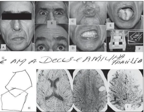
A 54-year-old right-handed (Edinburgh Inventory=100) waiter with 8 years of formal education lost the ability to speak and swallow on the evening before consultation. His ability to swallow rapidly returned to normal, but he re-mained speechless and unable to spontaneously generate and repeat even single words, although he retained the ability to vocalize a sound resembling an “Ah…” Although he strived to articulate consonant and other vowel sounds, he was absolutely unsuccessful. He did not present verbal stereotypies or recurring utterances. He nonetheless com-municated by gestures and writing, and by soliciting the aid of his wife and daughters to convey what he meant. He was oriented to time and place, and understood oral and written language perfectly. He could write meaningful sen-tences, both spontaneously and on dictation, with correct orthography and spelling. Figure 1 (bottom) shows a sam-ple of the patient’s handwriting and a freehand copy of two intersecting pentagons. He could sit, stand, and walk, and execute complex orofacial and limb movements in re-sponse to verbal and gestural (visuo-imitative) commands [Florida Apraxia Screening Test = 15/153]. At rest,
sponta-neous blinking was symmetric, but the lower face deviat-ed slightly to the left (Fig 1A). On command, he contractdeviat-ed the frontalis, corrugator, and orbicularis occuli (Fig 1B). His ability to pucker the lips (Fig 1C) contrasted with his inabil-ity to retract or lift the right corner of the mouth (Fig 1D). The tongue was trophic and without fasciculations (Fig 1E). The soft palate and tongue did not deviate at rest or
dur-ing movement. There was no emotional incontinence. Fun-doscopy and eye movements were normal. He had no his-tory of hypertension or diabetes, the heart rate was regular and the blood pressure was 130x80 mmHg. He denied fe-ver, headache, visual symptoms, dizziness, incontinence, and fainting. He fared normally on the Mini-Mental State Exam (28/30), Token Test (33/36), Right-Left Orientation (20/20), 3D Block Construction (29/29) (Fig 1F), Visual Form Discrimina-tion (28/32), Judgment of Line OrientaDiscrimina-tion (19/30), and the Visual Organization (26/30) tests4. On the Wisconsin Card
Sorting test he completed one category and committed 23 perseverative errors. He scored 24/36 (normal ≥25/36) on the Tower of London task5. Figure 1K shows a sample of
spon-taneous handwriting, Figure 1H shows a freehand copy of intersecting pentagons. CT (10 mm slices, parallel to the or-bito-meatal line) showed a hyperdense egg-shaped lesion surrounded by a thin hypodense hallo in the subcortex of the left lower precentral gyrus exerting a slight mass effect on surrounding regions (Fig 1I). The lesion spared the in-sula, the temporal lobe, and the medial hemispheric wall, suggestive of a lobar hematoma in the subcortex of Brod-mann’s areas (BA) 6 and 446. Written informed consent was
obtained from the patient to publish his pictures. He died a few days later of an acute myocardial infarction before completion of further tests.
DISCUSSION
The relevant fi ndings of this case were (i) the loss of speech with preservation of voice, language and cognition, with (ii) relative sparing of faciolingual mo-tility and praxis, and (iii) the location of the lesion. Our patient presented loss of speech in the absence of proportional faciolingual paralysis and aphasia. He did not develop agraphia, alexia, and apraxia – in
Arq Neuropsiquiatr 2007;65(4-B)
1222
Broca’s aphemia Oliveira-Souza et al.
ticular, faciolingual praxis was preserved, provided he would not attempt to speak. The clinical manifesta-tions of this case represent a typical instance of aphe-mia as described by Broca on patient Lelong2. Thus
defi ned, aphemia must be differentiated from a host of conditions that compromise fl uent speech. At the outset we would like to emphasize that, since our pa-tient could still produce vocal sounds, we did not con-sider him to be “mute”, a term that usually implies an inability to produce articulation and voice. The facial deviation could lead to an erroneous impression of
Bell’s palsy, which was discarded by the dispropor-tional affection of speech, the bilateral preservation of spontaneous blinking and the ability to close the eyes.Suprabulbar paralysis consists of impairment of voice, articulation, lower face motility, and swallow-ing with preservation of trophism and refl exes in the affected territories, most often caused by multiple cerebral infarcts. The latter differentiate suprabulbar from the bulbar paralysis of motor neuron disease, marked by lingual and masticatory atrophy and ar-reflexia. Thus, even conceding that the dysphagia of our patient might have represented a fragment of a suprabulbar paresis at the onset of symptoms, the severe speech defi cit could not be attributed to interruption of corticobulbar fascicles. A non-fl uent aphasia was altogether discarded by the intactness of language, as defi ned by the ability to comprehend and express ideas and thoughts by means other than by articulated speech. Finally, the typical “misarticu-latory symptoms among areas of fl uent speech” de-scribed in cases of damage to the precentral gyrus of the insula7 were conspicuously absent.
The lesion responsible for aphemia is typically seated in the opercular division of the inferior frontal gyrus (“Broca’s area”, BA 44)8. Lesions of the
opercu-lar cortex or of the short cortico-cortical fi bers issuing from it lead to “true” aphemia because they impair speech without compromising language, faciolingual motility, and the ability to produce vocal sounds. This was the case in our patient and in two others report-ed in the recent literature9,10. In contrast, lesions of
the lower precentral gyrus, where the cranial mo-tor terrimo-tories are represented, that spare the frontal operculum impair speech due to “anarthria”11 [Case
2], a term that should be reserved for paralysis from interruption of corticobulbar pathways12. In practice,
lesions are seldom small enough to produce pure aphemia or pure anarthria. Most often, variable com-binations of aphasia, aphemia and anarthria produce complex impairments of oral language output that may be inadvertently taken as unique. A systematic
assessment of such cases will often show that they fi t current concepts of the anatomical organization of the anterior language zone.
The left frontal operculum is composed of hetero-modal (“association”) cortex lying at the interface of language and speech. It sends short projections to the adjacent motor cortex, where the corticonuclear neurons responsible for the integration between articulation and voice are ultimately recruited. The frontal operculum contains the kinetic formula ( Be-wegungsformel) responsible for the automatic con-version of verbal language, a cognitive phenomenon, into speech, the product of the motor innervation of the articulatory muscles. The aphemia in our patient probably resulted from a lobar hematoma in the left frontal lobe. Coincidentally, aphemia in Lelong may likewise have resulted from an identical hemorrhage, both in size and in shape (“Il s’agit donc d’un ancien foyer apoplectique”2). Ruff and Arbit10 described a
15-year-old girl who developed aphemia after evacuation of a hematoma in the left precentral gyrus and frontal operculum. To our knowledge, these are the only in-stances of aphemia due to intracerebral hemorrhage. The diagnostic value of the status of faciolingual motility in the differentiation of aphemia (frontal operculum) and anarthria (lower precentral gyrus and its efferent pathways) has seldom been emphasized. Nevertheless, the relative preservation of faciolingual motility constituted the most interesting fi nding in our patient, as it provided a fresh insight into the mechanism of aphemia. The impairment of learned motor actions that manifests itself in certain behav-ioral contexts but not in others is a core feature of apraxia13,14. Conceptually, then, the aphemia of our
patient represented a particular instance of apraxia, namely, an apraxia of speech. The relationship be-tween aphemia due to injury of the left precentral gyrus of the left frontal lobe and the articulatory syn-drome that results from lesion of the left precentral gyrus of the insula7 remains to be determined15. The
Arq Neuropsiquiatr 2007;65(4-B)
1223 Broca’s aphemia Oliveira-Souza et al.
Acknowledgments – The authors are indebted to Dr. Dayse Gusmão for referral of the patient and to Profes-sor Omar da Rosa Santos (Head of the Internal Medicine Service of HUGG) for helpful comments. Mr. José Ricardo Pinheiro and Mr. Jorge Baçal (Instituto Oswaldo Cruz Li-brary, Rio de Janeiro) provided invaluable help in retriev-ing classical texts.
REFERENCES
1. Broca P. Remarques sur le siège de la faculté du langage articulé; suiv-ies d’une observation d’aphémie (perte de la parole). Bull Soc Anat (Par-is) 1861;6:330-357. Translated as “Remarks on the seat of the faculty of articulated language, following an observation of aphemia (loss of speech), by Mr. Paul Broca (186)” by C. D. Green on hĴ p://psychclassics. yorku.ca/Broca/aphemie-e.htm. Accessed July 25, 2005.
2. Broca P. Nouvelle observation d’aphémie par une lésion de la moitié postérieure des deuxième et troisième circonvolutions frontales. Bull Soc Anat (Paris) 1861;6:398-407.
3. Ochipa C, Rothi LJG, Heilman KM. Ideational apraxia: a defi cit in tool selection and use. Ann Neurol 1989;25:190-193.
4. Lezak MD. Neuropsycological assessment, 3.Ed. New York: Oxford, 1995. 5. Oliveira-Souza R, Ignácio FA, Cunha FC, Oliveira D LG, Moll J. The neu-ropsychology of executive behavior: performance of normal individuals on the Tower of London and Wisconsin Card Sorting tests. Arq Neurop-siquiatr 2001;59:526-531.
6. Damasio H, Damasio AR. Lesion analysis in neuropsychology. New York: Oxford, 1989.
7. Dronkers NF. A new brain region for coordinating speech articulation. Nature 1996;384:159-161.
8. Schiě HB, Alexander MP, Naeser MA, Galaburda AM. Aphemia: clini-cal-anatomic correlations. Arch Neurol 1983;40:720-727.
9. Fox RJ, Kasner SE, ChaĴ erjee A, Chalela JA. Aphemia: an isolated dis-order of articulation. Clin Neurol Neurosurg 2001;103:123-126. 10. Ruě RL, Arbit E. Aphemia resulting from a leĞ frontal hematoma.
Neu-rology 1981;31:353-356.
11. Tonkonogy J, Goodglass H. Language function, foot of the third frontal gyrus, and rolandic operculum. Arch Neurol. 1981;38:486-490. 12. Lecours AR, LhermiĴ e F. The “pure form” of the phonetic
disintegra-tion syndrome (pure anarthria): anatomo-clinical report of a historical case. Brain Lang 1976;3:88-113.
13. Liepmann H. The leĞ hemisphere and action (1905). In: D Kimura (Ed). Translations of Liepmann’s essays on apraxia. London, Ontario: DK Consultants, 1980:17-50.
14. Moll J, Oliveira-Souza R, Passman LJ, Cunha FC, Lima FS, Andreiuo-lo PA. Functional MRI correlates of real and imagined tool-use panto-mimes. Neurology 2000;54:1331-1336.
15. Hillis AE, Work M, Barker PB, Jacobs MA, Breese EL, Maurer K. Re-ex-amining the brain regions crucial for orchestrating speech articulation. Brain. 2004;127:1479-1487.