Arq Neuropsiquiatr 2003;61(2-A):269-273
LUMBOSACRAL ANGIOLIPOMA
Case report
Antonio Aversa do Souto
1, Flavio S. Domingues
1, Leila Chimelli
2, Armando M. Lemos
1ABSTRACT - We present a case of a 46-year old woman with a ventral epidural angiolipoma at the lumbosacral level with erosion of the sacrum. About ninety cases of spinal angiolipomas have been previously described in the literature, most of them situated on the thoracic region, dorsal to the dural sac. Angiolipomas can be radically excised with a good prognosis even in the presence of bone erosion. We did not find any other angiolipoma at the sacral level surgically explored in the review of the literature.
KEY WORDS: angiolipoma, spinal tumors, sacral tumors.
Angiolipoma lombosacral: relato de caso Angiolipoma lombosacral: relato de caso Angiolipoma lombosacral: relato de caso Angiolipoma lombosacral: relato de caso Angiolipoma lombosacral: relato de caso
RESUMO - Descrevemos o caso de uma mulher de 46 anos com um angiolipoma lombosacral ventral ao saco dural que erodia o sacro. Cerca de noventa casos de angiolipomas foram publicados na literatura, sendo na maioria localizados na região torácica e dorsal ao saco dural. Angiolipomas podem ser ressecados de maneira radical, com bom prognóstico mesmo na presença de infiltração óssea. Não encontramos, em revisão da literatura, nenhum outro caso semelhante de angiolipoma lombosacral com erosão do sacro abordado cirurgicamente.
PALAVRAS-CHAVE: angiolipoma, tumores medulares, tumores sacrais.
Hospital Universitário Clementino Fraga Filho, Universidade Federal do Rio de Janeiro (UFRJ), Rio de Janeiro RJ, Brasil: 1Serviço de
Neurocirurgia; 2Departamento de Patologia.
Received 23 July 2002, received in final form 23 October 2002. Accepted 6 November 2002.
Dr. Antonio Aversa do Souto Serviço de Neurocirurgia Hospital Universitário UFRJ Avenida Brigadeiro Trompowski s/n/10º andar -21941-590 Rio de Janeiro RJ - Brasil. E-mail: [email protected]
Spinal angiolipoma is a benign tumor of the epi-dural space. It is a rare cause of spinal cord compres-sion, accounting for 0.14% to 1.2% of the spinal tumors1. It is considered a distinct clinical and
patho-logical entity traditionally grouped as a variant of lipo-ma1. Characteristically the tumor lies over the dorsal
aspect of the dura at the thoracic level2-7. Its
port-wi-ne color or dark brown appearance contrasts very well with the normal epidural fat1,8,9. Sometimes the
tu-mor can be tu-more aggressive and invade the contiguous bone and adjacent soft tissues10,11. We report a patient
with a lumbosacral angiolipoma with bone erosion associated with a L4-L5 left sided disc herniation.
CASE
A 46-year old female with a history of ten years of low back pain had a worsening of the symptoms in the three months before diagnosis. The pain radiated down the pos-terior aspect of the left thigh, calf and ankle, and increased with walking and physical strength. The patient also re-ferred a progressive numbness of the perineum. A
neuro-logical examination demonstrated a mild paresis of the plantar flexion of the left toe and hypoactive left jerk reflex. A positive straight leg-raising test at ten degree at the left side could be elicited. Superficial hypoesthesia at the late-ral aspect of the left foot, buttocks and perineum was noted. An X-ray of the lumbar spine and sacrum showed erosion of the posterior aspect of the sacrum and widening of the sacral canal. A MRI scan revealed an epidural mass displacing the dural sac posteriorly, eroding the bone and projecting to the anterior sacral foramina with the sacral root. The mass was isointense in T1-weighted and hype-rintense in T2-weighted and showed a homogeneous and intense enhancement with gadolinium infusion. There was also a left sided disc herniation associated with the upper limit of the tumor at the level of L4-L5 space (Fig 1).
The herniated disc was dissected from the inferior aspect of the left L5 root and resected. Subcutaneous fat was harvest and then used in hemostasis and for filling the large epidural space ventral to the dural sac.
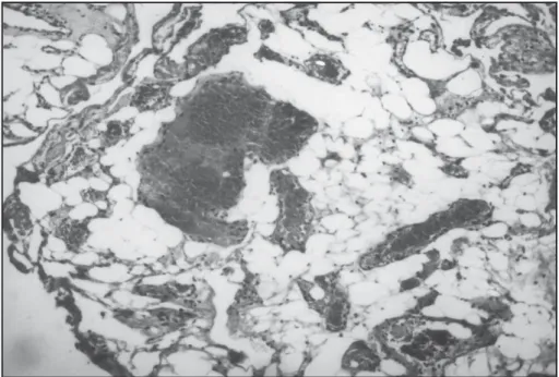
The patient recovered completely of the previous symp-toms and could return to her previous activities. Two repea-ted MRI studies six and eighteen months after the surgical procedure confirmed the total resection and did not show any evidence of tumor recurrence (Fig 3). Histopathological analysis was characteristic of an angiolipoma and showed that the lesion was composed by adult fatty tissue inter-mingled with many thin walled blood vessels (Fig 4).
DISCUSSION
Berenbruch in 1890 described the first case of angiolipoma in a sixteen-year-old male confirmed in
an autopsy12. Since then about ninety cases of
angio-lipoma have been reported at the literature. In the past, the finding of fatty tumors in the epidural space would be considered a matter of little interest and many cases would be unreported13. With the advance
of MRI and the widespread use of operating microsco-pe, even those small tumors have been distinguished from the epidural fat, and the number of cases being reported is increasing13. Otherwise those slow growing
tumors would be unrecognized by many surgeons1,13.
In the review of Preul1 spinal angiolipomas
acco-unt for 0.14% to 1.2% of all spinal tumors and 2% to 3% of extradural spinal tumors. Males and females are almost equally affected. The mean age of patients is 41.6 years (the youngest was 6 years and the oldest
Fig 1. Preoperative MRI scans. A, sagittal T1-weighted scan with gadolinium infusion of the lumbosacral spine showing a ventral mass from L4 to S2 with homogeneous contrast enhancement. B, axial T1-weighted scan, without contrast at the S1 level. C, axial T1-weighted scan with contrast showing erosion of the sacrum and extension of the tumor to the sacral foramina. D, axial T1-weighted scan displaying the upper limit of the tumor associated with a lumbar disc herniation at L4-L5 level.
A
C
B
Arq Neuropsiquiatr 2003;61(2-A) 271
Fig 2. A, intraoperative view of the surgical field showing a large epidural mass ventral to the dural sac (arrow) encasing the sacral roots. B, same view as in A after the tumor resection. The dural sac was displaced to show the L4-L5 left side disc herniation.
72 years old)1. If the lipomas secondary to prolonged
high corticosteroid intake or following epidural injec-tions are excluded, most of the symptomatic fatty tumors in the epidural space are angiolipomas4,14.
Spinal angiolipoma typically presents as a slow gro-wing mass causing compression of the spinal cord. Lower-extremity numbness, back pain and leg weak-ness are frequent initial complaints 1. The mean
dura-tion of the symptoms is usually longer than one
year4,15,16. There were rare described cases of sudden
installation of the symptoms or abrupt neurological deterioration17. Increasing of the body weight and
pregnancy appear to exacerbate the symptoms due to changes in the tumor mass and volume1,4,16. The
concomitant presence of spinal tumors and discal hernia is probably rare. However, before the
introduc-tion of CT and MRI studies, spinal tumors might be misdiagnosed as herniated intervertebral disk and vice-versa13. In the present case the simultaneous
occur-rence of a lumbar disc herniation could be crucial in the development of the symptoms of radicular compression. The sacral location of the tumor in this case explained the atypical clinical presentation. The association of spinal angiolipoma and lumbar disc her-niation was found in two previous reported cases9,13.
Histopathologically angiolipomas are characteri-zed by mature adipose tissue containing copious vas-cular elements that vary from sinusoids, thin-walled vessels or thick-walled vessels with proliferation of the smooth muscle layer4. Mitotic figures are
infre-quent and malignant changes have not been
identi-Fig 3. Postoperative MRI scans. A, sagittal T2-weighted scan. B,C axial T1-weighted scans with gadolinium infusion showing complete resection of the tumor.
A B
fied18. They have a gross aspect that vary in color
from whitish-yellow to a grayish-purple and can be usually well distinguished from the normal epidural
fat1,9,13,19. The pathogenesis of angiolipomas is
un-known. They may result from abnormal development of the primitive, pluripotential mesenchymal cells from which adipose tissue and vascular endothelium arise or may be hamartomatous in nature1,16,17.
Almost all-spinal angiolipomas are located at the epidural space. The great majority of the epidural angiolipomas are located in the thoracic region at the posterior surface of the dura1,4,13,16,20-23. There have
been described eight cases of angiolipomas involving the lumbar spine16,24. They were frequently located
anteriorly in the spinal canal in contrast with the regular dorsally located angiolipomas at the thoracic levels1,15, 17. Kasper et al. in 1929 reported an autopsy
study with angiolipomas in the lumbosacral area (from L3-S3) and in the cervical spine canal in a 6-years old boy25. Nishiura et al. described a case of
left L5 and S1 radicular compression pre-operatively diagnosed as disc herniations that was explored thro-ugh foraminotomies9. They found a hemorrhagic
mass similar to our case compressing the L5 and S1 roots which was subtotally removed and the final diagnosis was hemangiolipoma. Palkovic et al.28
re-ported the first case of intramedullary angiolipoma and Preul et al.1 described another similar case.
In 1966 Gonzalez-Crussi et al described the first case of infiltrating spinal angiolipoma27. They
consi-dered that bone infiltration was associated with a
more aggressive behavior and a worse prognosis. Subsequent studies failed to demonstrate differences in prognosis in the infiltrating angiolipomas in com-parison with the non-infiltrating group16. These
in-vasive angiolipomas are more common at the ven-tral aspect of the dura in the lumbar spinal canal13,27.
Total resection is the goal of the surgical treatment and could be achieved less frequently in the infiltra-ting angiolipomas specially if they are anterior and anterolateral in location1,28. However, angiolipomas
sub-totally resected have also a good prognosis with rare recurrences described even in the infiltrating va-riant12. Almost all the patients have improvement of
the neurological deficits after the surgery with total or near-total recovery17.
Plain X-rays are normal in the majority of the ca-ses. Computed tomography studies are not conclu-sive in angiolipomas, with the epidural mass more often isodense or slightly hyperdense with light or absent homogeneous contrast enhancement1,29. In
MRI studies the extradural components of angioli-pomas are isointense or hyperintense in T1-weighted images, probably due to the fat component and usually hyperintense in T2-weighted images. Those areas show an early enhancement after gadolinium administration and are considered the vascular com-ponent1,16. Slow-growing masses dorsally located in
the thoracic spinal canal with these MRI findings should be suspected of angiolipoma. With the wides-pread availability of MRI, spinal angiolipomas have been more frequently recognized and diagnosed.
Arq Neuropsiquiatr 2003;61(2-A) 273
In conclusion, spinal angiolipomas are rare tumors with a benign behavior. When localized in the thoracic spinal canal they have usually a well-defined clinical-radiological presentation with MRI studies. The authors describe a case of a lumbosacral angiolipoma diagnosed in vivo and totally resected with a good outcome. To the best of our knowledge we could not find any other angiolipoma with erosion of the sacrum reported in the literature.
REFERENCES
1. Preul MC, Leblanc R, Tampieri D, Robitaille Y, Pokrupa R. Spinal angiolipomas: report of three cases. J Neurosurg 1993;78:280-286. 2. Aguiar PH, Plese JP, Rosemberg S, et al. Thoracic spinal angiolipoma:
case report. Arq Bras Neurocirurg 1996;15:103-107.
3. Balbo RJ, Araujo JF, Melro CA, Iafigliola MG, Valvassore FR. Thoracic epidural angiolipoma: case report. Arq Neuropsiquiatr 1995;53:659-661.
4. Haddad FS, Abla A, Allam CK. Extradural spinal angiolipoma. Surg Neurol 1986;26:473-486.
5. Obrador S, Villarejo F, Deblas A. Angiolipoma extradural causa infrequente de compression medullar. Rev Clin Esp 1977;146:395-396. 6. Padovani R, Tognetti F, Speranza S, Pozzati E. Spinal extrathecal hemangiolipomas: report of two cases and review of the literature. Neurosurgery1982;11:674-677.
7. Turgut M. Spinal angiolipomas: report of a case and review of the ca-ses published since the discovery of the tumor in 1890. Br J Neurosurg 1999;13:30-40.
8. Machado JAF, Fabião OM Neto, Ordovas CA, Hack I, Gallo P. Spinal angiolipoma: case report. Arq Bras Neurocirurg 1993;12:141-144. 9. Nishiura I, Kubo Y, Koyama T. Spinal haemangiolipoma: three case
reports. Neurochirurgia (Stuttg) 1986;29:63-66.
10. Rivkind A, Margulies JY, Lebenstart P, Sherman Y, Robin GC. Anteri-or approach fAnteri-or removal of spinal angiolipoma: a case repAnteri-ort. Spine 1986;11:623-625.
11. Sakaida H, Waga S, Kojima T, Kubo Y, Matsubara T, Yamamoto J. Thoracic spinal angiolipoma with extracanal extension to the thoracic cavity: a case report. Spine 1998;23:391-394.
12. Stookey B. Intradural spinal angiolipoma: report of case and symptoms for 10 years in child aged 11: review of literature. Arch Neurol Psychiatry 1927;18:16-43.
13. Pagni CA, Canavero S. Spinal epidural angiolipoma: rare or unreported? Neurosurgery 1992;31:758-764.
14. Von Hanwehr R, Apuzzo MLJ, Ahmadi J, Chandrasoma P. Thoracic spinal angiomyolipoma: case report and literature review. Neurosur-gery 1985;16:406-411.
15. Pearson J, Stellar S, Feigin I. Angiolipoma: long term cure following radical approach to malignant-appearing benign intraspinal tumor. J Neurosurg 1993;78:280-286.
16. Trabulo A, Cerqueira L, Monteiro P, Roque P, Reis FC, Coelho MR. Spinal angiolipomas revisited: two case repoerts. Acta Neurochir (Wien) 1996;138:1311-1319.
17. Labram EK, el-Shunnar K, Hilton DA, Robertson NJ. Spinal angiolipoma: three additional cases. Br J Neurosurg1999;13:25-29. 18. Lin JJ, Lin F. Two entities in angiolipoma: a study of 459 cases of lipomas with
review of literature on infiltrating angiolipoma. Cancer 1974;34:720-727. 19. Boockvar JA, Black K, Malik S, Stanek A, Tracey KJ. Subacute
paraparesis induced by venous thrombosis of a spinal angiolipoma: a case report. Spine1997;22:2304-2308.
20. Bender JL, Van Landingham JH, Manno NJ. Epidural lipoma producing spinal cord compression. Report of two cases. J Neurosurg 1974;41:100-103. 21. Bouramas D, Korres D, Roussos L, Mantzilas T, Anagnostopoulos D.
Spinal extradural angiolipoma. J Spinal Disord 1995;8:324-327. 22. Costa FA, Balaguez HM, Ferreira LC, et al. Dorsal spinal epidural
angiolipoma: case report. Arq Bras Neurocirurg 1998;17:48-50. 23. Rubin G, Gornish M, Sandbank J, Shevach I, Rappaport ZH. Spinal
extradural angiolipoma: case report and review of the literature. Spine 1992;17:719-723.
24. Provencale JM, McLendon RE. Spinal angiolipomas: MR features. AJNR 1996;17:713-719.
25. Kasper GA, Cowan A. Extradural lipoma of the spinal cord. Arch Pathol 1929;8:800-802.
26. Palkovic S, Wassman H, Bonse R, Kashab M. Angiolipoma of the spinal cord. Magnetic resonance imaging and microsurgical management. Surg Neurol1988;29:243-245.
27. Gonzales-Cruzzi F, Ennecking WF, Arean WM. Infiltrating angioli-poma. J Bone Joint Surg 1966;48:1111-1124.
28. Kuroda S, Abe H, Akino M, Iwasaki Y, Nagashima K. Infiltrating spinal angiolipoma causing myelopathy: case report. Neurosurgery 1990;27:315-318. 29. Michilli R, Tzonos P, Iglesias-Rozas JR. Spinal extradural angiolipoma: case