NORMAL FINDINGS ON CHEST X-RAYS OF NEONATES*
Beatriz Regina Álvares1, Inês Carmelita Minniti Rodrigues Pereira1, Severino Aires de Araújo Neto2,Emerson Taro Inoue Sakuma3
The purpose of this study is to describe the normal findings of the newborn chest radiography, the criteria utilized for evaluating the quality radiographs and the correct catheter and tube positions, emphasizing the peculiarities inherent to the patient’s age. In the neonatal period changes in the fetal circulation contribute to an increase in cardiac size, skin folds and variations in the thymic silhouette may simulate diseases, the evaluation of catheter and tube positions avoids iatrogenic complications, the abdominal gas pattern must be correlated with the patient’s age and the presence of the secondary ossifications centers in the upper humerus and scapula is associated with the term newborn, providing a radiological sign for normal skeletal maturation. The knowledge of the peculiarities and normal radiological findings of the newborn chest radi-ography avoids ambiguous diagnosis, reduces iatrogenic complications and represents a valuable support in the diagnosis and clinical follow-up of these patients.
Keywords: Newborn; Normal findings; Chest radiography.
Achados normais no exame radiológico de tórax do recém-nascido.
O objetivo deste trabalho é descrever os achados normais na radiografia de tórax do recém-nascido, os cri-térios usados para avaliar a qualidade técnica do exame, assim como o posicionamento correto de sondas, cânulas e cateteres, enfatizando as especificidades dos achados radiológicos relacionados à faixa etária do paciente. No período neonatal, a imagem cardíaca é mais proeminente em virtude da conversão da circula-ção fetal, as dobras de pele e as variações da imagem tímica podem simular doenças, a avaliacircula-ção do posi-cionamento adequado de sondas e cateteres evita iatrogenias, o padrão gasoso intestinal apresenta mudan-ças relacionadas ao número de horas de vida do paciente e a presença dos núcleos de ossificação secundá-rios na extremidade proximal dos úmeros e processo coracóide está associada com a idade gestacional a termo do recém-nascido, representando, portanto, um sinal radiológico de desenvolvimento ósseo normal. O conhecimento das particularidades e dos aspectos radiológicos normais no tórax do recém-nascido evita diagnósticos equivocados, reduz as iatrogenias e representa um valioso suporte no diagnóstico e no acom-panhamento clínico destes pacientes.
Unitermos: Recém-nascido; Achados normais; Radiografia de tórax. Abstract
Resumo
* Study developed in the Department of Radiology – Faculty of Medical Sciences of Universidade Estadual de Campinas, Campinas, SP, Brazil.
1. Doctors, Assistant Professors at Faculty of Medical Sciences of Universidade Estadual de Campinas.
2. MD, Assistant at Service of Radiology – Centro de Atenção Integral à Saúde da Mulher, Universidade Estadual de Campinas. 3. MD, Resident for Department of Radiology at Faculty of Medical Sciences of Universidade Estadual de Campinas.
Mailing address: Profa. Dra. Beatriz Regina Álvares. Departa-mento de Radiologia, FCM/Unicamp, Cidade Universitária “Ze-ferino Vaz”, Barão Geraldo. CP 6111. Campinas, SP, Brazil 13081-970. E-mail: [email protected]
Received October 28, 2004. Accepted after revision Febru-ary 16, 2005.
INTRODUCTION
The chest x-ray is one of the most fre-quently requested radiological examina-tions in neonatal intensive care units (ICU), representing an essential tool in the diag-nosis of pulmonary diseases in preterm and term neonates. In these patients, the chest x-ray also allows the evaluation of nasogas-tric probes, endotracheal tubes, arterial and
venous umbilical catheters positioning, as well as detection of bone structures and abdominal alterations usually included in chest x-rays of neonates(1–4).
Since the neonatal radiography is essen-tial for the work of the clinician(1), it is
important for him/her to have a broad knowledge about singular aspects of this examination, from the examination perfor-mance itself to morphological aspects of the neonate chest anatomic structures which are absent in older children and adults.
Aiming at highlighting such specifici-ties, the authors of the present study have made a detailed review of the literature concerned with chest radiological exami-nation in neonates, illustrated with x-ray films from the teaching files of Centro de Atenção Integral à Saúde da Mulher (Caism) Radiology Service at Universidade Estadual de Campinas (Unicamp).
CHEST X-RAY OF NEONATES
The chest x-ray in neonates, especially the preterm ones, should preferably be per-formed in the neonatal ICU, with a portable x-ray equipment. The radiology technician must wash his/her hands before perform-ing any procedure for each neonate, aim-ing at reducaim-ing the neonatal infections in-cidence, since newborns usually present poor immunological defense mechanism(5).
In order to reduce the radiation dose for these small patients, only one anteroposte-rior view of the chest should be taken, since, in most of cases, this is enough to supply the necessary diagnostic informa-tion(6).
to cause respiratory symptoms. In subse-quent x-rays, lateral chest and abdomen views only should be included in cases where there is a clinical indication or ne-cessity of evaluating umbilical probes and catheters localization(1,4).
TECHNICAL FACTORS
Chest x-rays of neonates are in compli-ance the technical standards when they meet the following criteria(1,2,4):
a) Visualization of dorsal intervertebral spaces through the cardiac silhouette (film density);
b) right hemi-diaphragm at the level of the posterior arc of the eighth rib (satisfac-tory level of pulmonary aeration);
c) caudal inclination of anterior costal arcs appearing underneath the posterior ones (adequate centralization of the central beam on the thoracic cage);
d) symmetry of bone structures on both sides of the thoracic cage (correct position-ing of the neonate);
Main technical problems that may mimic pathological alterations inducing misdiagnosis are the following:
a) X-ray beam underpenetration, reduc-ing the density differences between in-trathoracic structures and simulating false pulmonary opacities;
b) pulmonary hypoaeration, resulting in horizontalization of costal arcs, false wid-ening of the cardiothymic silhouette and reduction of the pulmonary transparency,
with possibility of, occasionally, simulat-ing pulmonary edema, hemorrhage, atelec-tasis and pneumonic consolidations;
c) x-ray beam overpenetration, darken-ing the radiographic film and possibly con-cealing pulmonary opacities, mainly the most subtle ones, like interstitial opacities of the neonate transitory tachypnea and the reticulogranular infiltrate of the hyaline membrane disease;
d) patient rotation, causing asymmetry of the chest and resulting in a false promi-nence of the cardiothymic image towards the deviation side;
e) inadequate centralization of the cen-tral ray over the neonate’s abdomen, caus-ing a lordotic configuration of the thoracic cage characterized by the cephalic orienta-tion of the anterior arcs, and possibly caus-ing widencaus-ing and distortion of the cardio-thymic image.
The knowledge of these criteria utilized for evaluating the technical quality of chest x-ray of neonates results in technically cor-rect x-rays, besides reducing the possibil-ity of misdiagnosis due inadequately per-formed examinations (Figures 1, 2 and 3).
INTRATHORACIC ANATOMIC STRUCTURES
The neonate chest undergoes significant birth-related changes during the first hours of life, and presents quite distinct aspects in its anatomic structures, so these normal, specific radiological features should be
taken into consideration during the neona-tal period.
During the first hours of a neonate’s life, transitory cardiomegaly may occur as a re-sult of additional blood inflow from the placenta into the umbilical cord before its cutting, and of the presence of a bidirec-tional shunt through the arterial duct and oval foramen before its closure. Also, a pro-minent pulmonary vascularization may be observed as a result of residual lung fluid absorption through the lymphatic-venous system. A still open arterial canal may be seen on a chest x-ray as a convex promi-nence to the left of the spine, between T3 and T4 vertebras, this configuration being denominated ductus bump, that is consid-ered as a typical radiological finding at the neonate first hours of life(1,2) (Figure 4).
The oval foramen and arterial canal clo-sure, the decrease in pulmonary vascular resistance and absorption of the remaining lung fluid in the hours subsequent to the birth reduce the cardiac dimensions and the chest vascular prominence.
In neonates, the thymus is radiologically characterized by a widening of the upper mediastinum, above the cardiac image, on the frontal view, and an increase in the supracardiac, retrosternal density on the profile incidence (Figure 5). On the fron-tal view, the normal width of the thymic image must be higher than the double-width of the third thoracic vertebra, shorter dimensions representing a sign of thymic involution(7) (Figure 6). Under stress —
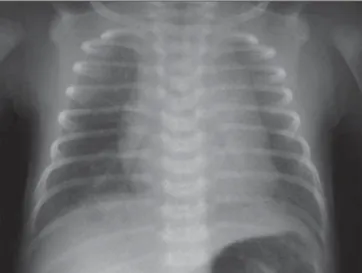
Figure 1. Normal chest x-ray of a two-hour-old newborn infant, in compliance with technical standards.
fever, infections, congenital cardiopathies, pulmonary diseases, malnutrition —, there may be a rapid thymic involution as a re-sult of the adrenal corticosteroid action, and yet its image may not be visualized on chest x-rays(8) (Figure 7). This accidental involution may revert once the stress situ-ation is overcome, and the thymus returns to its normal dimensions. Also, the thymus may present peculiar features, including the “wave sign” corresponding to a gentle un-dulation on the thymus surface produced by costal arcs compression, more frequently to the left; the “notch sign”, where the infe-rior border of the normal thymus blends
with the border of the cardiac silhouette; and the “sail sign” resulting from a pecu-liar shape of the thymus appearing like a normal anterior mediastinal sail shaped structure, more frequently to the right(1,2)
(Figures, 8, 9 and 10).
EXTRATHORACIC STRUCTURES
Soft tissues, the skeletal structure and the abdomen may provide relevant infor-mation for clinical management of neo-nates.
The thickness of the thoracic wall soft tissues reflects the nutritional condition,
and may be decreased in light-weight new-born infants(9).
Secondary ossification nuclei of the proximal humeral extremity and coracoid apophysis may be visualized on chest x-rays, and a relation is established between the presence of these ossification nuclei and the term gestational age of the neonate, therefore representing a sign of fetal matu-rity(10) (Figure 11).
Typically, the presence of air may be observed in the stomach right at birth, small bowel with three hours of live, and in the rectum, six to eight hours after birth, so it is always important to correlate
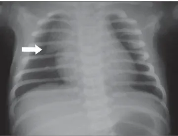
radiologi-Figure 3. Two-hour-old newborn infant chest x-ray with x-ray tube misalign-ment. Anterior costal arcs present cephalic orientation, projecting themselves above their posterior segments.
Figure 4. One-day-old newborn infant chest x-ray demonstrating the ductus bump (arrow).
Figure 5. Chest x-ray an-teroposterior and lateral views, demonstrating normal cardiothymic image.
Figure 6. Scheme showing cri-teria for evaluation of thymic image dimensions.
Figure 8. Nine-day-old newborn infant x-ray demonstrating “wave-sign” (ar-row).
Figure 9. Twenty-two-day-old newborn infant x-ray demonstrating “notch-sign” (arrow).
Figure 7. Chest x-ray of (A) three-hour-old, and (B) four-day-old newborn infant, showing thymus involution.
A B
cal findings with the neonate’s number of hours of life(2).
CATHETERS, CANNULAS AND PROBES
In the analysis of chest and abdomen x-rays, it is very important to describe the
localization of cannulas, probes and cath-eters, since the incorrect positioning of these tubes may cause iatrogenies(2,11).
The umbilical venous catheter follows its course through the umbilical vein, venous duct and inferior vena cava, pre-senting a straight course at the right side of the thoracic-lumbar spine. The correct site
for its extremity is the inferior vena cava, nearby the right atrium entry at right of T8-T9(2,12) (Figure 12). The umbilical arterial
level of iliac arteries bifurcation at L3-L5 — low localization—, or in the thoracic aorta under the arterial canal between T7 and T9 — high localization(2,12) (Figures13
and 14). Umbilical catheters should not be situated in the origin of smaller caliber vascular trunks under the risk of precipitat-ing spasms and thrombosis.
In patients under assisted respiration, the endotracheal intubation cannula should be placed in the medium third of the tra-chea, above the carina, and is visualized at
T4 level and below the medial clavicle(2,4).
In patients with gastric probing, the probe should be visualized at the right of the tra-cheal cannula and its end should be located in the stomach (Figure 15).
IMAGE ARTIFACTS
Artifacts must be identified, since ignor-ing their peculiarities may induce the diag-nosis of inexistent diseases by the inter-preter-physician. One of the most frequent
image artifacts are the skin folds projected over the thoracic cavity, and may simulate pneumothorax. The differential diagnosis is made by observing this artifact as a dense, linear image presenting an obliquity as opposed to the lung border, extending below the thoracic cavity(1,2,4) (Figure 16).
Another eventual image artifact is the pro-jection of the neonatal incubator access ports producing lower density round im-ages which may be confused with cystic lesions(1,2,4) (Figure 17).
Figure 12. Twenty-four-hour old newborn infant chest and abdomen x-ray presenting venous um-bilical catheter localized in the inferior vena cava (arrow).
Figure 13. Newborn infant x-ray demonstrating low-localization of arterial umbilical catheter at the L4 level (arrow).
Figure 14. Newborn infant x-ray showing high-lo-calization of arterial umbilical catheter (arrow).
Figure 10. Three-hour-old newborn infant x-ray presenting the “sail sign” (arrow).
Figure 16. Newborn infant chest x-ray showing skin fold at left (arrow). Figure 17. One-hour-old newborn infant x-ray demonstrating artifact related to projection of neonatal incubator access port (arrow).
Figure 15. Three-day-old newborn infant x-ray show-ing endotracheal cannula placed above the carina (asterisk) and the naso-gastric probe with its end lo-calized in the stomach (ar-row).
CONCLUSION
The chest x-ray is a valuable support in the diagnosis and clinical follow-up of neo-nates, especially those requiring intensive care. The knowledge on particularities and normal radiological aspects of the neonate’s chest avoids misdiagnosis, reduces iatro-genies and is of help in the diagnosis and clinical follow-up of these patients.
REFERENCES
1. Swischuk LE. Radiologia do recém-nascido, do lactente e da criança pequena. 5ª ed. Rio de Ja-neiro: Revinter, 2006.
2. Wesenberg RL. The newborn chest. New York: Harper & Row, 1973.
3. Grupo de Hospitales Castrillo. Estúdio prospec-tivo sobre el empleo de catéteres umbilicales en el recién nacido. An Esp Pediatr 2000;53:470–478. 4. Radiology in the nursery – indications, positio-ning and safety. Disponível em: www.paclac.org/
Manuals_Guidelines/Radiology_in_the_Nursery_ Final_6.1.98.pdf. Acessado em: 20 de junho de 2004.
5. Calil R, Tresoldi AT, Veiga JFFS. Controle de infecção hospitalar. In: Marba STM, Mezzacappa Filho F, editores. Manual de neonatologia. Uni-camp. Caism. Rio de Janeiro: Revinter, 1998; 289–293.
6. Gibson AT, Steiner GM. Review. Imaging the neo-natal chest. Clin Radiol 1997;52:172–186. 7. Dutz W, Kohout E, Rossipal E, Vessal K.
Infan-tile stress, immune modulation and disease pat-terns. Path Ann 1976;11:415–453.
8. Álvares BR. Avaliação do timo e coração, em ra-diografias de tórax de crianças eutróficas e des-nutridas de 1º, 2º e 3º graus, na faixa etária de 0 a 24 meses. (Tese de Doutorado). Rio de Janeiro: UFRJ, 1994.
9. Álvares BR. Avaliação dos tecidos moles e das estruturas ósseas do tórax de crianças eutróficas
e desnutridas na faixa etária de 0 a 5 anos com ênfase particular no estudo radiológico. (Tese de Mestrado). Rio de Janeiro: UFRJ, 1988. 10. Keats TE, Fletcher BD. The bones: normal and
variants. In: Kuhn JP, Slovis TL, Haller JO, edi-tors. Caffey’s pediatric diagnostic imaging. 10th ed. Philadelphia: Mosby, 2003;2035–2053. 11. Valdes-Dapena M. Iatrogenia no período
neona-tal. Clín Pediátr Am Norte 1989;1:71–98. 12. Hogan MJ. Neonatal vascular catheters and their