Freeware medical image viewers: can we rely only
on them?*
Visualizadores de imagens médicas gratuitos: é possível trabalhar apenas com eles?
Filipe Ramos Barra1, Renato Ramos Barra2, Alaor Barra Sobrinho2
OBJECTIVE: To search in the internet for freeware medical image viewers capable of running as a PACS (picture archiving and communication system) client, and to evaluate their main functions as well as the feasibility of their use in personal computers. MATERIALS AND METHODS: The Google search engine and specialized sites were utilized in the search for freeware softwares for Windows. The authors have found about 70 and among them 11 were able to run as PACS clients. Six were selected for analysis: ClearCanvas Workstation, KPACS, Onis, Synedra View Personal, Mito and Tudor DicomViewer. Sixteen functions selected according to the authors’ needs were evaluated. RESULTS: Among the six applications, two presented 10 of the 16 functions, and one of them presented only two. Three perform MPR (multiplanar reconstruction), one performs MIP (maximum intensity projection), two perform VR (volume rendering), two can run as a PACS server, two can create CDs, one performs images fusion, three allow the use of multiple monitors and only one is not compatible with Windows 7. CONCLUSION: Although several freeware applications are available, no one of them is complete. It is up to the users to analyze and select the software that best suits their needs. However, Onis, Synedra and ClearCanvas stand out because of their own peculiarities. The use of freeware image viewers is entirely feasible in the radiologists’ daily routine.
Keywords: DICOM; PACS; Medical images viewer; Teleradiology; Radiology information systems.
OBJETIVO: Pesquisar visualizadores de imagens médicas gratuitos disponíveis na internet capazes de funcio-nar como cliente PACS (picture archiving and communication system) e avaliar suas principais funções e a viabilidade do uso em computadores pessoais. MATERIAIS E MÉTODOS: Foi feita pesquisa, no Google e em sites especializados, por programas gratuitos disponíveis para o Windows. Foram encontrados cerca de 70, sendo 11 capazes de funcionar como cliente PACS, e selecionados seis destes para análise: ClearCanvas Workstation, KPACS, Onis, Synedra View Personal, Mito e Tudor DicomViewer. Com base nas necessida-des dos autores, 16 funções foram avaliadas. RESULTADOS: Dos seis programas avaliados, dois possuem 10 das 16 funções avaliadas e um possui apenas duas. Três realizam MPR (reconstrução multiplanar), um realiza MIP (reconstrução por projeção de intensidade máxima), dois realizam VR (renderizações volumétri-cas), dois funcionam como servidor PACS, dois geram CDs, um realiza fusão de imagens, três permitem utilizar múltiplos monitores e apenas um não é compatível com Windows 7. CONCLUSÃO: Diversos progra-mas gratuitos estão disponíveis e não existe nenhum completo. Cabe ao usuário analisar e selecionar o pro-grama que melhor se enquadra nas suas necessidades, porém, os propro-gramas Onis, Synedra e ClearCanvas se destacam, cada um com suas peculiaridades. É totalmente viável o uso de programas gratuitos para o dia-a-dia do radiologista.
Unitermos: DICOM; PACS; Visualizador de imagens médicas; Telerradiologia; Sistemas de informação em radiologia.
Abstract
Resumo
* Study developed at IMEB – Imagens Médicas de Brasília, Brasília, DF, Brazil.
1. MD, Resident in Radiology and Imaging Diagnosis at IMEB – Imagens Médicas de Brasília, Brasília, DF, Brazil.
2. Nuclear Physicians, Department of Nuclear Medicine, IMEB – Imagens Médicas de Brasília, Brasília, DF, Brazil.
Mailing address: Dr. Filipe Ramos Barra. SHIN, QI 04, con-junto 05, casa 24, Lago Norte. Brasília, DF, Brazil, 71510-250. E-mail: [email protected]
Received May 9, 2010. Accepted after revision September 13, 2010.
DICOM (digital imaging and communica-tions in medicine) format, a global standard for medical images, and cannot be visual-ized or manipulated in a Windows environ-ment or through Power Point, demanding the use of specific applications.
There are softwares that convert DICOM images into known formats, simple CD viewers and more complex viewers that run as clients in a network PACS (picture archiving and communica-tion system). Considering that most mod-ern imaging diagnosis centers have at least
Barra FR, Barra RR, Barra Sobrinho A. Freeware medical image viewers: can we relay only on them? Radiol Bras. 2010; 43(5):313–318.
imaging modalities are being integrated. Computed tomography (CT), magnetic resonance imaging (MRI), positron-emis-sion tomography (PET-CT) as well as ul-trasonography, angiography, plain radiog-raphy and even modern fluoroscopy gen-erate digital images to be analyzed on workstations.
Such images are eventually utilized for cases recording, classes, conferences and, scientific meetings among other activities. A difficulty faced in these cases lies on the fact that such images are available in the
INTRODUCTION
a small network connecting the CT or MRI equipment to a workstation, the use of viewers running as a PACS client is much more practical and fast.
The utilization of proprietary PACS may not be feasible because of their high cost. Several freeware softwares for DICOM images visualization are available at internet, but only a few run as PACS cli-ents.
As freeware and open-source softwares are analyzed, one realizes that such soft-wares require a very frequent updating, to an extent that upon searching for softwares that were previously evaluated, one ob-serves that several of those applications are no longer available, or are no longer being updated, and some are no longer freeware, with few alternatives remaining currently available(1–4). On the other hand, new and powerful softwares have been developed, incorporating more complex functions.
The freeware considered as the gold standard among the medical viewers is the OsiriX that, among the currently available applications, presents the highest number of functions and tools. However, it is only available for the MacOS X(5)operating sys-tem.
In Brazil, there are some research groups with good softwares and results; however some of them are proprietary. Among these applications, those developed by Univer-sidade Federal de Santa Catarina (UFSC), Faculdade de Medicina de Ribeirão Preto of Universidade de São Paulo (FMRP-USP) and Pontifícia Universidade do Rio Grande do Sul (PUC-RS)(6–8) should be highlighted. The Cyclops Medical Station developed by UFSC is a powerful tool, however it is no longer available on the internet.
The objective of this study is evaluating and testing several softwares, selecting those capable of running as PACS clients, besides evaluating the feasibility of using them in personal computers. The present study is not aimed at advertising and attrib-uting value to any software in particular.
MATERIALS AND METHODS
A search was performed at Google by utilizing the keywords “free dicom viewer” and “free dicom viewing”. Additionally, a
search was carried out at the specialized medical freeware Idoimaging, RTstudents and Medfloss websites(9–11). Some soft-wares that were known and used by the authors were also included. Considering that some softwares did not comprise de-tailed descriptions of their different func-tions, the authors have opted for testing those Windows-compatible capable of run-ning as viewers.
Approximately 70 softwares were found. Eleven of them were capable of searching and recovering DICOM images (DICOM Q/R), while running as a client in a PACS network. Five applications were excluded for several reasons (Table 1) and six were finally selected (ClearCanvas Workstation 2.0, KPACS 1.6.0, Onis 2.1, Synedra View Personal 1.0.12.3, Mito 1.0, Tudor DicomViewer 1.3).
Considering that one of the proposals of the present study was evaluating the pos-sibility of using medical viewers in per-sonal computers, the authors have opted for testing the softwares in a computer with baseline configurations: Intel Celeron 1.6 GHz with 2 GB of RAM memory, 40 GB of storage capacity, and on-board graphics accelerator, running Windows XP SP2. Another computer (AMD Athlon 64 Dual Core 2.7 GHz with 4 GB RAM memory RAM, 1 TB of storage capacity and ATI Radeon HD 4350 graphics accelerator) was also utilized running Windows 7, in order to test compatibility of the softwares with this operating system.
Some functions that were considered as the most essential ones in the radiologists’ daily practice were evaluated taking the services’ needs into consideration, as fol-lows: multiplanar reconstruction (MPR); maximum intensity projection (MIP) re-construction; volume rendering (VR) and image fusion capabilities; capacity of syn-chronizing different image series and showing reference lines with possibility of
adding comments on the studies; capacity of showing images in a sequential mode (cine); working with multiple monitors; running as a PACS server; recording CDs with DICOM images; making the studies data anonymous; exporting JPG, BMP and DICOM images, and compatibility with Windows 7.
RESULTS
The results of the software analyses are shown on Tables 2 and 3. The following topics include comments on the softwares highlighting their strengths and weak-nesses.
ClearCanvas Workstation – It is a part of an open-source solutions package that also comprises an imaging server and a RIS (radiology information system). Among the applications evaluated in the present study, it is the only accredited software (Canada Department of Health) for use with diag-nosis purposes. The current version (2.1) allows MPR, however it is not possible to change image display on the screen neither saving the reconstructed images. It syn-chronizes two images series, allowing the localization of a specific point on the dif-ferent series (Figure 1). It does not export the DICOM images neither generate CDs, however it runs as a PACS server. As a server, it is quite heavy, using fixed 96 Mb of memory. It allows the utilization of user-created plugins. One of them (study tag-ging), allows the addition of comments to the studies and later searches, which is very useful for filing cases, however it runs only on the version 1.2, which does not perform MPR.
KPACS – Among the evaluated softwares, this is the oldest and is the only one that is non-Windows 7-compatible. It is a freeware version of a more complete commercial package. The function buttons are large and located at the bottom of the
Table 1 Excluded softwares and reason for exclusion.
Software
DicomScope Mipav Cyclops Medical Station
DicomVista Medwx
Reason for exclusion from the analysis
Table 2 Results from analyzed functions and softwares. MPR MIP VR PACS server Gernerate CD Make exams anonymous
Images export Videos export Export in DICOM
Windows 7 Images fusion Comments Multiple monitors Cine function Reference lines Series synchronization ClearCanvas Workstation 2.0 Yes No No Yes No Yes Yes Yes No Yes No * No Yes Yes Yes KPACS 1.6.0 No No No Yes Yes Yes Yes Yes Yes No No No Yes Yes No No Onis 2.2 Yes No 3D-MIP No Yes No Yes No Yes Yes No No No Yes Yes No Synedra View Personal 1.0.12.3 Yes Yes No No No Yes Yes No Yes Yes No No Yes Yes Yes Yes Mito 1.0 No No Yes No No Yes No No Yes Yes Yes No No No No No Tudor DicomViewer 1.3 No No No No No † ‡ No No Yes No No Yes No No No
* Requires specific plugin that only works with version 1.2; † Requires the selection of DICOM files, not utilizing the exam in use; ‡ One image at a time.
Table 3 Information on the size and website of the software.
ClearCanvas Workstation 2.0 KPACS 1.6.0
Onis 2.2
Synedra View Personal 1.0.12.3 Mito 1.0
Tudor DicomViewer 1.3
Require register Yes Yes No No No No Size 46 MB 7 MB 21 MB 57 MB 11 MB 15 MB Download time* 9 minutes 1 minute 4 minutes 11 minutes 2 minutes 3 minutes
Site for download
http://www.clearcanvas.ca http://www.k-pacs.net/ http://www.onis-viewer.com http://www.synedra.com http://amico.icar.cnr.it/mito.php http://santec.tudor.lu/project/optimage/dicom/start
* Calculated time with a 1 Mb/s connection.
Figure 1. ClearCanvas software screen. Two series are displayed, with identification of the reference line and the spatial locator (arrowhead). There is also a region of interest (ROI) on the right lobe of the thyroid (arrow).
screen, with a different interface that lacks a nice appearance (Figure 2). It is quite simple, does not perform MIP, MPR or VR, however it runs as a PACS server, allow-ing the use of multiple monitors, besides generating CDs with a built-in viewer. As a PACS client it is a little slow, sometimes giving the impression that the system has locked up.
techniques and allows the generation of CDs with a built-in, lightweight (9 Mb) and simple viewer (without the MPR function). This software is quite fast, in spite of all its capabilities.
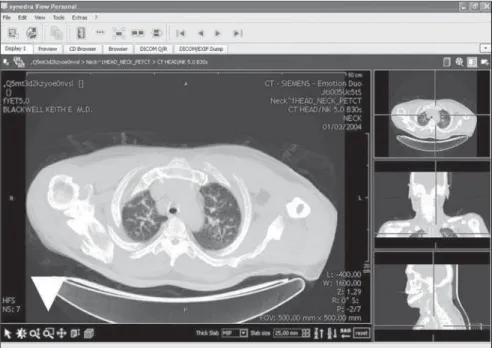
Synedra View Personal – Apowerful PACS client. The interface is quite unique, with the buttons located at the bottom of
the screen, which, initially, may make its use difficult. It is ISO 9001 and ISO 13485 certified, this latest one being a specific certification for medical devices. It allows the utilization of two monitors and per-forms both MPR and MIP, with the possi-bility of adjusting the slices thickness and also the way they are grouped, MIP,
me-dium or mini-MIP (minimum intensity pro-jection) (Figure 4). Its most important shortcoming is the fact that it lacks a data bank, i.e., it does not save the visualized exams, which can only be done manually for later visualization. It is also possible to save the performed reconstructions, an in-teresting feature. This is a rapid and friendly software.
Mito – Mito (medical imaging toolkit) is an open source software with some in-teresting features. Among the evaluated softwares, it is the only one capable of forming images fusion (Figure 5). It per-forms VR and MIP. It does not allow im-ages to be saved in the JPG and BMP for-mats and cannot open more than one series at a time. As compared with others, it is a little bit slow.
Tudor DicomViewer 1.3 – A simple software with few functions. It requires Java Runtime Environment® installation. It does not perform MPR, MIP or VR. It is ca-pable of saving images in JPG or BMP, however, only one image at a time (Figure 6). In spite of its simplicity, it allows the use of three monitors. The exams can be opened with the ImageJ, a software that is included, and presents innumerable re-sources and plugins, MPR and VR, for example.
DISCUSSION
Over the past few years, notebooks and personal computers have become very popular and widely available, and are be-coming increasingly powerful. A digital transformation also occurred in radiology services. With the availability of digital apparatuses (CT, MRI, PET-CT, ultra-sonography) older X-ray and fluoroscopy apparatuses were converted, allowing the direct and indirect acquisition of digital images.
Thanks to the use of RIS and PACS sys-tems and report workstations, films and paper have been abandoned in the whole process, from the images acquisition to the final report issuing. For this purpose, it is necessary to utilize updated softwares compatible with such changes, which make the use of freeware more difficult, considering that they do not generate a return on the investment made by their
Figure 2. KPACS software screen. Notice that the function buttons are large and located at the bottom of the screen (arrowhead).
developers, since their updating processes take a long time to be completed or are not performed at all.
Several tools are utilized in the radiolo-gists’ daily practice. Current tomography apparatuses are capable of acquiring vol-umes and, with the use of MPR,
reconstruc-tions can be made, sometimes isotropic, in the desired plane, being useful in the im-ages interpretation. MIP is a powerful tool for the analysis of CT angiography, MR an-giography, and MR cholanan-giography, but it also facilitates the identification of pulmo-nary nodules at chest CT. With the use of
VR, more agreeable and easily visualizable both by the laymen and the assisting phy-sician who have requested the study, com-ing closer to reality, thus enhanccom-ing the value of the imaging method. The PET/CT fusion image is useful in the accurate ana-tomic identification of PET uptake areas. The possibility of adding comments on the images facilitates later searches for find-ings or diseases, considering that with the greater storage capacity, personal comput-ers have been increasingly utilized to file cases. In cardiac radiology, the “cine” tool is essential, as it provides dynamic images of the functioning heart. Synchronizing different imaging series and showing ref-erence lines help in the identification of lesions or specific points, a very useful capability at MRI.
The utilization of medical viewers for diagnostic purposes is somewhat contro-versial. The Agência Nacional de Vigilân-cia Sanitária (Anvisa)(the Brazilian agency for health surveillance) does not provide specific regulations on this matter, there-fore the authors resource to there-foreign agen-cies. Among the evaluated programs, ClearCanvas is certified by the Canadian Department of Health (Health Canada MDR) and both ClearCanvas and Synedra are certified under ISO 13485:2003 (spe-cific international standard for medical equipment).
CONCLUSION
The internet is a vast source of informa-tion and softwares. Several DICOM view-ers, many of them freeware, are currently available These are excellent work tools, representing good alternatives to commer-cial softwares.
The selection of the appropriate soft-ware is many times difficult. The softsoft-ware with more functions is not always the best. Most of times, the best option is the one that best suits the user’s needs, whether be it MPR, image fusion, CD generation with a built-in viewer or a simple and rapid PACS client. In cases where the software will be permanently linked to the server network, visualizing a single image at a time, Synedra and Onis are indicated for being fast and not highly demanding in terms of computer memory; the difference
Figure 4. Synedra View Personal software screen. The multiplanar reconstruction function is being used, showing the three orthogonal planes. On the main picture, the maximum intensity projection function (MIP) is being used, showing the reconstruction of a 25 mm-thick section. The function buttons are lo-cated at the bottom of the screen (arrowhead).
between them is that the first one performs MIP and the second, VR. In cases where the exams are interpreted far from the network server or even in cases where there is the need to store them in files or for later analy-sis, ClearCanvas is indicated.
At IMEB, for example, two softwares are currently in use, one of them more com-plete and fast for CT, MRI and ultrasonog-raphy that does not perform MIP (ClearCanvas), and another simpler one for PET-CT with fusion capability (Mito).
Clear Canvas has been chosen because of its higher number of functions required in the IMEB daily routine and for its easier utilization. One should remind that it is important to analyze the personal needs and then select a software that best suits such needs, and if at all possible testing an ad-ditional alternative.
REFERENCES
1. Liao W, Deserno TM, Spitzer K. Evaluation of free non-diagnostic DICOM software tools. vol. 6919. SPIE; 2008. [cited 2010 Apr 10]. Available
Figure 6. Tudor Dicom Viewer screen. Two series are shown. Notice the absence of reference lines or synchronization.
from: http://spie.org/x648.html?product_id= 770431
2. Zeman RK, Lyshkow H, Garra BS, et al. View-ing DICOM-compliant CT images on a desktop personal computer: use of an inexpensive DICOM receive agent and freeware image display applications. AJR Am J Roentgenol. 1999;172: 305–8.
3. Escott EJ, Rubinstein D. Free DICOM image viewing and processing software for your desk-top computer: what’s available and what it can do for you. Radiographics. 2003;23:1341–57.
4. Varma DR. Free DICOM browsers. Indian J Radiol Imaging. 2008;18:12–6.
5. Rosset A, Spadola L, Ratib O. OsiriX: an open-source software for navigating in multidimen-sional DICOM images. J Digit Imaging. 2004;17: 205–16.
6. The Cyclops Group. Research on Medical Imag-ing Software [Internet]. Florianópolis, Brasil: The Cyclops Group; c1992-2010; [cited 2010 Apr 10]. Available from: http://cyclops.telemedicina.ufsc. br/
7. Caritá EC, Matos ALM, Azevedo-Marques PM. Ferramentas para visualização de imagens médi-cas em hospital universitário. Radiol Bras. 2004; 37:437–40.
8. Andrade MA, Costa MVS, Silva AMM. Java-based plugin for tomographic reconstruction for SPECT data. Med Phys. 2006;33:2015. 9. Crabb A. Free DICOM and medical image viewer
/ Converter software, open source DICOM con-version [Internet]. Baltimore, USA; c2002-2010; [revised 2009 Nov 19; cited 2010 Apr 10]. Avail-able from: http://www.idoimaging.com/
10. RTstudents.com. Radiology for students and pro-fessionals [Internet]. USA: c2004-2010; [revised 2009 Nov 19; cited 2010 Apr 10]. Available from: http://www.rtstudents.com/pacs/free-dicom-viewers.htm