Contents lists available atScienceDirect
Acta Tropica
j o u r n a l h o m e p a g e :w w w . e l s e v i e r . c o m / l o c a t e / a c t a t r o p i c a
Trypanosoma cruzi
nucleoside triphosphate diphosphohydrolase 1
(TcNTPDase-1) biochemical characterization, immunolocalization and
possible role in host cell adhesion
Christiane Mariotini-Moura
a,b, Matheus Silva e Bastos
a,b, Felipe Freitas de Castro
a,1,
Mellina Lanna Trindade
a, Raphael de Souza Vasconcellos
a,b,
Myrian Augusta Araújo Neves-do-Valle
a,b, Bernardo Pereira Moreira
a,1,
Ramon de Freitas Santos
a,b,1, Claudia Miranda de Oliveira
a,b,
Luana Celina Seraphim Cunha
a,3, Xênia Macedo Souto
a,2, Gustavo Costa Bressan
a,
Abelardo Silva-Júnior
g, Munira Muhammad Abdel Baqui
c,
Maria Terezinha Bahia
d, Márcia Rogéria de Almeida
a, José Roberto Meyer-Fernandes
e,f,
Juliana Lopes Rangel Fietto
a,b,∗aDepartamento de Bioquímica e Biologia Molecular, Universidade Federal de Vic¸osa, Vic¸osa, CEP 36570-000 MG, Brazil
bInstituto Nacional de Biotecnologia Estrutural e Química Medicinal em Doenc¸as Infecciosas – INBEQMeDI, Brazil
cDepartamento de Biologia Celular e Molecular e Bioagentes Patogênicos, Faculdade De Medicina de Ribeirao Preto, Universidade de São Paulo, Ribeirão
Preto, CEP 14090-900 SP, Brazil
dNúcleo de Pesquisa em Ciências Biológicas – NUPEB, Universidade Federal de Ouro Preto, Ouro Preto, CEP 35400-000 MG, Brazil
eInstituto de Bioquímica Médica, Universidade Federal do Rio de Janeiro, Rio de Janeiro, CEP 21941-590 RJ, Brazil
fInstituto Nacional de Biologia Estrutural e Bioimagem, IMBEB, Brazil
gDepartamento de Veterinária, Universidade Federal de Vic¸osa, Vic¸osa, CEP 36570-000 MG, Brazil
a r t i c l e
i n f o
Article history: Received 3 January 2013
Received in revised form 5 November 2013 Accepted 11 November 2013
Available online 19 November 2013
Keywords:
Recombinant protein Trypanosoma cruzi Nucleoside triphosphate diphosphohydrolase Immunolocalization Adhesion
a b s t r a c t
Previous work has suggested thatTrypanosoma cruzi diphosphohydrolase 1 (TcNTPDase-1) may be involved in the infection of mammalian cells and serve as a potential target for rational drug design. In this work, we produced recombinant TcNTPDase-1 and evaluated its nucleotidase activity, cellular localiza-tion and role in parasite adhesion to mammalian host cells. TcNTPDase-1 was able to utilize a broad range of triphosphate and diphosphate nucleosides. The enzyme’sKmfor ATP (0.096 mM) suggested a capabil-ity to influence the host’s ATP-dependent purinergic signaling. The use of specific polyclonal antibodies allowed us to confirm the presence of TcNTPDase-1 at the surface of parasites by confocal and electron microscopy. In addition, electron microscopy revealed that TcNTPDase-1 was also found in the flagellum, flagellum insertion region, kinetoplast, nucleus and intracellular vesicles. The presence of this enzyme in the flagellum insertion region and vesicles suggests that it may have a role in nutrient acquisition, and the widespread distribution of TcNTPDase-1 within the parasite suggests that it may be involved in other biological process. Adhesion assays using anti-TcNTPDase-1 polyclonal antibodies as a blocker or purified recombinant TcNTPDase-1 as a competitor revealed that the enzyme has a role in parasite–host cell adhesion. These data open new frontiers to future studies on this specific parasite–host interaction and other unknown functions of TcNTPDase-1 related to its ubiquitous localization.
© 2013 The Authors. Published by Elsevier B.V.
Abbreviations:NTPDas, nucleoside triphosphate diphosphohydrolase; TcNTPDase-1,T.cruziNTPDase-1; NDP, nucleoside diphosphate; NTP, nucleoside triphosphate.
∗Corresponding author at: Departamento de Bioquímica e Biologia Molecular, Universidade Federal de Vic¸osa, Av. P.H. Rolfs, s/n, Vic¸osa, CEP-36570-000 MG, Brazil. Tel.: +55 031 31 38993042; fax: +55 031 31 38992374.
E-mail addresses:[email protected],[email protected](J.L.R. Fietto).
1 Present address: Faculdade De Medicina de Ribeirão Preto, Universidade de São Paulo, Ribeirão Preto, CEP 14090-900 SP, Brazil. 2 Present address: Instituto Oswaldo Cruz-IOC-FIOCRUZ, Rio de Janeiro, CEP 21040-360 RJ, Brazil.
3 Present address: Faculdade de Sergipe, FaSe – Faculdade de Sergipe, Aracaju, CEP 49020-490 SE, Brazil.
0001-706X © 2013 The Authors. Published by Elsevier B.V. http://dx.doi.org/10.1016/j.actatropica.2013.11.008
Open access under CC BY-NC-ND license.
1. Introduction
Trypanosoma cruziis a flagellate protozoan known to be the eti-ological agent of Chagas disease (Chagas, 1909). The World Health Organization estimates that 8 million people are infected withT. cruziworldwide, predominantly in Latin America (WHO, 2010). The disease is expanding to non-Latin American countries and remains a serious health problem because it is difficult to diagnose and to treat the chronic form of the disease, and there is no vaccine.
Ecto-Nucleoside Triphosphate Diphosphohydrolases (NTP-Dases) are enzymes that hydrolyze ATP and other tri- and diphosphate nucleosides (Plesner, 1995; Zimmermann, 1999). Extracellular nucleotides act as signaling molecules in the immune response of mammalian hosts, and they may be hydrolyzed by par-asite ectonucleotidases. This hydrolysis could interfere with several events, such as ADP-dependent platelet aggregation and the ATP-dependent inflammatory response (Bours et al., 2006; de Almeida Marques-da-Silva et al., 2008; de Souza et al., 2010; Maioli et al., 2004; Sansom et al., 2008).
T. cruzi has ectonucleotidase activity on its surface, and an NTPDase gene was identified and cloned (TcNTPDase-1); subse-quently, the recombinant protein was expressed in a bacterial system (Fietto et al., 2004; Santos et al., 2009). In these previous studies, we demonstrated a positive correlation between extracel-lular ATP hydrolysis and the infectivity and virulence ofT. cruzi, and we suggested that TcNTPDase-1 would be a good target for ratio-nal drug design for Chagas disease chemotherapy, mainly because anti-TcNTPDase-1 antibodies decreased the infection (Santos et al., 2009). Other authors believe that high ecto-ATPase activity in pathogens is an adaptive parasitic behavior, and it has made these organisms more virulent because it could interfere with extra-cellular purinergic signals (Bisaggio et al., 2003; Sansom et al., 2007; Silverman et al., 1998). Based on the typical function of this family of proteins, it has been proposed that these enzymes can modulate biological responses induced by extracellular nucleotides and metabolites (Sansom et al., 2008). Furthermore, trypanoso-matids are unable to synthesize purine ringsde novo(Cohn and Gottlieb, 1997), depending instead on the salvage pathway (Borst and Fairlamb, 1998), in which NTPDases are suggested to have a role in extracellular purine acquisition (Berredo-Pinho et al., 2001).
Previously, we demonstrated that polyclonal antiserum against TcNTPDase-1 significantly decreased rates of T. cruzi in vitro infection. It did not, however, inhibit the enzymatic activity (nucleotidase activity) of the recombinant TcNTPDase-1 protein, suggesting a possible non-activity-dependent role for this enzyme in in vitro infection (Santos et al., 2009), possibly at an initial infection step, such as adhesion. In the present work, to bet-ter understand the role of TcNTPDase-1 inT. cruziinfection and in parasite biology, we expressed, purified and characterized the recombinant enzyme by its substrate preference and used it to investigate its immunolocalization and role in host cell-adhesion.
2. Materials and methods
2.1. Bacterial heterologous expression and TcNTPDase-1 purification
The recombinant form of T. cruzi NTPDase-1 (Accession no. AY540630) was expressed in a bacterial heterologous system as described previously (Santos et al., 2009). The purification and protein refolding were performed following previously described protocols (Areas et al., 2002) with few modifications. The lysis buffer contained 50 mM Tris pH 8.0, 100 mM NaCl and a protease inhibitor cocktail (aprotinin [1g/mL], pepstatin [1g/mL] and leupeptin [1g/mL]). Lysozyme (1 mg/mL) was added to facilitate
cell lysis, which was conducted using sonication (6 pulses of 10 s with 10 s intervals between each pulse of 20 Hz amplitude). The centrifugation steps were performed at 12,500×g, and the pel-let was solubilized and suspended in buffer (50 mM Tris pH 8.0, 500 mM NaCl) and stored at 4◦C for 24 h before use. A second purifi-cation step was performed using nickel affinity chromatography Ni-NTA-agarose (GE-Healthcare®). The equilibrium/wash buffer
contained 50 mM Tris pH 8.0, 100 mM NaCl, 10 to 20 mM imidazole, 8 M urea and 10 mM-mercaptoethanol. The elution buffer con-tained 250 mM imidazole and decreasing concentrations of urea, with fixed concentrations for the other constituents.
2.2. TcNTPDase-1 activity
The enzymatic activity was measured using the malachite green method (Ekman and Jager, 1993) with modifications. The assays were conducted in a total reaction volume of 80L, including the activity buffer (50 mM Tris, pH 8.0; 50 mM HEPES, pH 8; 2.5 mM MgCl2; 116 mM NaCl; 5.4 mM KCl; and 2.5 mM nucleotide) and 0.5g of purified TcNTPDase-1 for 30 min at 37◦C. TcNTPDase-1 presents linear hydrolysis until TcNTPDase-1 h (data not shown). After the addition of the colorimetric reagent, the reactions were read at 650 nm. To determine the Km and VMAX values, 0.5g of puri-fied TcNTPDase-1 were incubated in the same reaction medium described above in the presence of varying concentrations of ATP. The ATPase activity was measured at different periods of time, and the ATP hydrolysis did not exceed 10%. In these experiments, the ATPase activity was determined by measuring the hydrolysis of [␥-32P] ATP (specific activity of approximately 104Bq/nmol ATP) (Lemos et al., 2000). To evaluate the stability of the refolded pro-tein, each of three samples was divided into three different aliquots. From each sample, one aliquot was stored at−22◦C, another one at 4◦C and the last one at 22◦C. Then, enzymatic activity (UDPase) assays were performed for 20 consecutive days starting from time “zero” after purification. Before each test, all samples were kept on ice for 5 min.
2.3. Anti-TcNTPDase-1 polyclonal antiserum production and the purification of specific antibodies
The recombinant TcNTPDase-1 purified by nickel affinity chro-matography was used to produce specific polyclonal antiserum as previously described (Santos et al., 2009). All of the procedures were performed according to the guidelines of the Brazilian Col-lege of Animal Experimentation (COBEA). The immune antiserum was used to purify specific anti-TcNTPDase-1 antibodies. To purify the specific IgGs against TcNTPDase-1 that were present in the polyclonal antiserum, the purified recombinant TcNTPDase-1 pro-tein was coupled to CNBr-Sepharose Fast Flow 4B according to the manufacturer’s instructions (GE®). Specific anti-TcNTPDase-1 IgGs
were purified as described previously (Chandler, 2007).
2.4. Parasites
We used aT. cruziY strain isolated from an acute human case. This strain leads to low parasitemia and high mortality in mice (Silva and Nussenzweig, 1953).T. cruziepimastigotes grown in LIT medium and frozen in liquid nitrogen were thawed and seeded in Grace’s medium (Sigma) containing 5% fetal bovine serum (FBS) at 28◦C.
2.5. Western blot
PBS. A 10% SDS-PAGE gel was loaded with 20g of total protein extract from non-infective axenic epimastigotes. The proteins were separated by electrophoresis and blotted on a nitrocellulose mem-brane. We used anti-TcNTPDase-1 purified antibodies (1:1.000) as primary antibodies and anti-rabbit-IgG conjugated with FITC (Sigma®) as secondary antibodies (1:10,000).
2.6. Adhesion assay and blocking with anti-TcNTPDase-1 antibodies
A modification of the method previously described (Santos et al., 2009) was used to determine whether epimastigotes attached to VERO cell monolayers. Briefly, the epimastigote form of the para-site was grown in Grace’s medium at 26◦C until the culture reached the mid-log phase of growth and was then resuspended in RPMI with 5% FBS. Epimastigotes (20:1 parasites per cell) were gently washed with PBS and placed in contact with VERO cells that had been previously cultured for 48 h in RPMI with 5% FBS, removed with trypsin and plated on sterile coverslips (13 mm) at 5×10⁵cells per coverslip. The parasites interacted with the cells for 30 min at 4◦C. The coverslips were gently washed with PBS at 4◦C, fixed withBouinsolution for 15 min, stained with Giemsa and analyzed by light microscopy (Santos et al., 2009). Anti-dog IgG (Santa Cruz Biotechnology®) was used as negative control, and VERO cells plus
epimastigotes were used as positive control. The percentage of adhered parasites was determined by counting 300 cells, in trip-licate, in the presence or absence of polyclonal anti-TcNTPDase-1 at a dilution ratio of 1:100. The adhesion assays were performed in three independent experiments.
2.7. Adhesion assays and inhibition by competition with recombinant TcNTPDase-1
The adhesion competition assays with TcNTPDase-1 were con-ducted at different protein concentrations (0.01, 0.05, 0.1, 0.2, 0.5, 1.0, 2.0 and 4.0g/mL). The protein concentrations were deter-mined by the Bradford method (Bradford, 1976) using 96-well microplates (Biorad®). VERO cells were cultured for 48 h on sterile,
round, glass coverslips in a 24-well tissue culture plate at a density of 5×10⁵cells per coverslip in RPMI with FBS 5% at 37◦C with 5% CO2. To study the inhibition of adhesion, VERO cells were incubated with the recombinant protein for 5 min. Epimastigotes were then centrifuged, counted and resuspended at the desired concentration in RPMI with 5% FBS and added to the cell monolayers as described above (Santos et al., 2009). Albumin (4g/mL), recombinant pro-tein elution buffer and denatured TcNTPDase-1 (0.5g/mL, 5 min boiled at 95◦C) were used as negative controls, and VERO cells plus epimastigotes was used as a positive control. The percent-age of adhered parasites was determined by counting 300 cells in triplicate in the presence or absence of TcNTPDase-1 at each recom-binant protein concentration. The adhesion assays were performed as three independent experiments.
2.8. Immunolocalization of TcNTPDase-1 in epimastigotes by confocal laser scanning microscopy
The immunolocalization of TcNTPDase-1 in epimastigotes was performed with epimastigotes obtained as described above. The parasites were washed twice in PBS and settled onto glass slides containing 1% poly-lysine. After one wash with PBS, they were directly fixed for 10 min at room temperature with PBS contain-ing 4% paraformaldehyde and then blocked in PBS plus 2% BSA. The samples were incubated with a purified polyclonal antibody against TcNTPDase-1 (dilution 1:50) in PBS plus 2% BSA for 1 h at room temperature. The slides were washed in blocking solution and sub-sequently incubated for 30 min at 37◦C with Alexa 488-conjugated
goat anti-rabbit IgG secondary antibody (Invitrogen Life Technolo-gies) at a dilution of 1:400. The glass slides were mounted with Prolong Gold Antifade Reagent containing DAPI (Molecular Probes) and examined by confocal microscopy (Leica, SP5) at the Faculdade de Medicina de Ribeirao Preto-USP, Ribeirao Preto, SP (Baqui et al., 2000).
2.9. Ultrastructural immunocytochemistry
For transmission electron microscopy analysis, epimastigotes were fixed in 4% paraformaldehyde, 0.5% glutaraldehyde, 5 mM cal-cium chloride, and 3.7% sucrose in a 100 mM sodium cacodylate buffer (pH 7.2). The samples were gradually dehydrated in alcohol at low temperatures, infiltrated, and finally set in LR White resin at 60◦C. Ultrathin sections were collected on nickel grids of 300 mesh and incubated for 20 min at room temperature in 50 mM ammo-nium chloride in PBS at pH 7.2. Next, the sections were incubated in PBS (pH 8.0) containing 1.5% albumin and 0.01% Tween 20 for 20 min at room temperature and then overnight in the presence of purified anti-TcNTPDase1 (1:100 or 1:50 as indicated) except for control grids. The grids were washed in PBS and finally incubated (1:30) with a secondary anti-rabbit IgG produced in goat and con-jugated with 10 nm gold particles for 60 min. The ultrathin sections were contrasted with solutions of 3% uranyl acetate and 0.2% lead citrate. All of the materials were observed and photographed in a transmission electron microscope (Zeiss EM 109) at the Núcleo de Microscopia e Microanálise at Universidade Federal de Vic¸osa, Minas Gerais, Brazil.
2.10. Statistical analysis
The data were statistically analyzed using the ANOVA Holm–Sidak method using SigmaPlot software, Version 11.0 2008, andp< 0.05 was considered statistically significant.
3. Results and discussion
3.1. TcNTPDase-1 heterologous expression, purification and biochemical characterization
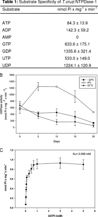
The TcNTPDase-1 gene was previously described by our group (Fietto et al., 2004), and its protein was demonstrated to be a vir-ulence factor and facilitator of infectivity (Santos et al., 2009). To expand on the TcNTPDase-1 studies and biochemical characteri-zation, we expressed the recombinant TcNTPDase-1 in a bacterial system and used it in biochemical and biological studies. The puri-fied recombinant protein presented only one protein band in a Coomassie blue-stained gel as previously shown (Santos et al., 2009). The recombinant protein was purified in its active form and showed a greater ability to hydrolyze diphosphate nucleosides over their respective triphosphate nucleosides (Fig. 1A and B). A sub-strate specificity characterization demonsub-strated that TcNTPDase-1 is a genuine apyrase enzyme. The hydrolysis intensity followed the order GDP = UDP > GTP = UTP > ADP = ATP (Fig. 1A), consistent with the results shown for ATP and ADP in a previous study from our group (Santos et al., 2009). The refolding and temperature stabil-ity tests using UDP as a substrate showed that the recombinant TcNTPDase-1 was more stable when stored at 4◦C than at room temperature (22◦C) or at a freezing temperature (−22◦C) (Fig. 1B). Furthermore, the activity increased until five days after purification, remained stable until approximately day 10, and then decreased after this time, descending to levels lower than 50% of the highest activity (obtained at day 5 after purification).
Fig. 1.Heterologous TcNTPDase-1 substrate specificity, stability andKm. Enzyme activity was assayed using the malachite green method in a reaction buffer contain-ing 50 mM HEPES, 50 mM Tris, 116 mM NaCl, 5.4 mM KCl, 2.5 mM MgCl2and 2.5 mM substrate, with the pH adjusted to 8.0. The results are the means±SD from at least three independent experiments. After TcNTPDase-1 purification, each of the three samples was aliquoted and stored to verify the ideal storage temperature. Activity assays were performed for 20 days using UDP as a substrate (B). (C) TheKmfor ATP. Activity was determined by measuring the hydrolysis of [␥-32P] ATP. The results are the means±SD from at least three independent experiments.
some distinctions. The human NTPDase 6 hydrolyzes GDP more effectively than UDP and GTP more effectively than UTP, whereas TcNTPDase-1 hydrolyzes GDP and UDP equally well; likewise, it hydrolyzes GTP and UTP equally well. Similar observations can be made from the study of human NTPDase 5 (Ivanenkov et al., 2003; Murphy-Piedmonte et al., 2005; Santos et al., 2009).
It is important to note that the hydrolytic capabilities assayed using an excess of substrate and a long time reaction (Fig. 1A) may not be directly related to the sensitivity to or affinity for the nucleotides, as demonstrated in the kinetic test with recombinant TcNTPDase-1, using ATP as the substrate. In this assay, we verified aKmof 0.096 mM (Fig. 1C), whereas theKmfor UDP was almost 10 times higher (data not shown), suggesting that we cannot rule out the importance of ATP hydrolysis, even though it had the low-est hydrolysis intensity in the substrate specificity assay (Fig. 1A). These data strengthen an idea proposed in our previous works (Fietto et al., 2004; Santos et al., 2009), in which we suggested that the hydrolytic activity of this enzyme might have a role in mod-ulating the host ATP-dependent purinergic signaling, such as that involved in the immune system. This supposition is based only on theKmfor ATP (e.g., CD39Kmfor ATP is 0.01–0.2 mM) (Zimmermann et al., 2012) and P2 receptors EC50, that would allow its desensiti-zation by TcNTPDase activity (e.g., P2X7 EC50for ATP is 0.1 mM) (Khakh et al., 2001). Nevertheless, because we used a purified pro-tein, the real role in physiological conditions needs to be better characterized in future studies. The importance of these receptors in triggering the immune response of the host is well known (Bours et al., 2006; Burnstock, 2007). Recent studies have demonstrated the importance of purine receptors in the elimination of intracellu-lar pathogens such asLeishmania. It has been shown that, during infection, the host cell increases its expression of these recep-tors, but they are inactive or have their activation blocked by an unknown mechanism (Chaves et al., 2009; Marques-da-Silva et al., 2011).
3.2. Immunodetection and immunolocalization of TcNTPDase-1 using specific antibodies
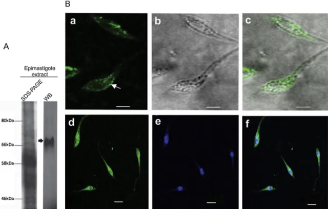
We purified specific antibodies using affinity chromatogra-phy, and these antibodies were able to detect the recombinant TcNTPDase-1. Western blotting analysis revealed a single band of approximately 70 kDa in epimastigotes (non-infective form) (Fig. 2A). This molecular weight is compatible with the weight of TcNTPDase-1 previously predicted by our group (Fietto et al., 2004). Assays ofin vitroenzymatic activity using the anti-TcNTPDase-1 antibody had not previously shown inhibitory effect against the recombinant protein, suggesting that this antibody might recog-nize a protein homologous to TcNTPDase-1 or that it might bind to TcNTPDase-1 in a region inessential for enzymatic activity (Santos et al., 2009). The antiserum, however, was able to inhibit thein vitro infectivity by approximately 50%, suggesting that binding to these other regions of TcNTPDase-1 still affects the infective ability of the trypomastigote.
Fig. 2.Immunodetection of TcNTPDase-1 inT. cruzi. (A) Western blot (WB) analysis of TcNTPDase-1 expression in epimastigote extract. After incubation with rabbit antibodies anti-NTPDase-1 (1:1000) and anti-IgG conjugated with FITC (1:10,000), nitrocellulose membranes were analyzed in FLA 5100 (Fujifilm®). The SDS-PAGE gel was stained
with Coomassie blue. (B) TcNTPDase-1 distribution in epimastigotes by confocal microscopy. Non-permeabilized parasites were fixed and stained with anti-TcNTPDase-1 (1:50) and visualized with Alexa 488-conjugated goat anti-rabbit IgG (a, d) (white arrow indicates the inner middle body), phase contrast (b) and merged (c, f). Nuclei and kinetoplasts were labeled with DAPI (e). Bar = 5m.
activities in epimastigotes (Field and Carrington, 2009; Landfear and Ignatushchenko, 2001).
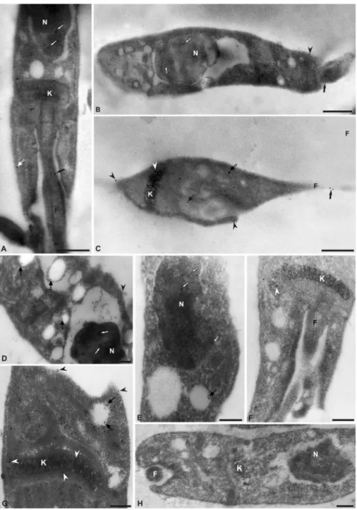
The subcellular localization of the enzyme in the epimastig-ote form was determined by immunoelectron microscopy using purified polyclonal antibodies against TcNTPDase-1. In an ultra-structural analysis, gold particles were visualized predominantly in the cell body, nucleus, flagellum and flagellar pocket region (Fig. 3). The presence of TcNTPDase-1 in the flagellum insertion region reca-pitulates the data suggested by the confocal images (Fig. 2B) and reinforces the possible role of this protein in nutrient acquisition by epimastigotes because they are a replicative form of the para-site, which has a metabolism that requires high levels of purines and derivatives to replicate DNA, transcribe RNA and execute other purine-dependent pathways (Berens et al., 1981).
Staining was also observed in the kinetoplast, in cytoplasmic vesicles and, to a lesser extent, on the external cell surface (white arrowhead) and inner cell surface (black arrowhead) (Fig. 3). No staining was observed in the control assay (e.g., Fig. 3H). The vesicular localization of this enzyme could be reservosomes. A recent and specific reservosome proteomic study revealed the pres-ence of TcNTPDase-1 in these organelles (Sant’Anna et al., 2009). These data reinforce the possible involvement of TcNTPDase-1 in the metabolic nutrition of epimastigotes because reservosomes are described as multivesicular bodies that are the main site for the storage of ingested proteins and lipids, as well as for secre-tory proteins that are synthesized by protozoans. Nevertheless, TcNTPDase-1 vesicular localization requires further investigation. In addition, TcNTPDase-1 has a signal peptide in its amino-terminal domain (Fietto et al., 2004). The peptide signal suggests TcNTPDase-1 may also be secreted, but this cannot be currently verified experimentally.
The observed cell surface localization is in accord with pre-vious data that described NTPDase activity on the cell surface of T. cruzi (Bernardes et al., 2000; Bisaggio et al., 2003; Fietto
et al., 2004; Meyer-Fernandes et al., 2004; Santos et al., 2009). Furthermore, immunolocalization using non-homologous anti-T. gondiiNTPDase1 antibodies revealed the presence of a NTPDase-homologous protein on the surface ofT. cruzi(Fietto et al., 2004). Our data support this observation with ultrastructural immunolo-calization using specific purified antibodies, and the lower level of surface detection is related to this post-inclusion technique.
The spread profile of TcNTPDase-1 subcellular localization is reinforced by recent findings showing similar ubiquitous local-ization of an NTPDase in Leishmania braziliensisand Leishmania amazonensispromastigotes, suggesting their involvement with var-ious biological process within these parasites (Detoni et al., 2013; Porcino et al., 2012).
3.3. TcNTPDase-1 may play a role in parasite adhesion to mammalian cells
Ectonucleotidases are very important for many biological pro-cesses, and their importance in infections caused by pathogens, including protozoa such as T. cruzi (Sansom et al., 2008), has been studied. We demonstrated that a high ratio of extracellu-lar ATP/ADP hydrolysis is important for the trypomastigote-stage parasite to maintain the ability to infect VERO cells (Santos et al., 2009). In that work, we found that the partial inhibition of ecto-ATPDase activity decreasedin vitroinfectivity and parasite virulence. Alternately, the antibodies produced against the recom-binant TcNTPDase-1 did not inhibit the ATPDase activity of the recombinant protein or the ecto-ATPDase activity in live para-sites. However, the same antibodies were able to inhibit anin vitro infection, suggesting a specific role for TcNTPDase-1 that would be partially independent of enzymatic activity.
Fig. 3.Ultrastructural analysis of TcNTPDase-1 subcellular localization in epimastigotes. (A–G) Electromicrographs using polyclonal anti-TcNTPDase-1 (1:110 A–C, and 1:50 D–G) and anti-IgG conjugated to 10 nm colloidal gold. TcNTPDase-1 is shown in the nucleus (N) (white arrows), kinetoplast (K) (white arrowhead), internal vesicles (dashed black arrow), flagellum (F) (black arrow), flagellum insertion region (panel F surrounding the flagellum), and outer and inner cell surface (black arrowheads and dashed white arrow, respectively). No staining was observed in the control (H). Bars: A, C–H = 0.2m; B = 0.5m.
stage of infection in mammalian cells, specifically in the adhesion step.
The adhesion assays were performed using epimastigotes at the log phase of growth. Direct adhesion assays were performed using VERO cells as a model. To avoid internal-ization, the assays were conducted at 4◦C for 30 min as previously described (Bisaggio et al., 2003). The adhesion assays were conducted either with cells that had been pre-viously incubated in the presence of purified recombinant protein in a competition assay (Fig. 4A) or with epimastigotes that had been previously incubated with purified anti-TcNTPDase-1 to directly block native TcNTPDase-anti-TcNTPDase-1 (Fig. 4B). In the presence of increasing amounts of the recombinant protein (Fig. 4A), we observed a significant dose-dependent inhibition of epimastigote adhesion, suggesting the presence of binding sites on the host cell that would be partially blocked by recombinant TcNTPDase-1.
Furthermore, the nucleotidase activity of the recombinant pro-tein could also influence this event. There was no inhibitory effect when the assay was performed in the presence of an unrelated pro-tein (albumin) or denatured TcNTPDase-1 (Fig. 4A). The parasites that had been previously incubated with purified polyclonal anti-TcNTPDase-1 antibodies (Fig. 4B) exhibited a significant decrease in motility (data not shown) and a significant decrease in adhe-sion (compared to the controls) in the absence of antibodies or in the presence of an unrelated antibody (anti-dog IgG). The effects of anti-TcNTPDase-1 are specific and independent of the conserved portion of IgGs, as shown by the fact that the control using unre-lated anti-IgG did not differ from the control without any antibodies (Fig. 4B). Although the decrease in motility could contribute to the observed decrease in adhesion.
Fig. 4.Participation of TcNTPDase-1 in cell adhesion. (A) Adhesion assays by competition with TcNTPDase-1. VERO cells were previously placed in contact with various concentrations of recombinant protein for 5 min (0.01, 0.05, 0.1, 0.2, 0.5, 1.0, 2.0 and 4.0g/mL). Epimastigotes were then added (20 parasites per cell). (B) Adhesion assays in the presence or absence of polyclonal anti-TcNTPDase-1. Epimastigotes were previously incubated with antibodies for 10 min and then placed in contact with the VERO cells (20:1). After incubation, coverslips were washed, fixed, stained and analyzed by light microscopy. Albumin (4g/mL BSA), the elution buffer of the recombinant protein (Buffer control), denatured TcNTPDase-1 (0.5g/mL), Anti-dog-IgG (Santa Cruz Biotechnology®) and VERO cells with epimastigotes only (VERO + Epi) were used as controls.
The percentage of adhered parasites was determined by counting 300 cells in triplicate on each coverslip tested. The results are the means±SD from at least three independent experiments.
this hypothesis: firstly, antibodies may bind to the modulatory regions of the enzyme, thus preventing it from responding to certain stimuli; alternately, the action of antibody binding could generate a steric hindrance on the surface of the parasite, impair-ing its adhesion to host cells; finally, there could be a synergic interaction between the enzyme and a host receptor, and the anti-TcNTPDase-1 would also be capable of inhibiting the adhe-sion to the host cell. All three hypotheses are currently under investigation.
4. Conclusions
Our results provide new information regarding TcNTPDase-1 nucleotidase activity, demonstrating its ability to use a broad range of triphosphate and diphosphate nucleosides. Additionally, we used specific antibodies to provide new data regarding several aspects of subcellular localization. The ubiquitous localization of TcNTPDase-1 in epimastigotes highlights the need to investigate the participation of this enzyme in other processes, in addition to
its possible roles in both purine acquisition and virulence. Further-more, we demonstrated that TcNTPDase-1could be involved in host cell adhesion. This specific parasite–host interaction and the pos-sible unknown functions of TcNTPDase-1 (related to its ubiquitous localization in replicative parasite cells) merit further study.
Acknowledgements
References
Areas, A.P., Oliveira, M.L., Ramos, C.R., Sbrogio-Almeida, M.E., Raw, I., Ho, P.L., 2002. Synthesis of cholera toxin B subunit gene: cloning and expression of a functional 6xhis-tagged protein inEscherichia coli. Protein Expr. Purif. 25, 481–487. Baqui, M.M., Milder, R., Mortara, R.A., Pudles, J., 2000. In vivo and in vitro
phos-phorylation and subcellular localization of trypanosomatid cytoskeletal giant proteins. Cell Motil. Cytoskeleton 47, 25–37.
Berens, R.L., Marr, J.J., LaFon, S.W., Nelson, D.J., 1981. Purine metabolism in Try-panosoma cruzi. Mol. Biochem. Parasitol. 3, 187–196.
Bernardes, C.F., Meyer-Fernandes, J.R., Saad-Nehme, J., Vannier-Santos, M.A., Peres-Sampaio, C.E., Vercesi, A.E., 2000. Effects of 4,4′-diisothyocyanatostilbene-2,2′ -disulfonic acid onTrypanosoma cruziproliferation and Ca(2+) homeostasis. Int. J. Biochem. Cell Biol. 32, 519–527.
Berredo-Pinho, M., Peres-Sampaio, C.E., Chrispim, P.P., Belmont-Firpo, R., Lemos, A.P., Martiny, A., Vannier-Santos, M.A., Meyer-Fernandes, J.R., 2001. A Mg-dependent Ecto-ATPase inLeishmania amazonensisand its possible role in adenosine acquisition and virulence. Arch. Biochem. Biophys. 391, 16–24. Bisaggio, D.F., Peres-Sampaio, C.E., Meyer-Fernandes, J.R., Souto-Padron, T., 2003.
Ecto-ATPase activity on the surface ofTrypanosoma cruziand its possible role in the parasite–host cell interaction. Parasitol. Res. 91, 273–282.
Borst, P., Fairlamb, A.H., 1998. Surface receptors and transporters ofTrypanosoma brucei. Annu. Rev. Microbiol. 52, 745–778.
Bours, M.J., Swennen, E.L., Di Virgilio, F., Cronstein, B.N., Dagnelie, P.C., 2006. Adenosine 5’-triphosphate and adenosine as endogenous signaling molecules in immunity and inflammation. Pharmacol. Ther. 112, 358–404.
Bradford, M.M., 1976. A rapid and sensitive method for the quantitation of micro-gram quantities of protein utilizing the principle of protein-dye binding. Anal. Biochem. 72, 248–254.
Burnstock, G., 2007. Purine and pyrimidine receptors. Cell. Mol. Life Sci. 64, 1471–1483.
Chagas, C., 1909. Nova tripanozomiaze humana: estudos sobre a morfolojia e o ciclo evolutivo doSchizotrypanum cruzin. gen., n. sp., ajente etiolojico de nova entidade morbida do homem. Mem. Inst. Oswaldo Cruz 1, 159–218.
Chandler, J.P., 2007. Purification and Characterization of Antibodies. Making and Using Antibodies: A Practical Handbook. CRC Press and Taylor & Francis Group, New York.
Chaves, S.P., Torres-Santos, E.C., Marques, C., Figliuolo, V.R., Persechini, P.M., Coutinho-Silva, R., Rossi-Bergmann, B., 2009. Modulation of P2x(7) puriner-gic receptor in macrophages byLeishmania amazonensisand its role in parasite elimination. Microbes Infect. 11, 842–849.
Cohn, C.S., Gottlieb, M., 1997. The acquisition of purines by trypanosomatids. Para-sitol. Today 13, 231–235.
de Almeida Marques-da-Silva, E., de Oliveira, J.C., Figueiredo, A.B., de Souza Lima Jr., D., Carneiro, C.M., Rangel Fietto, J.L., Crocco Afonso, L.C., 2008. Extracellular nucleotide metabolism inLeishmania: influence of adenosine in the establish-ment of infection. Microbes Infect. 10, 850–857.
de Souza, M.C., de Assis, E.A., Gomes, R.S., Marques da Silva Ede, A., Melo, M.N., Fietto, J.L., Afonso, L.C., 2010. The influence of ecto-nucleotidases onLeishmania amazonensisinfection and immune response in C57b/6 mice. Acta Trop. 115, 262–269.
Detoni, M.L., Fessel, M.R., Maia, A.C., Porcino, G.N., Quellis, L.R., Faria-Pinto, P., Mar-ques, M.J., Juliano, M.A., Juliano, L., Diniz, V.A., Côrte-Real, S., Gonc¸alves-da-Costa, S.C., Souza, C.S., Vasconcelos, E.G., 2013. An antigenic domain of theLeishmania amazonensisnucleoside triphosphate diphosphohydrolase (NTPDase 1) is asso-ciated with disease progression in susceptible infected mice. Parasitol. Res. 112 (8), 2773–2782.
Ekman, P., Jager, O., 1993. Quantification of subnanomolar amounts of phosphate bound to seryl and threonyl residues in phosphoproteins using alkaline hydrol-ysis and malachite green. Anal. Biochem. 214, 138–141.
Field, M.C., Carrington, M., 2009. The trypanosome flagellar pocket. Nat. Rev. Micro-biol. 7, 775–786.
Fietto, J.L., DeMarco, R., Nascimento, I.P., Castro, I.M., Carvalho, T.M., de Souza, W., Bahia, M.T., Alves, M.J., Verjovski-Almeida, S., 2004. Characterization and
immunolocalization of an NTP diphosphohydrolase of Trypanosoma cruzi. Biochem. Biophys. Res. Commun. 316, 454–460.
Ivanenkov, V.V., Murphy-Piedmonte, D.M., Kirley, T.L., 2003. Bacterial expression, characterization, and disulfide bond determination of soluble human NTPDase6 (CD39l2) nucleotidase: implications for structure and function. Biochem. 42, 11726–11735.
Khakh, B.S., Burnstock, G., Kennedy, C., King, B.F., North, R.A., Seguela, P., Voigt, M., Humphrey, P.P., 2001. International Union of Pharmacology. XXIV. Current sta-tus of the nomenclature and properties of P2X receptors and their subunits. Pharmacol. Rev. 53, 107–118.
Landfear, S.M., Ignatushchenko, M., 2001. The flagellum and flagellar pocket of try-panosomatids. Mol. Biochem. Parasitol. 115, 1–17.
Lemos, A.P., Peres-Sampaio, C.E., Guimaraes-Motta, H., Silva, J.L., Meyer-Fernandes, J.R., 2000. Effects of naturally occurring polyols and urea on mitochondrial F0F1 ATPase. Z. Naturforsch. C 55, 392–398.
Maioli, T.U., Takane, E., Arantes, R.M., Fietto, J.L., Afonso, L.C., 2004. Immune response induced by new worldLeishmaniaspecies in C57bl/6 mice. Parasitol. Res. 94, 207–212.
Marques-da-Silva, C., Chaves, M.M., Chaves, S.P., Figliuolo, V.R., Meyer-Fernandes, J.R., Corte-Real, S., Lameu, C., Ulrich, H., Ojcius, D.M., Rossi-Bergmann, B., Coutinho-Silva, R., 2011. Infection withLeishmania amazonensisupregulates purinergic receptor expression and induces host-cell susceptibility to UTP-mediated apoptosis. Cell. Microbiol. 13, 1410–1428.
Meyer-Fernandes, J.R., Saad-Nehme, J., Peres-Sampaio, C.E., Belmont-Firpo, R., Bis-aggio, D.F., Do Couto, L.C., Fonseca De Souza, A.L., Lopes, A.H., Souto-Padron, T., 2004. A Mg-dependent ecto-ATPase is increased in the infective stages of Trypanosoma cruzi. Parasitol. Res. 93, 41–50.
Murphy-Piedmonte, D.M., Crawford, P.A., Kirley, T.L., 2005. Bacterial expression, folding, purification and characterization of soluble NTPDase5 (CD39l4) ecto-nucleotidase. Biochim. Biophys. Acta 1747, 251–259.
Plesner, L., 1995. Ecto-ATPases: identities and functions. Int. Rev. Cytol. 158, 141–214.
Porcino, G.N., Carvalho-Campos, C., Maia, A.C., Detoni, M.L., Faria-Pinto, P., Coimbra, E.S., Marques, M.J., Juliano, M.A., Juliano, L., Diniz, V.A., Corte-Real, S., Vas-concelos, E.G., 2012. Leishmania (Viannia) braziliensis nucleoside triphosphate diphosphohydrolase (NTPDase 1): localization and in vitro inhibition of pro-mastigotes growth by polyclonal antibodies. Exp. Parasitol. 132, 293–299. Sansom, F.M., Newton, H.J., Crikis, S., Cianciotto, N.P., Cowan, P.J., d’Apice, A.J.,
Hart-land, E.L., 2007. A bacterial ecto-triphosphate diphosphohydrolase similar to human CD39 is essential for intracellular multiplication ofLegionella pneu-mophila. Cell. Microbiol. 9, 1922–1935.
Sansom, F.M., Robson, S.C., Hartland, E.L., 2008. Possible effects of microbial ecto-nucleoside triphosphate diphosphohydrolases on host–pathogen interactions. Microbiol. Mol. Biol. Rev. 72, 765–781.
Sant’Anna, C., Nakayasu, E.S., Pereira, M.G., Lourenco, D., de Souza, W., Almeida, I.C., Cunha, E.S.N.L., 2009. Subcellular proteomics ofTrypanosoma cruzi reservo-somes. Proteomics 9, 1782–1794.
Santos, R.F., Possa, M.A., Bastos, M.S., Guedes, P.M., Almeida, M.R., Demarco, R., Verjovski-Almeida, S., Bahia, M.T., Fietto, J.L., 2009. Influence of ecto-nucleoside triphosphate diphosphohydrolase activity onTrypanosoma cruziinfectivity and virulence. PLoS Negl. Trop. Dis. 3, e387.
Silva, L.H., Nussenzweig, V., 1953. Sobre uma cepa deTrypanosoma cruzivirulenta para o camundongo branco. Folia Clin. Biol. 20, 191–207.
Silverman, J.A., Qi, H., Riehl, A., Beckers, C., Nakaar, V., Joiner, K.A., 1998. Induced activation of theToxoplasma gondiinucleoside triphosphate hydrolase leads to depletion of host cell ATP levels and rapid exit of intracellular parasites from infected cells. J. Biol. Chem. 273, 12352–12359.
WHO, 2010. Chagas disease: control and elimination, sixty-third world health assembly, April, 22 ed.
Zimmermann, H., 1999. Two novel families of ectonucleotidases: molecular struc-tures, catalytic properties and a search for function. Trends Pharmacol. Sci. 20, 231–236.