Contents lists available atScienceDirect
International Immunopharmacology
journal homepage:www.elsevier.com/locate/intimp
Short communication
Evaluation of vaccinal e
ff
ectiveness of preparations containing membrane
antigens of
Leishmania
(
L.
)
amazonensis
in experimental cutaneous
leishmaniasis model
João G. Ribeiro
a,b, Amália S. Ferreira
a, Sharon R.A. Macedo
a, Norton R.D.L.P. Rossi
a,
Mayara C.P. da Silva
c, Rosane N.M. Guerra
c, Neuza B. de Barros
a, Roberto Nicolete
a,d,⁎aLaboratório de Biotecnologia Aplicada à Saúde, Fundação Oswaldo Cruz (Fiocruz Rondônia), Porto Velho, RO, Brazil bInstituto Federal do Amazonas (IFAM), Campus Humaitá, Humaitá-AM, Brazil
cLaboratory of Immunophysiology, Department of Pathology, Center for Biological and Health Sciences, Federal University of Maranhão (UFMA), São Luís, MA, Brazil dFundação Oswaldo Cruz (Fiocruz Ceará), Fortaleza, CE, Brazil
A R T I C L E I N F O
Keywords: L. amazonensis
Membrane fraction Vaccines
PLGA micro/nanoparticles Protection
A B S T R A C T
American tegumentary leishmaniasis (ATL) is considered a neglected disease, for which an effective vaccine or an efficient diagnosis is not yet available and whose chemotherapeutic arsenal is threatened by the emergence of resistance by etiological agents such asLeishmania amazonensis. ATL is endemic in poor countries and has a high incidence in Brazil. Vaccines developed from native parasite fractions have led to the identification of defined antigenic subunits and the development of vaccine adjuvant technology. The purpose of the present study was to develop and compare preparations based on membrane antigens fromL. amazonensis, as a biotechnological prototype for the immunoprophylaxis of the disease in a murine experimental model. For this purpose, batches of biodegradable polymeric micro/nanoparticles were produced, characterized and compared with other parasite's antigens in solution. All preparations containing membrane antigens presented low toxicity on murine macrophages. Thein vivo evaluation of immunization efficacy was performed against a challenge withL. amazonensis, along with an evaluation of the immune response profile generated in BALB/C mice. The animals were followed for sample processing and quantification of serum-specific cytokines, nitrites and antibodies. The sera of animals immunized with the non-encapsulated antigen formulations showed higher intensities of nitrites and total IgGs. This approach evidenced the importance of the biological studies involving the immune response of the host against the parasite being interconnected and related to the subfractionation of its proteins in the search for more effective vaccine candidates.
1. Introduction
American tegumentary leishmaniasis (ATL) is a zoonotic disease that affects man and several species of wild and domestic animals[1]. It affects around 12 million people in 88 countries, with 350 million people living in at-risk areas. It has an annual incidence of approxi-mately 2 million new cases; of these 1.5 million cases are cutaneous leishmaniasis and 500 thousand are visceral leishmaniasis[2].
One of the strongest correlations with the polarized response in mice is the subclass profile of IgG in the antigen-specific response. Non-self-cured mice assemble an IgG1- and IgE-mediated humoral response promoted by TH2 cells, in contrast to mice from resistant phenotype lines that assemble a dominant humoral response and IgG2a promoted by TH1 cells. In mice, IgG2a antibodies, which are effective in
complement opsonization andfixation, can act by individually target-ing amastigotes released from infected macrophages[3].
Although there are chemotherapeutic agents for leishmaniasis, the drugs used are expensive, limited, toxic and threatened by drug resistance mechanisms by parasites. The prevention of leishmaniasis is superior to treatment due to the patient's health impairment and the disfigurement caused during the clinical manifestations of leishmania-sis. A prophylactic vaccination may be the most effective strategy to control the infection and spread of this group of diseases[4]. However, to date there are no reports of an effective vaccine for any form of human leishmaniasis. The fact that infected people acquire immunity against reinfections indicates the feasibility of developing a vaccine[5]. With regard to theseLeishmania (L.) amazonensismembrane anti-gens, vaccine candidates for efficient activation of the immune system
http://dx.doi.org/10.1016/j.intimp.2017.04.014
Received 30 January 2017; Received in revised form 12 April 2017; Accepted 13 April 2017
⁎Corresponding author at: Fundação Oswaldo Cruz (Fiocruz Ceará), Av. Santos Dumont, 5051, sl.1303, 60175-047 Fortaleza, CE, Brazil. E-mail address:[email protected](R. Nicolete).
International Immunopharmacology 47 (2017) 227–230
Available online 25 April 2017
1567-5769/ © 2017 Elsevier B.V. All rights reserved.
have recently been described, including the effect of a protein-enriched membrane fraction with glycosyl-phosphatidyl inositol-GPI anchors, and proteoliposomes constructed from this fraction, in reducing the infection percentage in murine peritoneal macrophages and the per-centage of intracellular amastigote cells [6]. The potential for the development of biotechnological products from micro/nanostructured antigen dispersion systems for the treatment and diagnosis of leishma-niasis was evidenced by these results. Of these protein fractions, 12 proteins were identified, requiring immunogenic characterization and identification of the other proteins present for the selection and screening of defined antigenic subunits. In this context, the purpose of this study was to develop a biodegradable polymeric micro/ nanostructured system containing membrane antigens fromLeishmania and compare it to other parasite's antigens in solution.
2. Material and methods
2.1. Extraction and fractionation of proteins
Aliquots containing from 1.3 to 1.9 × 10⁷promastigotes in RPMI culture medium, subcultured for up to eight passages, were centrifuged at 1200 ×gfor 10 min to remove the culture medium, washed with Tris–HCl buffer (5 mM, pH 7.25) containing 0.2 mM CaCl2 (calcium chloride) and 1 mM NaCl (sodium chloride) and centrifuged again. The pellets formed were resuspended in 1 mL of Tris–HCl buffer and 5μL of
a mixture of protease inhibitors (GE-Healthcare) and sonicated. The samples were frozen and stored at−20 °C. The pellets were subjected to ultracentrifugation at 100,000 ×gfor 1 h at 4 °C; the supernatant was reserved in another tube, subjected to ultrafiltration to exchange the buffer for PBS; the pellet was resuspended in PBS[6]. The fraction concentrations were measured in the presence of 2% SDS using the BCA Proten Assay kit (Thermo), according to the manufacturer's instruc-tions.
2.2. Production and characterization of PLGA micro/nanoparticles
Each batch of particles was obtained by the emulsion and solvent evaporation method as described[7,8], with modifications. Briefly, the stirring system and the other materials were sterilized and mounted in a laminarflow hood to ensure sterile conditions during the preparation of the particles. One hundred mg of the polymer were dissolved in each batch using PLGA 50:50 (PURASORB® 5002) in 2 mL of dichloro-methane, sonicated for 1 min and thirty-six seconds at 50 W of power, added to a glass tube containing 9 mL of 1.5% polyvinyl alcohol solution, subjected for an ultrasound again for 7 min and 12 s at 50 W of power[6]. The resulting supernatant was discarded and the precipitate was resuspended in deionized water and centrifugation was repeated three times. Thefinal precipitate was resuspended in 5 mL of deionized water for further lyophilization and storage at−80 °C.
The distribution of the mean diameters of the micro/nanoparticu-late system and the Zeta particle potential analysis was performed using a Nano Zeta Sizer laser diffractometer (Malvern instruments, England), according to the manufacturer's instructions. Briefly, 50μL of particu-late suspension was dispersed in 1 mL of water and injected into the cuvette with a syringe for analysis.
The viability of the macrophages (1 × 10⁵cells/well) exposed to the preparations was assessed using the MTT colorimetric method. Cells were 24 h incubated at 25 °C with the micro/nanoparticles (0.5 mg/ well). After this period, 10μL of MTT solution was added to all wells and 4 h later, 50μL of SDS (20%, w/v) was added to each well. Then, after an overnight period, the plates were gently shaken and read spectrophotometrically at 570 nm. The cytotoxicity index was calcu-lated using the following equation:
Cytotoxicity = (1 − mean OD test mean OD of the negative control) × 100.
2.3. Immunization of mice with the preparations containing antigens from L. amazonensis and evaluation of the response
Male BALB/C mice, 8–10 weeks old, weighing 20–28 g, obtained
from the IPEPATRO/FIOCUZ-RO vivarium were used. The animals were kept under standard animal vivarium conditions. The experiments were carried out according to the rules established by the Ethical Committee on Animal Use (CEUA) at IPEPATRO-FIOCRUZ-RO, upon approval of the research project under number 2013/3.
Groups of eight male BALB/C mice were immunized subcutaneously (3 times at 3 weeks intervals) with bovine serum albumin (BSA), L. amazonensissoluble antigens (SLA),L. amazonensismembrane antigens (MAg), PLGA micro/nanoparticles with no protein content (P_CNT), PLGA micro/nanoparticles containing SLA (P_SLA) and PLGA nanopar-ticles containing MAg (P_MAg). After two weeks, mice received booster doses and one week later, they were challenged with stationary growth phaseL. amazonensis1 × 106promastigotes. The challenge was sub-cutaneously applied in the right hind paw of each of the mice.
In immunization groups with non-encapsulated protein antigens, the proteins were mixed with Freund's complete adjuvant in thefirst dose and with Freund's incomplete adjuvant in the booster dose. In the immunization groups with the micro/nanoparticles, they were sus-pended in sterile saline buffer (PBS) at both doses. The volume applied was 300μL, containing 40μg of particles. Two groups of eight mice each, which were not immunized, are part of the experimental controls, one in which the mice were only infected to control the challenge (INFEC) and one group in which the mice were not infected (NORMAL).
2.4. Nitric oxide and specific antibody production in immunized mouse serum
Half of the animals in each group were sacrificed 72 h after the challenge. The amount of nitric oxide produced and released in the serum of the mice was determined by the Griess assay using the procedure described[9].
Serum samples were used to determine the concentration of total IgG, IgG1 and anti-Leishmania IgG2a antibodies by enzyme-linked immunosorbent assay (ELISA) as described[10], using 40μg/ml ofL. amazonensisantigens at a ratio of 1:1000 with anti-Mouse IgG ALDRICH M8642). Biotinylated anti-Leishmania anti-IgG1 (SIGMA-ALDRICH A3562), IgG1 (SIGMA-(SIGMA-ALDRICH SAB3701172) or anti-IgG2a (SIGMA-ALDRICH SAB3701179), conjugated to alkaline phos-phatase diluted 1:500 in 10% PBS + FBS, were used for detection.
2.5. Statistical analysis
Statistical analyses of the results were performed considering the means ± standard deviation obtained. The analysis of variance was performed by determining the significance level forp< 0.05, applying a multiple comparison test (One-way ANOVA, followed by Tukey's post-test). GraphPad prism 5 Demo 5.03 software was used; SPSS Inc. GPW5-614601-RAG-1147, 2013.
3. Results
The membrane (0.54 mg/mL) and soluble Leishmania proteins (1.14 mg/mL) obtained were used for the production of micro/nano-particles. Table 1 shows PLGA control micro/nanoparticles with diameters of 512 nm ± 264.7 and Zeta potential of−16 mV ± 4.21; PLGA micro/nanoparticles containing SLAg with diameters of 637 nm ± 177.3 and Zeta potential of −18.4 mV ± 5.38; PLGA micro/nanoparticles containing MAg with diameters of 514.9 ± 215.8 nm and Zeta potential with a value of−15.3 mV ±
4.14.
All preparations containing Leishmania antigens presented low toxicity on murine macrophages. Particles containing SLA and MAg
J.G. Ribeiro et al. International Immunopharmacology 47 (2017) 227–230
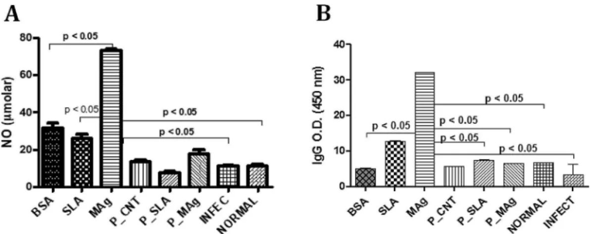
showed 4.3% and 19.2% of cytotoxicity, respectively (Table 1). Experimental groups composed of animals that were immunized with non-encapsulated protein antigens showed higher NO serum production compared to encapsulated ones after the challenge withL. amazonensis. MAg caused the highest NO production (about 80μM). Of the micro/nanoencapsulated formulations, P_MAg showed the highest level (about 20μM) (Fig. 1A).
The sera of animals immunized with the non-encapsulated antigen formulations (SLA and MAg) showed higher intensity of total IgGs compared to all other immunized groups (Fig. 1B). Interestingly, although lower levels of IgGs were detected in the sera of the mice that were immunized with the micro/nanoparticulated preparations, the IgG2a/IGg1 ratio was 2 × higher for P_SLA (1) compared to SLA (0.5) and 10 × higher for P_MAg (2) compared to MAg (0.2) immunized group.
4. Discussion
The parasites were grouped by passages in order to make extracts with homogeneous protein composition representative of infective promastigotes, ensuring that the material used in immunizations with and without adjuvants and/or micro/nanostructured antigen dispersion systems are defined and reproducible as to the ability for immune response modulation, since the successive transfer in culture can cause a change in the virulence of parasites of the genusLeishmania[11].
The polymeric particles' production method employed allowed for the production of micro/nanoparticles with desired diameter and Zeta potential characteristics for a vaccinal approach. Microscopic particles can establish a long-term antigen release profile, enhancing the exposure of the antigens to antigen-presenting cells in the animals and consequently the induction of immune memory after challenge with the parasite[12].
The procedure for particle production was reassessed and
standar-dized to obtain batches of particles with diameters from 512 nm to 637 nm. Particles smaller than 1 mm may have a larger surface area, influencing the release rate of the antigens. Microparticles induce polarization of the immune response to the TH2 pathway and nano-particles to the TH1 pathway[13].
In this study, we also speculated about three components of signaling pathways, especially proinflammatory ones, which were mostly activated during the macrophages' incubation (24 h) with the micro/nanoparticulate preparations. The encapsulated membrane anti-gen formulation (P_MAg) was able to increase 1.46, 1.16 and 1.03 (in arbitrary units) for NF-κB p65, phospho p38 and STAT3 signaling pathways, respectively. These results are also consistent with the cytokine profile produced by the stimuli with P_MAg, showing high levels of TNF-α(500 pg/mL) and IL-6 (130 pg/mL) in the cell super-natants.
According to these results, it is known that activation of STAT3 signaling pathway would indicate a reaction of inflammatory processes, such as lymphocyte proliferation and maturation and inflammation regulation, being activated by IL-6[14], corroborating the suggestion that micro/nanostructured preparations, especially that containing MAg, was able to stimulate macrophages at the molecular level for the production of an inflammatory response.
The protection or progression of the disease in murine ATL models is related to the TH1 and TH2 immune response profiles, respectively, which can be characterized by the cytokine profile in each pathway, markedly IL-12, IFN-γand TNF-αin the TH1; IL-4 and IL-6 pathways in the TH2 pathway [15], as well as the production of chemokines responsible for the polarization of T helper cells and the coordination mechanisms of nitric oxide production in macrophages [16]. IFN-γ induces the isotype exchange from IgM to IgG2a, whereas IL-4 promotes the exchange of IgM isotypes for IgG1 and IgE[17]. Infection control as a function of a TH1 response occurs through the elimination of parasites in macrophages by microbicidal mechanisms mediated by reactive oxygen species, such as nitric oxide (NO). IFN-γand TNF-αact synergistically in the induction of NO production in macrophages [18,19].
The results obtained from the immunization experiments revealed that animals immunized with the free or encapsulated membrane fraction (MAg) were able to produce high amounts of NO. However, encapsulated parasite fractions proved to be better inducers of a specific TH1 response (IGg2a) as opposed to free ones. More studies will be conducted in order to clarify these opposite behaviors of the parasites' antigens and the role of ach formulation in stimulating the host cellular and/or humoral immune response.
Table 1
Results of particles' size, Zeta potential and % of cytotoxicity.
Formulations Size (nm) Zeta potential (mV) Cytotoxicity (%)
P_CNT 512 ± 264 −16.0 ± 4.21 n.d. P_SLA 637 ± 167.3 −18.4 ± 5.38 4.35 P_MAg 514.9 ± 214.8 −18.4 ± 5.38 19.22
Size (nm), Zeta potential (mV) and cytotoxicity assay of control particles (P_CNT), those containing soluble antigens (P_SLA) and those containing membrane antigens fromL. amazonensis(P_MAg). The values were obtained from two different batches of particles conducted in triplicate.
n.d. = not detected.
Fig. 1.(A) Measuring NO concentration and (B) anti-Leishmaniatotal IgG isotyping in immunized BALB/C mice serum after the challenge withL. amazonensis. BSA: bovine serum albumin; SLA: soluble antigen fromL. amazonensis; MAg: membrane antigens fromL. amazonensis; P_CNT: particle with no antigens; P_SLA: particle loaded with SLA; P_Mag: particle loaded with MAg; INFEC: non-immunized infected mice; NORMAL: non-immunized non-infected mice.p-Values were calculated using One-way ANOVA, followed by Tukey's post-test. Two set of experiments were conducted.
J.G. Ribeiro et al. International Immunopharmacology 47 (2017) 227–230
Acknowledgements
The authors express their gratitude to Johnny Ramos from the Laboratory of Immunophysiology (UFMA) for his technical contribution in CBA experiments. Fundação Oswaldo Cruz (FIOCRUZ), Conselho Nacional de Desenvolvimento Científico e Tecnológico (CNPq, n. 470455/2013-6) and Coordenação de Aperfeiçoamento de Pessoal de Nível Superior (CAPES) were responsible forfinancial support.
References
[1] Brasil. Ministério da Saúde, Manual de Vigilândia em Leishmaniose Tegumentar americana, second ed., Ministério da Saúde, Brasília, DF, 2007.
[2] P. Mitropoulos, P. Konidas, M. Durkin-Konidas, New World cutaneous leishmania-sis: updated review of current and future diagnosis and treatment, J. Am. Acad. Dermatol. 63 (2010) 309–322,http://dx.doi.org/10.1016/j.jaad.2009.06.088.
[3] M.J. Day, Immunoglobulin G subclass distribution in canine leishmaniosis: a review and analysis of pitfalls in interpretation, Vet. Parasitol. 147 (2007) 2–8,http://dx.
doi.org/10.1016/j.vetpar.2007.03.037.
[4] N. Dunning, Leishmania vaccines: from leishmanization to the era of DNA technology, Biosci. Horiz. 2 (2009) 73–82,http://dx.doi.org/10.1093/biohorizons/
hzp004.
[5] R. Nagill, S. Kaur, Vaccine candidates for leishmaniasis: a review, Int. Immunopharmacol. 11 (2011) 1464–1488,http://dx.doi.org/10.1016/j.intimp.
2011.05.008.
[6] M.C. Colhone, I. Silva-Jardim, R.G. Stabeli, P. Ciancaglini, Nanobiotechnologic approach to a promising vaccine prototype for immunisation against leishmaniasis: a fast and effective method to incorporate GPI-anchored proteins ofLeishmania amazonensisinto liposomes, J. Microencapsul. 32 (2015) 143–150,http://dx.doi.
org/10.3109/02652048.2014.958203.
[7] T. Niwa, H. Takeuchi, T. Hino, N. Kunou, Y. Kawashima, Preparations of biodegradable nanospheres of water-soluble and insoluble drugs withD,L-lactide/ glycolide copolymer by a novel spontaneous emulsification solvent diffusion method, and the drug release behavior, J. Control. Release 25 (1993) 89–98,http://
dx.doi.org/10.1016/0168-3659(93)90097-O.
[8] R. Nicolete, K.M. Lima, J.M.R. Júnior, M.D. Baruffi, A.I. de Medeiros, M.V.L.B. Bentley, C.L. Silva, L.H. Faccioli, In vitro and in vivo activities of leukotriene B4-loaded biodegradable microspheres, Prostaglandins Other Lipid Mediat. 83 (2007) 121–129,http://dx.doi.org/10.1016/j.prostaglandins.2006.10.
007.
[9] N.B. De Barros, S.R.A. Macedo, A.S. Ferreira, M.P. Tagliari, F.B. Zanchi, A.M. Kayano, A.M. Soares, R. Nicolete, Liposomes containing an ASP49-phospho-lipase A2 fromBothrops jararacussusnake venom as experimental therapy against cutaneous leishmaniasis, Int. Immunopharmacol. 36 (2016) 225–231,http://dx.
doi.org/10.1016/j.intimp.2016.04.025.
[10] R. Thorpe, A. Johnstone, Immunoassays, in: A. Johnstone, R. Thorpe (Eds.), Immunochemistry in Practice, second ed., Blackwell Scientific Publications, London, 1987, pp. 241–260.
[11] B. Espiau, V. Vilhena, A. Cuvillier, A. Barral, G. Merlin, Phenotypic diversity and selection maintainLeishmania amazonensisinfectivity in BALB/C mouse model, Mem Inst Oswaldo Cruz Rio Janeiro 112 (2017) 44–52,http://dx.doi.org/10.1590/
0074-02760160280.
[12] S. Gordon, J.C. Unkeless, Z.A. Cohn, Induction of macrophage plasminogen activator by endotoxin stimulation and phagocytosis, J. Exp. Med. 140 (1974) 995–1010.
[13] I. Gutierro, R.M. Hernández, M. Igartua, A.R. Gascón, J.L. Pedraz, Size dependent immune response after subcutaneous, oral and intranasal administration of BSA loaded nanospheres, Vaccine 21 (2002) 67–77,
http://dx.doi.org/10.1016/S0264-410X(02)00435-8.
[14] T. ZARUBIN, J. HAN, Activation and signaling of the p38 MAP kinase pathway, Cell Res. 15 (2005) 11–18,http://dx.doi.org/10.1038/sj.cr.7290257.
[15] J. Ji, J. Sun, L. Soong, Impaired expression of inflammatory cytokines and chemokines at early stages of infection withLeishmania amazonensis, Infect. Immun. 71 (2003) 4278–4288,http://dx.doi.org/10.1128/iai.71.8.4278-4288.2003.
[16] S. Bhattacharyya, S. Ghosh, B. Dasgupta, D. Mazumder, S. Roy, S. Majumdar, Chemokine induced leishmanicidal activity in murine macrophages via the generation of nitric oxide, J. Infect. Dis. 185 (2002) 1704–1708,http://dx.doi.org/
10.1086/340820.
[17] M.R. Mohammadi, M. Zeinali, S.K. Ardestani, A. Kariminia, Identification of novel
Leishmania majorantigens that elicit IgG2a response in resistant and susceptible mice, Korean J. Parasitol. 44 (2006) 43–48,http://dx.doi.org/10.3347/kjp.2006.
44.1.43.
[18] F.Y. Liew, S. Millott, C. Parkinson, R.M. Palmer, S. Moncada, Macrophage killing of Leishmaniaparasite in vivo is mediated by nitric oxide fromL-arginine, J. Immunol. 144 (1990).
[19] D.M. Santos, M.W. Carneiro, T.R. de Moura, K. Fukutani, J. Clarencio, M. Soto, S. Espuelas, C. Brodskyn, A. Barral, M. Barral-Netto, C.I. de Oliveira, Towards development of novel immunization strategies against leishmaniasis using PLGA nanoparticles loaded with kinetoplastid membrane protein-11, Int. J. Nanomedicine 7 (2012) 2115–2127,http://dx.doi.org/10.2147/IJN.S30093.
J.G. Ribeiro et al. International Immunopharmacology 47 (2017) 227–230