239 239239 239 239 Mem Inst Oswaldo Cruz, Rio de Janeiro, Vol. 99(3): 239-251, M ay 2004
Clinical and Immunopathological Spectrum of American
Cutaneous Leishmaniasis with Special Reference to the D isease in
Amazonian Brazil - A Review
Fernando T Silveira/+, Ralph Lainson, Carlos EP Corbett*
Departamento de Parasitologia, Instituto Evandro Chagas, Secretaria de Vigilância em Saúde do Ministério da Saúde, Av. Almirante Barroso 492, 66090-000 Belém, PA, Brasil *Departamento de Patologia, Faculdade de Medicina, Universidade de São Paulo, São
Paulo, SP, Brasil
The wide variety of Leishmania species responsible for human American cutaneous leishmaniasis combined with the immune mechanisms of the host results in a large spectrum of clinical, histopathological, and immunopathologi-cal manifestations. At the middle of this spectrum are the most frequent cases of loimmunopathologi-calized cutaneous leishmaniasis (LCL) caused by members of the subgenera Leishmania and Viannia, which respond well to conventional therapy. The two pathogenicity extremes of the spectrum generally recognized are represented at the hypersensitivity pole by mucocutaneous leishmaniasis (MCL) and at the hyposensitivity pole by anergic diffuse cutaneous leishmaniasis (ADCL). Following the present study on the clinical, histopathological and immunopathological features of cuta-neous leishmaniasis in Amazonian Brazil, we propose the use of the term “borderline disseminated cutacuta-neous leishmaniasis” for the disseminated form of the disease, due to parasites of the subgenera Leishmania and Viannia, which might be regarded as intermediate between LCL and the extreme pathogenicity poles MCL and ADCL.
Key words: cutaneous leishmaniasis - clinical classification - Amazonian Brazil
American cutaneous leishmaniasis (ACL) is a para-sitic protozoal disease, widely spread in most countries of Latin America, and caused by different species of the genus Leishmania. There are at present fourteen recog-nized species of Leishmania within the subgenera Leish-mania and Viannia, which may produce a variety of cuta-neous and mucocutacuta-neous lesions in man (Lainson & Shaw 1972, 1987, 1998). In Amazonian Brazil, ACL is re-garded as a zoonotic infection of silvatic mammals, among which the parasites are transmitted by the bite of natu-rally infected species of phlebotomine sand flies (Diptera: Psychodidae) (Lainson & Shaw 1992, Lainson et al. 1994). In this region, the disease may be the result of infection due to seven recognized species of Leishmania, six of them within the subgenus Viannia and one within the subgenus Leishmania (Silveira et al. 2002) (Table). Fol-lowing infection of man by these parasites, there will be some naturally resistent individuals (asymptomatic) and others with different degrees of susceptibility to infec-tion (symptomatic). Depending on the species of the in-fecting Leishmania and the infected person’s cell-medi-ated immune response, there develops a spectrum of clini-cal forms of disease, conventionally known as loclini-calized cutaneous leishmaniasis (LCL), mucocutaneous leishma-niasis (MCL), and anergic diffuse cutaneous
leishmania-Financial support: Secretaria de Vigilância em Saúde, Ministry of Health, Brazil (FTS) and the Wellcome Trust, London (RL). +Corresponding author. Fax: +55-91-226.1284. E-mail:
[email protected] Received 18 September 2003 Accepted 29 April 2004
sis (ADCL) (Castes et al. 1983, Carvalho et al. 1985, Silveira et al. 1997a, Lainson & Shaw 1998). Recently, a new clinical feature was added to the spectrum during an at-tempt to characterize some cases presenting with dissemi-nated lesions desigdissemi-nated as borderline dissemidissemi-nated cu-taneous leishmaniasis (BDCL) (Silveira et al. 1997b). There is some evidence indicating that the genetic background of the human host may have a major influence in deter-mining the outcome of the disease (Blackwell 1985, 1999, Petzl-Erler et al. 1991, Lara et al. 1991). In considering this complex situation regarding the etiology of ACL, and the accompanying dificulties in interpreting this spectrum of clinical and immunopathological features, we present a brief review of these together with our own observations on cutaneous leishmaniasis in Amazonian Brazil, where Leishmania (Leishmania) amazonensis and Leishmania (Viannia) braziliensis are of principal medical interest.
LOCALIZED CUTANEOUS LEISHMANIASIS
240 240240
240240 Am erican Cutaneous Leishm aniasis • Fernando T Silveira et al.
and other species of the subgenus Viannia, where there is a more modest infiltration in the skin bordering the ul-cerated lesion, and in which macrophages and parasites are generally scanty: in contrast, lymphocytes and plasma cells are more frequent in the infiltrate, which has the char-acteristics of an epithelioid granuloma (Magalhães et al. 1986) (Fig. 1 a-d). In a smaller number of patients other types of cutaneous manifestations may appear in addi-tion to the ulcerated lesion. They may appear as verru-cose vegetative lesions, papules, nodules, and infiltra-tions in the skin which, together, have led to the consider-ation of LCL as a polymorphic skin disease (Silveira et al. 1997a).
With regards to the immunology of LCL, investiga-tions have been principally on the profile of CD4+ and CD8+ T cell subsets and the cytokines produced by these cells in the lesions of patients, with special interest in interferon-gamma (INF-γ) and interleukin (IL)-4. It is be-lieved that these play a crucial role in determining resistence (CD4+ Th1) or susceptibility (CD4+ Th2), re-spectively, to leishmanial infections (Ribeiro-de-Jesus et al. 1998, Barral-Neto et al. 1998). As a result, and depend-ing on the frequency of memory T cells CD4+ and CD8+ in the cellular infiltrate of the lesion, and the balance of CD4+ Th1/Th2 immune responses, the lesions of some patients may heal spontaneously. In most cases, how-ever, some kind of treatment is required to end the dis-ease. In general, it has been considered that LCL patients present an adequate cell-mediated immunity with a pre-dominance of CD4+ Th1 immune response (Cáceres-Dittmar et al. 1993, Pirmez et al. 1993, Carvalho et al. 1995). There is not yet, however, a consensus of opinion as to which type of memory T cell (either CD4+ or CD8+ sub-sets) is the more prevalent in the infiltrate of lesions (Modlin et al. 1985, Barral et al. 1987, Pirmez et al. 1990, Martinez-Arends et al. 1991, Esterre et al. 1992, Isaza et al. 1996, Vieira et al. 2002). In view of the complex etiology of ACL in Amazonian Brazil, the cell-mediated immunity of the disease needs to be studied not only in terms of the clinical features of the disease but also with regards to the specific leishmanial parasite involved. Thus, although an immunocytochemistry analysis (Silveira et al. unpub-lished data) has shown a higher prevalence of CD8+ than CD4+ T cells in LCL in patients infected with L. (V.) braziliensis or L. (L.) amazonensis (Fig. 2), the CD4+ Th1
immune response was found to be more intense in pa-tients with LCL due to species of the subgenus Viannia [e.g. L. (V.) braziliensis] than in those infected by species of the subgenus Leishmania [e.g. L. (L.) amazonensis]. Using a semi-quantitative reverse transcription-poly-merase chain reaction (RT-PCR), Silveira et al. (unpub-lished data) demonstrated an increased mRNA expression of IFN-γ in biopsies of cutaneous lesions of patients in-fected with L. (V.) braziliensis, whereas no expression was observed for mRNA to IL-4 in the same samples. On the other hand, patients infected with L. (L.) amazonensis showed an increased mRNA expression of IL-4 in their lesions (Fig. 3). The levels of this cytokine were, however, strikingly lower than those of IFN-γ and corresponded to only about a tenth part of the levels of IFN-γ. This sug-gests that even low levels of IL-4 may be able to decrease the CD4+ Th1 immune response in these patients. More-over, it must be emphasized that the cytokines IFN-γ and IL-4 were also demonstrated in L. (V.) braziliensis-stimu-lated peripheral blood mononuclear cells (PBMC) from LCL patients before and after treatment of infections due to L. (V.) braziliensis and L. (L.) amazonensis (data not shown). Interestingly, no significant differences were re-corded in the levels of IFN-γ expression in the L. (V.) braziliensis stimulated PBMC among LCL patients, be-fore or after treatment, in cases infected by L. (V.) brazi-liensis or L. (L.) amazonensis. The levels of IFN-γ were, however, significantly higher (≥ 4 times) than those of IL-4 in the same samples (data not shown), again indicating that there were very low levels of IL-4 in the peripheral blood of LCL patients. These data, together with the clini-cal and cellular immune evaluations explain why patients infected with L. (V.) braziliensis present a higher preva-lence of positive reactions to the delayed hypersensitiv-ity skin-test reaction (DTH) to Leishmania antigen and to the lymphocyte proliferation assay, respectively, than those infected with L. (L.) amazonensis (Silveira et al. 1991, 1998).
With regards to the IgG antibody response of LCL patients, measured by the indirect fluorescent antibody test (IFAT) and the enzyme linked immune assay (ELISA), low to moderate levels (mean titre 160) of specific anti-Leishmania IgG antibodies may be found in cases of in-fection due to L. (V.) braziliensis (Guimarães et al. 1983, Valli et al. 1999, Corrêa et al. 2003). However, in cases of TABLE
The neotropical Leishmania species, etiological agents of American cutaneous leishmaniasis
Subgenus Viannia Lainson & Shaw, 1987 Subgenus Leishmania Ross, 1903
L. (V.) braziliensis a Vianna, 1911 L. (L.) mexicana Biagi, 1953
L. (V.) peruviana Velez, 1913 L. (L.) pifanoi Medina & Romero, 1959
L. (V.) guyanensis a Floch, 1954 L.(L.) amazonensis a
Lainson & Shaw, 1972
L.(V.) panamensis Lainson & Shaw, 1972 L. (L.) garnhami Scorza et al. 1979
L. (V.) lainsoni a Silveira et al. 1987 L.(L.) venezuelensis Bonfante-Garrido, 1980 L. (V.) naiffi a Lainson & Shaw, 1989
L. (V.) shawi a Lainson et al. 1989 L.(V.) colombiensis Kreutzer, 1991
L.(V.) lindenbergi a Silveira et al. 2002
241 241 241 241 241 Mem Inst Oswaldo Cruz, Rio de Janeiro, Vol. 99(3), M ay 2004
LCL patients infected with L. (L.) amazonensis from Ama-zonian Brazil it has been shown that there is a significant increase in the level of these antibodies (mean titre 640) as measured by IFAT (Chagas et al. 1999, 2001). This sup-ports the idea that this parasite has a greater ability to stimulate the CD4+ Th2 imune response than has L. (V.) braziliensis.
From the middle of the spectrum, infections not to-tally controlled by cell-mediated immune mechanisms may evolve to one of two polar forms of the disease; either to the cellular hypersensitivity pole, represented by muco-cutaneous leishmaniasis (MCL), or to the cellular hy-posensitivity pole in cases of anergic diffuse cutaneous leishmaniasis (ADCL). This deviation in the course of in-fection to one or other of these diseases is also influ-enced by the type of antigen that is stimulating the im-mune system of the host: in other words, by the species of parasite concerned. Thus, although L. (V.) braziliensis and L. (L.) amazonensis may be isolated from the mucous membrane tissue of patients suffering from MCL and ADCL, respectively, the immunopathological features of these two kinds of tegumentary leishmaniasis are totally different. MCL patients present a vigorous T cell immune response against L. (V.) braziliensis and the parasite may be isolated from the mucosal tissue as early as one year after a cutaneous infection. On the other hand, ADCL patients have a deficient T cell immune response against L. (L.) amazonensis: only in a few cases, and only after a very prolonged evolution of this disease (10 years, or more) has the parasite been isolated from mucosal tissue. This was the situation we observed in 2 of 12 ADCL pa-tients from the state of Pará, in the Amazon region of Bra-zil. In addition, L. (V.) braziliensis may be found in mu-cosal tissue from MCL patients with no apparent cutane-ous lesion, whereas L. (L.) amazonensis in the mucosae is always associated with active and older cutaneous le-sions on the face of ADCL patients. This suggests that unlike L. (V.) braziliensis, which disseminates via the blood, L. (L.) amazonensis utilizes a contiguous mecha-nism for dissemination from the skin to the mucosal tis-sue.
MUCOCUTANEOUS LEISHMANIASIS
Although some patients may simultaneously exhibit skin and mucosal lesions, it has been observed that in the majority of cases (59%) from Amazonian Brazil the mucosal disease has resulted from an old, prolonged, and untreated (self-healing) cutaneous infection with L. (V.) braziliensis (Silveira et al. 1999). This parasite is recog-nized as the most important etiologic agent of MCL in the New World (Lainson 1983, Grimaldi et al. 1987, 1989, Lainson & Shaw 1998).
In this form of the disease (Fig. 1e), necrosis of the nasopharyngeal mucous tissue is associated with a strong T cell immune response, as evidenced by the exacerbated DTH to Leishmania antigens and the lymphocyte prolif-eration assay (Silveira et al. 1998). In this respect, it should be stressed that DTH reactions elicited by homologous antigen – L. (V.) braziliensis – are significantly higher (≥ 3 times) than those elicited by a heterologous antigen – L. (L.) amazonensis – (Silveira, personal observation). This
indicates, conclusively, the antigen-specific host immune response against L. (V.) braziliensis. The marked cellular hypersensitivity response seen in skin-tested MCL pa-tients may also be demonstrated in histological sections of the mucosal tissue where, in some patients, there may be a tuberculoid granulomatous reaction: this might be regarded as the extreme expression of the cellular hyper-sensitivity pole seen in this form of disease. Sections also show the presence of an abundant infiltrate of lympho-cytes and plasma cells with few histiolympho-cytes and scanty parasites. Necrosis of the cartilaginous structures is the main sequel (Magalhães et al. 1986) (Fig. 1f).
242 242242
242242 Am erican Cutaneous Leishm aniasis • Fernando T Silveira et al.
243 243 243 243 243 Mem Inst Oswaldo Cruz, Rio de Janeiro, Vol. 99(3), M ay 2004
γ in cutaneous lesions, and IFN-γ and TNF-α in mucosal lesions). In this way, it is possible that the cellular hyper-sensitivity immune response recorded in these patients was largely the result of a prolonged antigen-specific CD4+ Th1 activation by L. (V.) braziliensis. This culminated in a high production of IFN-γ in the mucous membrane le-sions and, consequently, the TNF-α. It is TNF-α that is considered to be the major cytokine responsible for dam-age to the mucosal tissue (Castes et al. 1993, Da-Cruz et al. 1996, Ribeiro-de-Jesus et al. 1998, Blackwell 1999). In this context, it is noteworthy that in a retrospective evalu-ation of 85 cases of MCL examined in Amazonian Brazil, it was concluded that the mean time between the beginning of mucosal symptoms and the inicial cutaneous lesion(s) was nearly five years (Silveira et al. 1999).
With regards IgG antibody response in MCL patients, in Amazonian Brazil there have been found, in general, moderate levels of anti-Leishmania IgG antibodies (mean titre 640) as measured by IFAT using L. (L.) amazonensis and L. (V.) shawi as antigens for this assay (Silveira et al. 1999, Corrêa et al. 2003). In the Southern region of Brazil, similar results have been obtained by other workers us-ing IFAT and ELISA assays to investigate the antibody response of MCL patients (Guimarães et al. 1974, Valli et al. 1999). These results suggest the presence of a weak CD4+ Th2 immune response in the peripheral blood of MCL patients, functioning together with a very highly activated CD4+ Th1 immune response, also in the periph-eral blood and particularly in the mucosal lesions of these patients. As a result, the therapy of MCL patients gives
Fig. 2: immunocytochemistry profiles of CD4+ and CD8+ T cells in cutaneous and mucosal lesions of American cutaneous leishma-niasis (ACL) from Amazonian Brazil. ADCL: anergic diffuse cuta-neous leishmaniasis; BDCL: borderline disseminated cutacuta-neous leish-maniasis; LCL: localized cutaneous leishleish-maniasis; MCL: mucosal leishmaniasis; each clinical group contained 5 to 8 patients: ADCL had the lowest number of cases (5); L.a.: Leishmania (L.) amazonensis; L.b.: Leishmania (V.) braziliensis; CD4+ T cell: lym-phocyte T CD4+; CD8+ T cell: lymlym-phocyte T CD8+. For recogni-tion of these two types of T cells, the following monoclonal antibodies were used: Anti-Human T cell CD45RO (clone OPD4) and Anti-Human T cell CD8 Supressor/Cytotoxic (clone DK25) (DAKO-Denmark), respectively. The technical procedures were as used by Corbett et al. (2001).
Fig. 3: semi-quantitative reverse transcription-polymerase chain reaction (RT-PCR) analysis for cytokines in American cutaneous leish-maniasis. a: gamma interferon mRNA and b: interleikin-4 mRNA in biopsies of cutaneous and mucosal lesions from ACL patients. LCL: localized cutaneous leishmaniasis; MCL: mucocutaneous leishmaniasis; ADCL: anergic diffuse cutaneous leishmaniasis. Patients and samples: each clinical group contained 5 to 8 patients (ADCL had the lowest number: 5). Skin biopsy specimens (4-mm punch) were put into cryoembedding medium, flash frozen, and stored at –70oC for RT-PCR. The sequence of technical procedures of RNA isolation,
Reverse Transcription(RT), cDNA synthesis(PCR),and Hybridization of PCR product followed that used by Moraes et al. (1999). For each cytokine, the mean concentration ± SD is shown for the four clinical groups of patients.
follows: In the cases of LCL and MCL due to L. (V.) braziliensis we found a very high antigen-specific CD4+ Th1 immune response activation at the lymph nodes. It is suggested that, in consequence, the CD4+ T cells re-cruited from the peripheral blood to the inflamatory infil-trate of cutaneous and mucosal lesions are preferentially primed to operate as cytokine Th1 – producing cells
244 244244
244244 Am erican Cutaneous Leishm aniasis • Fernando T Silveira et al.
satisfactory results mainly in cases of short evolution, in which there is no large area of ulceration (necrosis) of the mucous tissue (Marsden et al. 1984, Marsden 1986).
ANERGIC DIFFUSE CUTANEOUS LEISHMANIASIS
ADCL is the most common form of the disease seen at the cellular hyposensitivity pole. It is a relatively rare form of New World cutaneous leishmaniasis caused by leish-manial parasites of the subgenus Leishmania. ADCL was first described in Venezuela (Convit & Lapenta 1946) as a bizarre form of ACL having the following characteristics: nodular lesions disseminated all over the body and rich in amastigotes, a negative Montenegro skin-test, and a fre-quent failure to respond to conventional antimony treat-ment. After its discovery, new cases of ADCL were re-corded in Venezuela and, subsequently, in other coun-tries of the Americas [Bolivia, Brazil, Colombia, the Do-minican Republic, Honduras, Mexico, United States of America (southern Texas) and Peru]. L. (L.) pifanoi, L. (L.) mexicana, and L. (L.) amazonensis are the causative agents involved (Lainson & Shaw 1998).
In Brazil, L. (L.) amazonensis is considered to be the only species causing ADCL (Lainson 1983, Silveira et al. 1997a, Lainson & Shaw 1998). The disease is clinically characterized by a diffuse infiltration of the skin, on which appear a large number of nodules, papules, tubercules, and infiltrated plaques that rarely become ulcerated (Fig. 1g). In older cases of the disease, disseminated lesions may cover much of the body, but are predominantly on the extremities and rarely involve the nasopharyngeal mucous membranes (Convit et al. 1972, 1993, Barral et al. 1995). In the dermis the histopathological feature is a se-vere infiltration of macrophages containing abundant amastigotes: lymphocytes and plasma cells are rare, giv-ing the infiltration the aspect of a macrophagic granuloma (Bittencourt & Guimarães 1968, Silveira et al. 1990, Bittencourt & Barral 1991, Moraes & Silveira 1994) (Fig.1h). In terms of immunology, the DTH to Leishmania anti-gen and the lymphocyte proliferation assay are always negative in ADCL cases, indicating that in these patients the cell-mediated immune mechanisms are incapable of specifically controlling the leishmanial infection (Petersen et al. 1982, Barral-Netto et al. 1998, Silveira et al. 1998). In support of this conclusion an immunocytochemistry as-say of five ADCL patients from Amazonian Brazil, showed the lowest level of CD4+ and CD8+ T cells seen in all forms of ACL studied (Fig. 2). This, together with the dem-onstration of the weakest expression of mRNA to IFN-γ and the strongest expression of mRNA to IL-4 (4 times more in some cases) in cutaneous lesions of the same patients (Fig. 3), clearly confirms that the CD+4 Th2 im-mune response is predominant in ADCL disease (Ribeiro-de-Jesus et al. 1998, Barral-Neto et al. 1998). Moreover, unlike LCL and MCL patients infected with L. (V.) braziliensis [in which there was demonstrated a much higher expression of mRNA to IFN-γ than that to IL-4 in the L. (V.) braziliensis- stimulated PBMC], ADCL patients showed a reverse situation of the cytokines; i.e., two times more expression of mRNA to IL-4 than that to IFN-γ (data not shown). These findings reinforce the argument that in ADCL patients due to L. (L.) amazonensis there is likely
to have been a very high antigen-specific CD4+ Th2 im-mune response activation at the lymph nodes, resulting in a proliferation of CD4+ T cells primed to operate as Th2 cytokine-producing cells (mainly IL-4 and IL-10) in the peripheral blood as well as in the cutaneous lesions. As a result, conventional therapy is very frequently accompained by relapses of cutaneous lesions of ADCL patients (Bonfim et al. 1996), due to the very poor CD4+ Th1 immune response in these cases. Although specific anti-Leishmania IgG antibodies are present in great con-centrations (mean titre 20.480 by IFAT) in the serum of these patients(unlike individuals with LCL and MCL due to L. (V.) braziliensis), there is no evidence of their influ-ence in controlling L. (L.) amazonensis infection (Chagas et al. 1999, 2001).
BORDERLINE DISSEMINATED CUTANEOUS LEISHMAN-IASIS
Together with cases of LCL, between the two poles of MCL and ADCL, a few patients may present disseminated forms of infections that have been referred to as BDCL, and in which it has been possible to determine the loca-tion of the primary skin lesion(s) and the secondary ones (Silveira et al. 1997b). In those cases with infection due to L. (V.) braziliensis or other species of the subgenus Viannia, the process of dissemination is relatively rapid. It may take place in two or three months, when a hundred or more erythematous-papules (acneiform lesions) and/ or ulcerated cutaneous lesions may appear (Fig. 4a). The histology of this picture normally shows a nodular infil-tration of lymphocytes and plasma cells in the dermis, with rare macrophages and parasites (Fig. 4b). This is the commonest situation seen in the acute phase of infection but, in cases of delayed evolution (over 1 year), some untreated patients may present with simultaneous cuta-neous and nasopharyngeal mucosal lesions. This indi-cates that active cutaneous infections may persist for pro-longed periods and that, in these cases, mucosal lesions represent the final result of infection. Among three of our patients with this condition acquired in Pará, a 62 years old man presented with disseminated ulcerated and infil-trated cutaneous lesions of about 15 years duration and a nasal mucosal lesion which appeared during the last 2 years of his disease (Fig. 4c). In these cases, cutaneous lesions may be accompained by an epithelioid granuloma in the dermis (Fig. 4d).
245 245 245 245 245 Mem Inst Oswaldo Cruz, Rio de Janeiro, Vol. 99(3), M ay 2004
246 246246
246246 Am erican Cutaneous Leishm aniasis • Fernando T Silveira et al.
Th2 cytokines from PBMC or from cutaneous lesions of these cases have not yet been studied, it is very likely, therefore, that the presence of these two types of T cells in the inflammatory infiltrate of cutaneous lesions, together with antimony therapy, may result in a resolution of the disease equal to that obtained by a restoration of the cell-mediated immune responses (e.g.positive DTH and lym-phocyte proliferation tests) of these patients. It would appear that in these BDCL patients the CD4+ Th1 immune response is at least partially preserved, and functions in such a way as to overcome the opposite CD4+ Th2 im-mune response. Supporting this hypothesis, is the fact that the serum of these patients shows a low to moderate level (mean titre 640) of specific anti-Leishmania IgG an-tibodies by IFAT (Silveira, unpublished observation), confirming the presence of a weak antibody response.
In the case of BDCL caused by L. (L.) amazonensis, differences can readily be indicated in relation to the dis-semination of the parasite and the outcome of disease. Firstly, we have recorded cases in which the dissemina-tion of L. (L.) amazonensis has taken place only after 6 months following the appearance of the primary cutane-ous lesion, and during this period it was limited to a maxi-mum of six detectable metastatic lesions seen in eight male, adult patients examined, almost all of them with 1 to 2 years of disease (Fig. 4e). In the dermis of these cases it is possible to find large collections of vacuolated and heavily parasitezed macrophages, surrounded by groups of lym-phocytes and plasma cells (Fig. 4f). The exceptional case was a 27 years old man who had been mistakenly treated for lepromatous leprosy during the past 7 years. The pa-tient had large area of skin compromised by an erythema-tous infiltration and some nodular lesions at the extremi-ties (Fig. 4g). The histology of his lesions was marked by an extensive infiltration of heavily parasitized macroph-ages with some surrounding groups of lymphocytes and plasma cells in the dermis (Fig. 4h). This case and the 3 cases of BDCL due to infections of L. (V.) braziliensis with a long period of evolution represent patients with the major characteristic of this form of disease, as they were on the verge of converting to ADCL and MCL, re-spectively. This suggests that without the intervention of therapy these leishmanial infections had been in a con-tinuous process of interaction with the host immune re-sponse for a long time. Secondly, lymphatic dissemina-tion of infecdissemina-tion was demonstrated in 7 of our 8 patients, with L. (L.) amazonensis recovered in the culture of mate-rial from enlarged lymph nodes in Difco B45 culture me-dium. This is an interesting finding which seems not to have been recorded in LCL or ADCL forms of L. (L.) amazonensis infection. The explanation of this remains uncertain. It might be regarded as an attempt of the host cell-mediated immune mechanisms to change the reper-toires of the antigen-specific CD4+ T cells’ activation at the lymph node for a beneficial CD4+ Th1 immune re-sponse or, on the contrary, an evasive mechanism of L. (L.) amazonensis in maintaining the pathogenic (CD4+ Th2) profile of the cellular immune response. The lesions of BDCL patients may be principally in the form of infil-trated plaques, localized at the extremities but absent in the nasopharyngeal mucous membrane.
In terms of cell-mediated immunity, it has been noted that the immune mechanisms of patients with BDCL due to L.(L.) amazonensis are more intensely inhibited than in the cases of infection due to L. (V.) braziliensis, resulting in a totally negative response of the DTH to Leishmania antigen and to the lymphocyte proliferation assay in all cases. This severe, but incomplete, inhibition of the cell-mediated immune mechanisms is also evidenced by im-munocytochemistry assay. This shows a smaller amount of CD4+ and CD8+ T cells in the dermal infiltrate of these patients than in those infected with L. (V.) braziliensis, but also at an intermediate level between LCL and ADCL (CD4+: LCL>BDCL>ADCL, CD8+: LCL>BDCL>ADCL) (Fig. 2). As a result, these patients have been cured after a period of nearly six months of conventional antimony therapy. Even without precise information on the cytokine (Th1/Th2) profiles, both for PBMC and for cutaneous le-sions, this suggests to us that, in part, the CD4+ Th1 immune response must have been preserved among the memory T lymphocytes infiltrating the cutaneous lesions of these patients. Favouring this hypothesis is the histo-pathological characteristic of the BDCL lesion. It differs from that of the seemingly incurable ADCL patients by the marked presence of lymphocytes and plasma cells in the dermal infiltrate, even though there may be a signifi-cant number of parasitized macrophages. In contrast to BDCL caused by L. (V.) braziliensis, which normally pro-duces low to moderate levels of anti-Leishmania IgG an-tibodies (mean titre 640), there have been demonstrated higher levels of these antibodies (mean titre 5.120) shown by IFAT in patients with BDCL due to L. (L.) amazonensis (Chagas et al. 1999, 2001). This indicates a lesser CD4+ Th1 immune response activation in these patients.
DISCUSSION
The spectrum of clinical and immunopathological mani-festations of ACL has been the subject of many investi-gations in attempts to fully understand the host immune mechanisms that are playing a crucial role in the patho-genesis of the disease. However, most of these investiga-tions have neglected two very important features: (1) the accurate identity of the specific leishmanial parasite that is stimulating the host immune response and, (2) the qual-ity and the magnitude of the host immune response stimu-lated by this specific leishmanial parasite. The spectrum of ACL proposed here is, therefore, based on character-ization of the leishmanial parasites responsible for ACL and the subsequent cellular and humoral immune re-sponses elicited by these parasites, with special refer-ence to the disease in the Amazon region of Brazil.
247 247 247 247 247 Mem Inst Oswaldo Cruz, Rio de Janeiro, Vol. 99(3), M ay 2004
predominance of CD4+ T cells but, in contrast, Modlin et al. (1985), Martinez-Arends et al. (1991), Isaza et al. (1996) and Vieira et al. (2002) showed that CD8+ T cells are the most frequent type of lymphocyte. In Amazonian Brazil, however, our results of immunocytochemistry analysis (Fig. 2) have conclusively shown that CD8+ T cells were present at a higher level in all forms of the disease (with the one exception of MCL) including cases of LCL due to both L. (V.) braziliensis and L. (L.) amazonensis. These results, then, seem to confirm the role of CD8+ T cells in a well balanced immune response to LCL and, probably, in the process of cure: this has also been confirmed by Coutinho et al. (1998). There is experimental evidence in-dicating the participation of CD8+ T cells in the process of cure of murine leishmaniasis, as well as the role of these cells in the production of IFN-γ (Chan 1993, Conceição-Silva et. al. 1994). In addition, a semi-quantitative RT-PCR (Fig. 3) has shown that the CD4+ Th1 immune response was more intensely associated with patients with LCL due to parasites of the subgenus Viannia (especially L. (V.) braziliensis) than with patients infected by L. (L.) amazonensis. These results, together with previous clini-cal and cell-mediated immune evaluations (Silveira et al. 1991, 1998), have confirmed that L. (V.) braziliensis has a greater ability to stimulate a CD4+ Th1 immune response than has L. (L.) amazonensis, and that the latter parasite is a very high stimulator of a CD4+ Th2 immune response. Supporting this is recent experimental evidence showing that amastigotes of L. (L.) amazonensis are able to condi-tion dendritic cells of BALB/c mice to promote a CD4+ Th2 immune response activation (Qi et al. 2001). When considering all these findings, dichotomy of the clinical and immunopathological spectrum of ACL proposed here is more readily understood: i.e., from the middle of this spectrum, and depending on the species of Leishmania involved, there are some patients in which the infection escapes from the cell-mediated immunity mechanisms and evolves to one or other of the two poles of disease. In cases of infection due to L. (V.) braziliensis and, more rarely, other species of the subgenus Viannia, this infec-tion generally leads to the cellular hypersensitivity pole represented by MCL (CD4+>CD8+ and Th1>Th2). On the opposite side of the spectrum, in cases of infection due to L. (L.) amazonensis and some other species of the subge-nus Leishmania, infection generally leads to the cellular hyposensitivity pole represented by ADCL (CD8+>CD4+ and Th1<Th2).
In the case of MCL, there are at least two aspects of major interest to discuss here regarding the immunology of this form of ACL. Firstly, it has been shown that MCL is characterized by a highly antigen-specific T cell immune response against L. (V.) braziliensis and that this strong reaction corroborates with deviation of the immune re-sponse of patients to the cellular hypersensitivity pole of the spectrum. This immunological status may be evi-denced at a clinical level by an exacerbated DTH, particu-larly to a homologous antigen of Leishmania [e.g. L. (V.) braziliensis] and, at in vitro evaluation, by the lympho-cyte proliferation assay (Cástes et al. 1983, Carvalho et al. 1985, Convit et al. 1993, Silveira et al. 1998). Secondly, an immunocytochemistry assay among our ACL patients from
Amazonian Brazil has indicated that MCL was the only clinical form of disease in which CD4+ T cells were more predominant than the counterpart CD8+ T cells. We feel it must be more than coincidental that this occurs only in MCL, which is closely associated with the cellular hyper-sensitivity pole within the ACL spectrum. Considering the pronounced cellular hypersensitivity immune re-sponse in MCL, one would expect to find high levels of CD4+ Th1 cytokines (IFN-γ, IL-2, and TNF-α, mainly) in mucosal lesions of these patients. On the contrary, how-ever, there is evidence that a “mixture” of CD+4 Th1 and Th2 immune responses occur in MCL patients from Ven-ezuela (Cáceres-Dittmar et al. 1993, Castes et al. 1993) and Southeast of Brazil (Pirmez et al. 1993). This differs from our results in Amazonian Brazil where a typical CD4+ Th1 immune response was found in MCL patients; i.e., a high expression of mRNA to IFN-γ and a complete lack of mRNA to IL-4 in mucous membrane lesions of these patients. In fact, these results were not very different from those of Cáceres-Dittmar et al. (1993), who found a high expres-sion of mRNA to IFN-γ and a low expression of mRNA to IL-4 in their cases of MCL. They differed very much, how-ever, from those of Pirmez et al. (1993), whose demon-strated the highest level of mRNA expression of IL-4 in cases of MCL, approximately 3 times more than was indi-cated by Cáceres-Dittmar et al. (1993). Moreover, as Pirmez et al. (1993) also found an increased mRNA expression of IL-10 in their samples of MCL, they concluded that there is a mixture of Th1 and Th2 immmune responses in MCL. In addition, it may be emphasized that in our study in the Amazon region of Brazil we also demonstrated both cytokines, IFN-γ and IL-4, in the L. (V.) braziliensis stimu-lated PBMC from MCL patients. Although this, too, we regard as “a mixture” of CD4+ Th1 and Th2 immune re-sponses, the level of mRNA expression of IFN-γ was very much higher (≥ 6 times) than that of IL-4, and this does characterize a functional CD4+ Th1 immune response. Supporting this suggestion is the finding of Barral et al. (1998) of a very much higher level of IFN-γ, which was over 10 times that of IL-10, in supernatants from cultures of Leishmania-antigen stimulated cells of MCL patients. Our conclusion, remains, therefore, that in MCL patients there is probably a very highly antigen-specific CD4+ Th1 immune activation at lymph nodes by antigen-presenting cells (e.g. cells of Langerhans) primed by L. (V.) brazi-liensis, and a minimal CD4+ Th2 immune activation: this results in high levels of gene expression of IFN-γ and minimal levels of IL-4 in PBMC. In consequence, T cells recruited from the peripheral blood to the inflamatory in-filtrate of mucosal lesions (the focus of leishmanial infec-tion) are preferentially primed to operate as Th1 cytokines (IFN-γ, IL-2 and TNF-α, principally). In this respect, Costa et al. (2003) have recently demonstrated the role of adhe-sion molecules (CD11a, CD11b and CD62L) in determin-ing the preferential address of T cells to inflamatory sites of leishmaniasis lesions, especially their effect on the high-est expression of CD11a in CD4+ T cells. This raises specu-lations regarding the possible role of adhesion molecules in promoting a CD4+ Th1 immune activation by L. (V.) braziliensis.
conflict-248 248248
248248 Am erican Cutaneous Leishm aniasis • Fernando T Silveira et al.
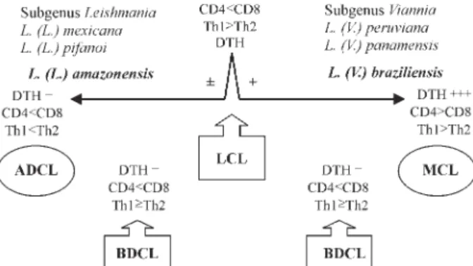
ing situation regarding its immunology. It has been shown that there is “mixture” of Th1 and Th2 cytokines in PBMC and cutaneous lesions of ADCL patients from the Ama-zon region, resulting in very low levels of mRNA expres-sion of IFN-γ and very high levels of mRNA expression of IL-4, respectively. There is general agreement, however, that there is a high predominance of CD4+ Th2 immune response in these patients, which has resulted in ADCL being regarded as a polar form of ACL. The view that has been expressed in most situations is that MCL is an inter-mediate form between LCL and ADCL and, consequently, the disease has been linked to a mixture of Th1 and Th2 immune responses (Convit et al. 2004). We feel it more correct, however, to regard MCL and ADCL as polar forms of ACL, in the hypersensitivity pole (high Th1 im-mune activation) and the hyposensitivity pole (high Th2 immune activation), respectively, with LCL (moderate Th1 immune activation) occupying the middle of this spec-trum. Accepting the existence of an immunological mix-ture of Th1 plus Th2 immune responses in the spectrum of ACL this would represent BDCL. In this way, then, between LCL and the extreme pathogenicity poles MCL and ADCL, there are a few patients presenting dissemi-nated forms of infections due to parasites of the subgen-era Viannia and Leishmania, respectively, which we re-gard as the intermediate form, BDCL. This form of the disease is characterized by an incomplete inhibition of the cellular immune response but with clinical, histopatho-logical and immunohistopatho-logical evidence suggesting that, at least partially, the CD4+ Th1 immune response of these patients is preserved (CD8+>CD4+ and probably Th1≥ Th2) (Fig. 5).
A number of previous publications have discussed some clinical features which may be related to BDCL. Thus, Bryceson (1969) was the first to use a clinical and histo-pathological classification, similar to that applied in lep-rosy, in order to study 33 cases of ADCL disease caused by L. (L.) aethiopica in Ethiopia. He considered five of these as being of the “intermediate histopathological pat-tern II” (i.e. a “borderline” form). Two of them gave a
positive DTH to Leishmania antigen, and two others had early evolution of the disease confined to the face. These 5 cases might be considered analogous to the cases of BDCL caused by L. (L.) amazonensis in Brazil.
Moriearty et al. (1978) first used the term “borderline” when they described a case of leishmaniasis from a re-gion with a high incidence of the mucocutaneous disease caused by L. (V.) braziliensis in the state of Bahia, Brazil. The patient had multiple nodular, papular and ulceroveg-etative skin lesions as well as a lesion in the nasal septum mucosae and, on the basis of Bryceson’s work, these au-thors regarded the case as borderline, between MCL and ADCL, because the patient had a negative DTH to leish-manial antigen (as in ADCL). The test was also negative using other antigens, including 2,4 dinitrochlorobenzene (DNCB), indicating, most likely, the existence of a non-specific immunodeficiency which might have been inter-fering with the patient’s immune response.
Convit et al. (1989) used the term “intermediate form” to designate a chronic (1 to 30 years of evolution) and cell-mediated hypersensitivity form of ACL recorded in 11 patients from Venezuela, most of them presenting with multiple verrucose and ulcerovegetative skin lesions caused by L. (V.) braziliensis (10 cases) and L. (V.) pana-mensis (1 case). After a treatment schedule using immu-notherapy alone (heat-killed L. (L.) amazonensis promastigotes plus BCG) for 6 cases, and immunotherapy plus chemotherapy (conventional antimonial Glucantime) for the other 5, all patients were clinically cured. Later, Convit et al. (1993) presented a review of the clinical and immunological spectrum of ACL in which they consid-ered LCL and DCL as immunologically reactive and nonreactive polar forms of the disease, respectively, and mucosal leishmaniasis (ML) as a hipereactive and inter-mediate form between the LCL and DCL forms in this spectrum.
Costa et al. (1986) described some clinical and immu-nological aspects of 8 cases of disseminated cutaneous leishmaniasis from the state of Bahia, in Northeast Brazil. Isolates of the parasites were made from 5 patients, with 4 identified as L. (V.) braziliensis and only 1 as L. (L.) amazonensis. These authors noted that all the patients, with one exception, had a negative DTH to Leishmania antigen before the treatment and converted to positive after cure of disease. All of them had presented moderate levels of circulating anti-Leishmania IgG antibodies and 5 responded well to pentavalent antimonial therapy, which suggested a functioning cellular (Th1) immune response. Finally, Carvalho et al. (1994) also presented some clini-cal and immunopathologiclini-cal aspects seen in 8 cases of disseminated cutaneous leishmaniasis from the north-east state of Bahia. Contrary to the patients of Costa et al. (1986), however, 5 of these individuals were infected with L. (L.) amazonensis. There were some interesting clinical aspects which differed from those seen in BDCL due to the same parasite in Amazonian Brazil. These included a very high number of cutaneous lesions (as many as 75 to 800), which appeared in only 1 to 6 months; papules and acneiform lesions, which were the most frequent cutane-ous manifestations; dissemination of infection, in 4 cases, occurring in a period as short as only 2 days; 3 patients
Fig. 5: American cutaneous leishmaniasis: clinical and immuno-pathological classification according to the species of Leishmania.
249 249 249 249 249 Mem Inst Oswaldo Cruz, Rio de Janeiro, Vol. 99(3), M ay 2004
presented mucosal lesions and 5 of the 8 cases had no adenopathy. Some features, however, did resemble BDCL due to L. (L.) amazonensis. Thus, in 4 cases the DTH to Leishmania antigen and the lymphocyte proliferation as-say were negative, and there was also a decreased level in CD4+ T cell markers. These immunological deficiencies were restored, after antimony therapy, in a manner similar to that seen in our BDCL patients from Amazonian Brazil. In summary, based on the clinical and immunopatho-logical aspects of ACL, BDCL would seem a immunopatho-logical term to clinically classify those forms of disseminated infec-tions, with parasites of the subgenera Viannia [e.g. L. (V.) braziliensis] and Leishmania [e.g. L. (L.) amazonensis] which appear to be intermediate between LCL and the extreme pathogenicity poles of MCL and ADCL respec-tively. Such infections have the potential to evolve, how-ever, to either one of these poles, depending on the spe-cies of Leishmania causing the disease.
ACKNOWLEDGMENTS
To Dr JM Blackwell for facilities given to the senior author in his iniciation of the RT-PCR analyses in her laboratory (Cam-bridge, UK), and Dr C Evans and Dr M-A Shaw for their tech-nical assistence.
REFERENCES
Barral A, Costa JML, Bittencourt AL, Barral-Neto M, Carvalho EM 1995. Polar and subpolar diffuse cutaneous leishma-niasis in Brazil: clinical and immunopathologic aspects. Int J Dermatol 34: 474-479.
Barral-Netto M, Brodskyn C, Carvalho EM, Barral A 1998. [email protected]. Braz J Med Biol Res 31: 149-155.
Barral A, Jesus AR, Almeida RP, Carvalho EM, Barral-Netto M, Costa JM, Badaro R, Rocha H, Jonhson JD 1987. Evalu-ation of T-cell subsets in the lesion infiltrates of human cutaneous and mucocutaneous leishmaniasis. Parasite Immunol 9: 487-497.
Bittencourt AL, Barral A 1991. Evaluation of the histopatho-logical classification of American cutaneous and mucocuta-neous leishmaniasis. Mem Inst Oswaldo Cruz86: 51-56. Bittencourt AL, Guimarães N 1968. Imunopatologia da
leishmaniose tegumentar difusa. Med Cutan Ibero Lat Am 2: 395-402.
Blackwell JM 1985. A murine model of genetically controlled host responses to Leishmania. In D Rollinson, RM Ander-son (eds), Ecology and Genetics of Host Parasite Interac-tions, Academic Press, New York, p. 147-157.
Blackwell JM 1999. Tumour necrosis factor alpha and muco-cutaneous leishmaniasis. Parasitol Today15: 73-76. Bonfim G, Nascimento C, Costa JML, Carvalho EM,
Barral-Neto M, Barral A 1996. Variation of cytokine patterns re-lated to therapeutic response in diffuse cutaneous leishma-niasis. Exp Parasitol 84: 188-194.
Bryceson ADM 1969. Diffuse cutaneous leishmaniasis in Ethio-pia. I. The clinical and histological features of the disease.
Trans R Soc Trop MedHyg63: 708-737.
Cáceres-Dittmar G, Tapia FJ, Sanchez MA, Yamamura M, Uyemura K, Modlin RL, Bloom BR, Convit J 1993. Deter-mination of the cytokine profile in American cutaneous leish-maniasis using the polymerase chain reaction. ClinExp Immunol 91: 500-505.
Carvalho EM, Barral A, Costa JML, Bittencourt A, Marsden PD 1994. Clinical and immunopathological aspects of dis-seminated cutaneous leishmaniasis. Acta Trop56: 315-325.
Carvalho EM, Correia-Filho D, Baccelar O, Lessa H, Rocha H 1995. Characterization of the immune response in subjects with self healing cutaneous leishmaniasis. Am JTrop Med Hyg 53: 273-277.
Carvalho EM, Johnson WD, Barreto E, Marsden PD, Costa JML, Reed S, Rocha H 1985. Cell mediated immunity in American cutaneous and mucosal leishmaniasis. J Immunol 135: 4144-4148.
Cástes M, Agnelli A, Verde O, Rondon AJ 1983. Characteriza-tion of the cellular immune response in American cutaneous leishmaniasis. Clin Immunol Imunopathol27: 176-186. Cástes M, Trujilho D, Calgano M, Cabrera M, Convit J 1993.
Response Th1/Th2 in human American cutaneous leishma-niasis: its possible relevance for the design of a vaccine.
Mem InstOswaldo Cruz88: 42-43.
Chagas EJP, Corrêa CZ, Silveira FT 1999. Avaliação da resposta imune humoral através do teste de imunofluorescência indireta na leishmaniose cutânea causada por Leishmania (L.) amazonensis na região Amazônica do Brasil. Rev Soc Bras Med Trop 32 (Supl. I): 26.
Chagas EJP, Ishikawa EA, Silveira FT 2001. Humoral response (IgG) in the borderline disseminated cutaneous leishmania-sis (BDCL) caused by Leishmania (L.)amazonensis in Pará State, Brazil. WOLRDleish 2, Crete, Greece, (P226) p. 118. Chan MMY 1993. T cell response in murine Leishmania mexicana amazonensis infection: production of interferon-γ by CD8+ cells. Europ J Immunol 23: 1181-1184. Conceição-Silva F, Perlaza BL, Louis JA, Romero P 1994.
Leish-mania major infection in mice primes for specific major histocompatibility complex class I-restricted CD8+ citotoxic T cell responses. Europ J Immunol 24: 2813-2817. Convit J, Lapenta P 1946. Sobre un caso de leishmaniose
tegumentaria de forma disseminada. Rev de la Policlinica (Caracas) 18: 153-158.
Convit J, Castelanos PF, Ulrich M, Cástes M, Rondon A, Pinardi ME, Rodriguez N, Bloom BR, Formica S, Valecilos L, Bretana A 1989. Immunotherapy of localized, intermediate and diffuse forms of American cutaneous leishmaniasis.
J Infec Diseases160: 104-115.
Convit J, Pinardi ME, Rondon AJ 1972. Diffuse cutaneous leishmaniasis: A disease due to an immunological defect of the host. Trans R Soc Trop Med Hyg 66: 603-610. Convit J, Ulrich M, Fernandez CT, Tapia FJ, Cáceres-Dittmar
G, Cástes M, Rondon AJ 1993. The clinical and immuno-logical spectrum of American cutaneous leishmaniasis. Trans R Soc Trop Med Hyg 87: 444-448.
Convit J, Ulrich M, Polegre MA, Avila A, Rodriguez N, Mazzedo MI, Blanco B 2004. Theraphy of Venezuelan patients with severe mucocutaneous or early lesions of dif-fuse cutaneous leishmaniasis with a vaccine containig pas-teurized Leishmania promastigotes and bacillus Calmet Guerin: preliminary report. Mem Inst Oswaldo Cruz 99: 57-62.
Corbett CEP, Ribeiro-Jr U, Prianti MG, Habr-Gama A, Okumura O, Gama-rodrigues O 2001. Cell-mediated immune response in megacolon from patients with chronic Chagas disease. J Dis Colon Rectum 44: 993-998.
Corrêa ZJC, Lima LVR, De-Jesus RCS, Everdosa D, Machado R, Martins AP, Brandão J, Barbosa RNP, Ikeda C, Jennings Y, Ishikawa EA, Silveira FT 2003. Comparação da reatividade entre antígeno de Leishmania (L.) amazonensis e Leishma-nia (V.)shawi na resposta humoral (IgG) da leishmaniose tegumentar americana, Estado do Pará, Brasil. Rev Soc Bras Med Trop 36 (Supl. I): 315.
250 250250
250250 Am erican Cutaneous Leishm aniasis • Fernando T Silveira et al.
a field clinic in Bahia, Brazil: a report of eight cases. An J Trop Med Hyg89: 319-321.
Costa RP, Gollob KJ, Machado PR, Bacellar OA, Almeida RP, Barral A, Barral-Netto M, Carvalho EM, Dutra WO 2003. Adhesion molecule expression patterns indicate activation and recruitment of CD4+ T cells from the lymph node to the peripheral blood of early cutaneous leishmaniasis pa-tients. Immunol Lett 90: 155-159.
Coutinho SG, Da-Cruz AM, Bertho AL, Santiago MA, De-Luca P 1998. Immunologic patterns associated with cure in human american cutaneous leishmaniasis. Braz JMed Biol Res 31: 139-142.
Da-Cruz AM, De-Oliveira MP, De-Luca PM, Mendonça SC, Coutinho SG 1996. Tumor necrosis fator-alpha in human American tegumentary leishmaniasis. Mem InstOswaldo Cruz 91: 225-229.
Esterre P, Dedet JP, Frenay C, Chevallier M, Grimaud JA 1992. Cell populations in the lesion of human cutaneous leishma-niasis: a light microscopical, immunocytochemical and ul-trastructural study. Virchows Arch A Pathol AnatHistopathol 421: 239-247.
Grimaldi Jr G, David JR, McMahon-Pratt D 1987. Identifica-tion and distribuIdentifica-tion of New World Leishmania species characterized by serodeme analysis using monoclonal anti-bodies. Ann Trop Med Hyg 36: 270-287.
Grimaldi Jr G, Tesh RB, McMahon-Pratt D 1989. A review of the geographic distribution and epidemiology of leishma-niasis in the New World. Am J Trop MedHyg 4: 687-725. Guimarães MCS, Celeste BJ, Camargo ME, Diniz JMP 1983.
Seroepidemiology of cutaneous leishmaniasis from Ribeira do Iguape Valley. IgM and IgG antibodies detected by means of an immunoenzymatic assay (ELISA). Rev Inst Med Trop São Paulo25: 108-112.
Guimarães MCS, Giovannini VL, Camargo ME 1974. Anti-genic standardization for mucocutaneous leishmaniasis im-munofluorescence test. Rev Inst Med Trop SãoPaulo 16: 145-148.
Isaza DM, Restrepo M, Restrepo R, Caceres-Dittmar G, Tapia FJ 1996. Immunocytochemical and histopathologic charac-terization of lesions from patients with localized cutaneous leishmaniasis caused by Leishmania panamensis. Am JTrop Med Hyg 55: 365-369.
Lainson R 1983. The American leishmaniases: some observa-tions on their ecology and epidemiology. Trans R Soc Trop Med Hyg 77: 569-596.
Lainson R, Shaw JJ 1972. Leishmaniasis of the New World: taxonomic problems. Br Med Bull 28: 44-48.
Lainson R, Shaw JJ 1987. Evolution, classification and geo-graphical distribution. In W Peters, R Killick-Kendrick (eds), The Leishmaniases in Biology and Medicine, Vol. 1, Academic Press, London, p. 1-120.
Lainson R, Shaw JJ 1992. A brief history of the genus Leishma-nia (Protozoa: Kinetoplastida) in the Americas with par-ticular reference to Amazonian Brazil. Ciência e Cultura 44: 94-106.
Lainson R, Shaw JJ 1998. New World Leishmaniasis – The Neotropical Leishmania Species. In FEG Cox, JP Kreier, D Wakelin (eds), Topley & Wilson’s Microbiology and Micro-bialInfections, 9th ed., Vol. 5, Parasitology, Arnold, Lon-don, p. 242-266.
Lainson R, Shaw JJ, Silveira FT, Souza AAA, Braga R, Ishikawa EAI 1994. The dermal leishmaniases of Brazil, with special reference to the eco-epidemiology of the disease in Amazonia. Mem Inst Oswaldo Cruz 89: 435-443. Lara ML, Layrisse Z, Scorsa JV, Garcia E, Stoikow Z, Granados
J, Bias W 1991. Immunogenetics of human American cuta-neous leishmaniasis. Study of HLA haplotypes in 24
fami-lies from Venezuela. Hum Immunol 30: 129-135.
Llanos-Cuentas EA, Marsden PD, Lago EL, Barreto AC, Cuba CC, Johnson WD 1984. Human mucocutaneous leishma-niasis in Três Braços, Bahia, Brazil. An area of L.braziliensis braziliensis transmission. II Cutaneous disease: presenta-tion and evolupresenta-tion. Rev Soc Bras Med Trop 17: 169-391. Magalhães AV, Moraes MAP, Raick NA, Llanos-Cuentas EA,
Costa JML, Cuba CC, Marsden PD 1986. Histopatologia da leishmaniose tegumentar por Leishmania braziliensis braziliensis. Rev Inst Med Trop São Paulo28: 253-262. Marsden PD 1986. Mucosal leishmaniasis (“espundia” Escomel,
1911). TransR Soc Trop Med Hyg80: 859-876.
Marsden PD, Llanos-Cuentas EA, Lago EL, Cuba CC, Barreta AC, Costa JML, Jones TC 1984. Human mucocutaneous leishmaniasis in Três Braços, Bahia-Brazil. An area of Leish-mania braziliensis braziliensis transmission. III. Mucosal disease presentation and initial evolution. Rev Soc Bras Med Trop17: 179-186.
Martinez-Arends A, Tapia FJ, Caceres-Dittmar G, Mosca W, Valecil L, Convit J 1991. Immunocytochemical character-ization of immune cells in lesions of American cutaneous leishmaniasis using novel T cell markers. Acta Trop 49: 271-280.
Modlin RL, Tapia FJ, Bloom BR, Gallinoto ME, Castes M, Rondon A, Rea TH, Convit J 1985. In situ characterization of the cellular immune response in American cutaneous leish-maniasis. Clin Exp Immunol 60: 241-248.
Moraes MAP, Silveira FT 1994. Histopatologia da forma localizada de leishmaniose cutânea por Leishmania (Leish-mania) amazonensis. Rev InstMed Trop São Paulo 36: 459-463.
Moraes MO, Sarno EN, Almeida AS, Saraiva BCC, Nery JAC, Martins RCL, Sampaio EP 1999. Cytokine mRNA expres-sion in leprosy: A possible role for interferon-γ and interleukin-12 in reactions (RR and ENL). Scand J Immunol 50: 541-549.
Moriearty PL 1978. Borderline cutaneous leishmaniasis: clini-cal, immunological and histological differences from muco-cutaneous leishmaniasis. Rev Inst Med Trop São Paulo 20: 15-21.
Petersen EA, Neva FA, Oster CN, Diaz HB 1982. Specific inhibition of lymphocyte proliferation response by adher-ent suppressor cells in diffuse cutaneous leishmaniasis. N Engl J Med 306: 387-391.
Petzl-Erler ML, Belich MP, Queiroz-Telles F 1991. Associa-tion of mucosal leishmaniasis with HLA. Hum Immunol 32: 254-260.
Pirmez C, Cooper C, Paes-Oliveira M, Schubach A, Torigian VK, Modlin RL 1990. Immunologic responsiveness in American cutaneous leishmaniasis lesions. J Immunol 145: 3100-3104.
Pirmez C, Yamamura M, Uyemura K, Paes-Oliveira M, Conceição-Silva F, Modlin RL 1993. Cytokine patterns in the pathogenegis of human leishmaniasis. J Clin Invest91: 1390-1395.
Qi H, Popov V, Soong L 2001. Leishmania amazonensis -den-dritic cell interactions in vitro and the priming of parasite-specific CD4+ T cells in vivo. J Immunol 167: 4534-4542. Ribeiro-de-Jesus A, Almeida RP, Lessa H, Baccelar O, Carvalho EM 1998. Cytokine profile and pathology in human leish-maniasis. Braz J Med Biol Res 31: 143-148.
251 251 251 251 251 Mem Inst Oswaldo Cruz, Rio de Janeiro, Vol. 99(3), M ay 2004
Immunol20: 19-26.
Silveira FT, Duarte ERL, De-Farias ECF, Ikeda CS, Lopes AP, Chagas EJP, Teixeira LM, Ishikawa EA 1999. Leishmaniose mucosa na Amazônia brasileira: Avaliação, retrospectiva, dos aspectos clínicos e epidemiológicos da doença, com ênfase ao estado do Pará. Rev Soc Bras Med Trop 32 (Supl. I): 9.
Silveira FT, Ishikawa EA, De Souza AAA, Lainson R 2002. An outbreak of cutaneous leishmaniasis among soldiers in Belém, Pará State, Brazil caused by Leishmania (Viannia) lindenbergi n. sp., a new leishmanial parasite of man in the Amazon region. Parasite 9: 43-50.
Silveira FT, Lainson R, De Brito AC, Oliveira MRF, Paes MG, De Souza AAA, Da Silva BM 1997a. Leishmaniose tegumentar americana. In RNG Leão, Doenças Infecciosas e Parasitárias: Enfoque Amazônico, CEJUP, Belém, PA, p. 619-630.
Silveira FT, Lainson R, Shaw JJ, De Souza AAA, Ishikawa EA, Braga RR 1991. Cutaneous leishmaniasis due to Leishma-nia (LeishmaLeishma-nia) amazonensis in Amazonian Brazil, and the significance of a Montenegro skin-test in human infec-tions. Trans R Soc Trop Med Hyg 85: 735-738.
Silveira FT, Moraes MAP, Lainson R, Shaw JJ 1990.
Leishmaniose cutânea experimental. III. Estudo do comportamento evolutivo da lesão cutânea produzida por
L. (V.)braziliensis, L. (V.) lainsoni e L. (L.) amazonensis no primata Cebus apella (Primates: Cebidae). Rev Inst Med Trop São Paulo 32: 387-394.
Silveira FT, Moraes MAP, Shaw JJ, Lainson R 1997b. Pathol-ogy and pathogenesis of cutaneous leishmaniasis of man in the Amazon Region of Brazil caused by Leishmania (Leish-mania) amazonensis. Acta Parasitol Turcica 21 (Supl. I): 97-98.
Stefani MMA, Martelli CMT, Gillis TP, Krahenbuhl JL 2003. In situ type 1 cytokine gene expression and mechanisms associated with early leprosy progression. J InfecDiseases 188: 1024-1031.
Valli LCP, Passos VMA, Dietze R, Callahan HL 1999. Humoral immune responses among mucosal and cutaneous leishma-niasis patients caused by Leishmania braziliensis. J Parasitol 85: 1076-1083.